



Obtustatin
Based on 4 publication(s) in Google Scholar
Obtustatin is a non-RGD disintegrin consisting of 41 residues. Obtustatin inhibits the adhesion of α1β1 integrin to type IV Collagen (HY-NP003), blocks α1β1 integrin signaling in endothelial cells, and suppresses FGF2-induced angiogenesis. Obtustatin inhibits tumor progression in mouse models and upregulates VEGF expression in sarcoma-bearing mice. Obtustatin can be used in research related to Lewis lung carcinoma and S-180 sarcoma.
Nur für Forschungszwecke. Wir verkaufen nicht an Patienten.
- Reinheit : 99.94%
- CAS. Nr.: 404882-00-4
- Formel: C184H284N52O57S8
- Molecular Weight:4393.06
-
Speicherung:
Sealed storage, away from moisture and light, under nitrogen.
Powder -80°C, 2 years , -20°C, 1 year* In solvent : -80°C, 6 months; -20°C, 1 month (sealed storage, away from moisture and light, under nitrogen)

To place orders, for customer services and technical support, please contact: MedChemExpress USA
Tel: 609-228-6898 E-mail: [email protected] [email protected]


-
 Biologische Aktivität
-
Chemical Information
-
Lösungsmittel & Löslichkeit
- Protokoll
- Reinheit & Dokumentation
- Verweise
-
 Help & FAQs


Publications Citing Use of MedChemExpress (MCE) Obtustatin
MoreAlle VEGFR Isoform-spezifische Produkte anzeigen
More

Biologische Aktivität
Beschreibung
In Vitro
Obtustatin potently inhibits the binding of soluble α1β1 integrin to type IV Collagen (HY-NP003), with an IC50 of 0.8 nM[1].
Obtustatin (0-18 nM; 30 min) inhibits the adhesion of α1K562 cells to type IV collagen, with an IC50 of 2 nM[1].
MedChemExpress (MCE) has not independently confirmed the accuracy of these methods. They are for reference only.
In Vivo
Obtustatin (5 mg/kg; i.p.; once every other day; for 1 week) significantly inhibits the growth of established Lewis lung carcinoma in syngeneic C57BL/6 mice[1].
Obtustatin (1 mg/kg; intratumoral injection; once daily for 5 consecutive days) inhibits the growth of S-180 sarcoma in mice via an anti-angiogenic mechanism associated with the upregulated expression of VEGF, with an inhibition rate of 33%[2].
MedChemExpress (MCE) has not independently confirmed the accuracy of these methods. They are for reference only.
-
Animal Model:embryos (10-day-old)[1]
-
Dosage:5 µg/disk
-
Administration:topical; single application at 24 hours after initial disk placement
-
Result:Reduced the FGF2-stimulated angiogenesis index (vessel branch points) from 179 to 79, corresponding to 84% inhibition of angiogenesis.
-
Animal Model:C57BL/6 (4 animals per group)[1]
-
Dosage:5 mg/kg
-
Administration:i.p.; every other day; 1 week
-
Result:Reduced tumor volume by approximately 50% after 1 week of treatment compared to control.
-
Animal Model:unspecified[2]
-
Dosage:1 mg/kg
-
Administration:intratumoral injection; daily; 5 days
-
Result:Reduced average S-180 sarcoma weight by 33% (average inhibitory rate 32.5%).
Showed no significant weight loss in treated mice.
Increased VEGF expression in treated tumors.
Left expression levels of caspase 8, procaspase 9, caspase 5, and cleaved caspase 3 unchanged relative to controls.


Chemical Information
-
CAS. Nr. 404882-00-4
-
Appearance Solid
-
Molecular Weight 4393.06
-
Formel C184H284N52O57S8
-
Color White to off-white
-
Sequence
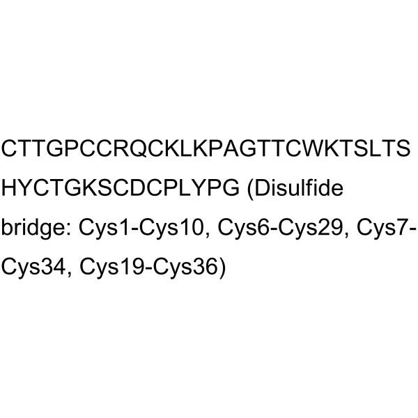
Cys-Thr-Thr-Gly-Pro-Cys-Cys-Arg-Gln-Cys-Lys-Leu-Lys-Pro-Ala-Gly-Thr-Thr-Cys-Trp-Lys-Thr-Ser-Leu-Thr-Ser-His-Tyr-Cys-Thr-Gly-Lys-Ser-Cys-Asp-Cys-Pro-Leu-Tyr-Pro-Gly (Disulfide bridge: Cys1-Cys10, Cys6-Cys29, Cys7-Cys34, Cys19-Cys36)
-
Sequence Shortening
CTTGPCCRQCKLKPAGTTCWKTSLTSHYCTGKSCDCPLYPG (Disulfide bridge: Cys1-Cys10, Cys6-Cys29, Cys7-Cys34, Cys19-Cys36)
-
Versand
Room temperature in continental US; may vary elsewhere.
-
Speicherung
Sealed storage, away from moisture and light, under nitrogen
Powder -80°C 2 years -20°C 1 year * In solvent : -80°C, 6 months; -20°C, 1 month (sealed storage, away from moisture and light, under nitrogen)


Publications (4)
-
Journal Impact Factor
-
Most Recent
-
Adv Sci (Weinh)
Targeting Microglial CD49a Inhibits Neuroinflammation and Demonstrates Therapeutic Potential for Parkinson's Disease. [Abstract]2025 Dec 29:e15138. PMID: 41462565 -
Acta Neuropathol Commun
JMJD6-driven epigenetic activation of COL4A2 reprograms glioblastoma vascularization via integrin α1β1-dependent PI3K/MAPK signaling. [Abstract]2025 Sep 24;13(1):194. PMID: 40993804 -
J Cell Sci
Bile canaliculi formation in primary hepatocytes requires α1β1 integrin-dependent adherens junction re-organization. [Abstract]2025 Dec 1;138(23):jcs264412. PMID: 41347643 -
Exp Eye Res
Therapeutic targeting of ITGA1 delayed retinoblastoma progression through suppression of STAT3 signaling. [Abstract]2025 Nov:260:110636. PMID: 40939856


Lösungsmittel & Löslichkeit
In Vitro:
DMSO : 100 mg/mL (22.76 mM; Need ultrasonic; Hygroscopic DMSO has a significant impact on the solubility of product, please use newly opened DMSO)
Please refer to the solubility information to select the appropriate solvent. Once prepared, please aliquot and store the solution to prevent product inactivation from repeated freeze-thaw cycles.
Storage method and period of stock solution: -80°C, 6 months; -20°C, 1 month (sealed storage, away from moisture and light, under nitrogen). When stored at -80°C, please use it within 6 months. When stored at -20°C, please use it within 1 month.
Please refer to the solubility information to select the appropriate solvent. Once prepared, please aliquot and store the solution to prevent product inactivation from repeated freeze-thaw cycles.
Storage method and period of stock solution: -80°C, 6 months; -20°C, 1 month (sealed storage, away from moisture and light, under nitrogen). When stored at -80°C, please use it within 6 months. When stored at -20°C, please use it within 1 month.
Konzentration (Stammlösung) × Volumen (Stammlösung) = Konzentration (Ziellösung) × Volumen (Ziellösung)
In Vivo:
Select the appropriate dissolution method based on your experimental animal and administration route.
- For the following dissolution methods, please ensure to first prepare a clear stock solution using an In Vitro approach and then sequentially add co-solvents:
- To ensure reliable experimental results, the clarified stock solution can be appropriately stored based on storage conditions. As for the working solution for In Vivo experiments, it is recommended to prepare freshly and use it on the same day.
- The percentages shown for the solvents indicate their volumetric ratio in the final prepared solution. If precipitation or phase separation occurs during preparation, heat and/or sonication can be used to aid dissolution.
Add each solvent one by one: 10% DMSO 40% PEG300 5% Tween-80 45% Saline
Solubility: ≥ 2.5 mg/mL (0.57 mM); Clear solution
This protocol yields a clear solution of ≥ 2.5 mg/mL (saturation unknown).
Taking 1 mL working solution as an example, add 100 μL DMSO stock solution (25.0 mg/mL) to 400 μL PEG300, and mix evenly; then add 50 μL Tween-80 and mix evenly; then add 450 μL Saline to adjust the volume to 1 mL.
Preparation of Saline: Dissolve 0.9 g sodium chloride in ddH₂O and dilute to 100 mL to obtain a clear Saline solution.
Add each solvent one by one: 10% DMSO 90% (20% SBE-β-CD in Saline)
Solubility: ≥ 2.5 mg/mL (0.57 mM); Clear solution
This protocol yields a clear solution of ≥ 2.5 mg/mL (saturation unknown).
Taking 1 mL working solution as an example, add 100 μL DMSO stock solution (25.0 mg/mL) to 900 μL 20% SBE-β-CD in Saline, and mix evenly.
Preparation of 20% SBE-β-CD in Saline (4°C, storage for one week): 2 g SBE-β-CD powder is dissolved in 10 mL Saline, completely dissolve until clear.
In Vivo Dissolution Calculator
Please enter the basic information of animal experiments:
-
-
-
-
Recommended: Prepare an additional quantity of animals to account for potential losses during experiments.
Please enter your animal formula composition:
-
%DMSO +
Recommended: Keep the proportion of DMSO in working solution below 2% if your animal is weak.
-
%+
-
+%Tween-80 + +
-
%Saline +
The co-solvents required include: DMSO, . All of co-solvents are available by MedChemExpress (MCE). , Tween 80. All of co-solvents are available by MedChemExpress (MCE).
Working solution concentration: 0.22 mg/mL
Method for preparing stock solution: mg drug dissolved in μL DMSO. Stock solution concentration: mg/mL. * In solvent : -80°C, 6 months; -20°C, 1 month (sealed storage, away from moisture and light, under nitrogen)
1. Take μL DMSO stock solution;
2. Add μL .
μL , mix evenly;
3. Then add μL Tween 80, mix evenly;
4. Then add μL
Please ensure that the stock solution in the first step is dissolved to a clear state, and add co-solvents in sequence. You can use ultrasonic heating (ultrasonic cleaner, recommended frequency 20-40 kHz), vortexing, etc. to assist dissolution.


Reinheit & Dokumentation
-
Data Sheet (291 KB)
-
SDS (254 KB)
- English - EN (254 KB)
- Français - FR (254 KB)
- Deutsch - DE (254 KB)
- Norwegian - NO (254 KB)
- Español - ES (254 KB)
- Swedish - SV (254 KB)
- Italian - IT (254 KB)
- Korean - KR (254 KB)
- Portuguese - PT (254 KB)
-
Handling Instructions (2659 KB)
Verweise
[1]. Marcinkiewicz C, et al. Obtustatin: a potent selective inhibitor of alpha1beta1 integrin in vitro and angiogenesis in vivo. Cancer Res. 2003 May 1;63(9):2020-3. [Content Brief]
[2]. Ghazaryan N, et al. The antitumor efficacy of monomeric disintegrin obtustatin in S-180 sarcoma mouse model. Invest New Drugs. 2019;37(5):1044-1051. [Content Brief]
[3]. Paz Moreno-Murciano M, et al. NMR solution structure of the non-RGD disintegrin obtustatin. J Mol Biol. 2003;329(1):135-145. [Content Brief]


Complete Stock Solution Preparation Table
Please refer to the solubility information to select the appropriate solvent. Once prepared, please aliquot and store the solution to prevent product inactivation from repeated freeze-thaw cycles.
Storage method and period of stock solution: -80°C, 6 months; -20°C, 1 month (sealed storage, away from moisture and light, under nitrogen). When stored at -80°C, please use it within 6 months. When stored at -20°C, please use it within 1 month.
| Optional Solvent | Concentration Solvent Mass | 1 mg | 5 mg | 10 mg | 25 mg |
|---|---|---|---|---|---|
| DMSO | 1 mM | 0.2276 mL | 1.1382 mL | 2.2763 mL | 5.6908 mL |
| 5 mM | 0.0455 mL | 0.2276 mL | 0.4553 mL | 1.1382 mL | |
| 10 mM | 0.0228 mL | 0.1138 mL | 0.2276 mL | 0.5691 mL | |
| 15 mM | 0.0152 mL | 0.0759 mL | 0.1518 mL | 0.3794 mL | |
| 20 mM | 0.0114 mL | 0.0569 mL | 0.1138 mL | 0.2845 mL |


Obtustatin Related Classifications
HY-P1408 Related Classifications
 Powered by Bioz