



Obtustatin triacetate
Based on 4 publication(s) in Google Scholar
Obtustatin triacetate is a 41-residue non-RGD disintegrin. Obtustatin triacetate can be isolated from the venom of Vipera lebetina obtusa. Obtustatin triacetate is a potent and selective inhibitor of integrin α1β1 adhesion to type IV collagen. Obtustatin triacetate inhibits angiogenesis and may be used in cancer research.
Nur für Forschungszwecke. Wir verkaufen nicht an Patienten.
- Reinheit : 99.97%
- Formel: C184H284N52O57S8.3C2H4O2
- Molecular Weight:4573.21
-
Speicherung:
Sealed storage, away from moisture.
Powder -80°C, 2 years , -20°C, 1 year* In solvent : -80°C, 6 months; -20°C, 1 month (sealed storage, away from moisture)

To place orders, for customer services and technical support, please contact: MedChemExpress USA
Tel: 609-228-6898 E-mail: [email protected] [email protected]


-
 Biologische Aktivität
-
Chemical Information
-
Lösungsmittel & Löslichkeit
- Protokoll
- Reinheit & Dokumentation
- Verweise
-
 Help & FAQs


Publications Citing Use of MedChemExpress (MCE) Obtustatin triacetate
More

Biologische Aktivität
Beschreibung


Chemical Information
-
Appearance Solid
-
Molecular Weight 4573.21
-
Formel C184H284N52O57S8.3C2H4O2
-
Color White to off-white
-
Sequence
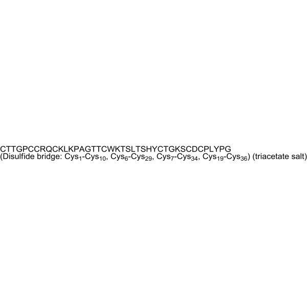
Cys-Thr-Thr-Gly-Pro-Cys-Cys-Arg-Gln-Cys-Lys-Leu-Lys-Pro-Ala-Gly-Thr-Thr-Cys-Trp-Lys-Thr-Ser-Leu-Thr-Ser-His-Tyr-Cys-Thr-Gly-Lys-Ser-Cys-Asp-Cys-Pro-Leu-Tyr-Pro-Gly (Disulfide bridge: Cys1-Cys10, Cys6-Cys29, Cys7-Cys34, Cys19-Cys36)
-
Sequence Shortening
CTTGPCCRQCKLKPAGTTCWKTSLTSHYCTGKSCDCPLYPG (Disulfide bridge: Cys1-Cys10, Cys6-Cys29, Cys7-Cys34, Cys19-Cys36)
-
Versand
Room temperature in continental US; may vary elsewhere.
-
Speicherung
Sealed storage, away from moisture
Powder -80°C 2 years -20°C 1 year * In solvent : -80°C, 6 months; -20°C, 1 month (sealed storage, away from moisture)


Publications (4)
-
Journal Impact Factor
-
Most Recent
-
Adv Sci (Weinh)
Targeting Microglial CD49a Inhibits Neuroinflammation and Demonstrates Therapeutic Potential for Parkinson's Disease. [Abstract]2025 Dec 29:e15138. PMID: 41462565 -
Acta Neuropathol Commun
JMJD6-driven epigenetic activation of COL4A2 reprograms glioblastoma vascularization via integrin α1β1-dependent PI3K/MAPK signaling. [Abstract]2025 Sep 24;13(1):194. PMID: 40993804 -
J Cell Sci
Bile canaliculi formation in primary hepatocytes requires α1β1 integrin-dependent adherens junction re-organization. [Abstract]2025 Dec 1;138(23):jcs264412. PMID: 41347643 -
Exp Eye Res
Therapeutic targeting of ITGA1 delayed retinoblastoma progression through suppression of STAT3 signaling. [Abstract]2025 Nov:260:110636. PMID: 40939856


Lösungsmittel & Löslichkeit
In Vitro:
DMSO : 100 mg/mL (21.87 mM; Need ultrasonic; Hygroscopic DMSO has a significant impact on the solubility of product, please use newly opened DMSO)
Please refer to the solubility information to select the appropriate solvent. Once prepared, please aliquot and store the solution to prevent product inactivation from repeated freeze-thaw cycles.
Storage method and period of stock solution: -80°C, 6 months; -20°C, 1 month (sealed storage, away from moisture). When stored at -80°C, please use it within 6 months. When stored at -20°C, please use it within 1 month.
Please refer to the solubility information to select the appropriate solvent. Once prepared, please aliquot and store the solution to prevent product inactivation from repeated freeze-thaw cycles.
Storage method and period of stock solution: -80°C, 6 months; -20°C, 1 month (sealed storage, away from moisture). When stored at -80°C, please use it within 6 months. When stored at -20°C, please use it within 1 month.
Konzentration (Stammlösung) × Volumen (Stammlösung) = Konzentration (Ziellösung) × Volumen (Ziellösung)


Reinheit & Dokumentation
-
Data Sheet (273 KB)
-
SDS (252 KB)
- English - EN (252 KB)
- Français - FR (252 KB)
- Deutsch - DE (252 KB)
- Norwegian - NO (252 KB)
- Español - ES (252 KB)
- Swedish - SV (252 KB)
- Italian - IT (252 KB)
- Korean - KR (252 KB)
- Portuguese - PT (252 KB)
-
Handling Instructions (2659 KB)
Verweise
[1]. Paz Moreno-Murciano M, et al. NMR solution structure of the non-RGD disintegrin obtustatin. J Mol Biol. 2003 May 23;329(1):135-45. [Content Brief]
[2]. Marcinkiewicz C, et al. Obtustatin: a potent selective inhibitor of alpha1beta1 integrin in vitro and angiogenesis in vivo. Cancer Res. 2003 May 1;63(9):2020-3. [Content Brief]


Complete Stock Solution Preparation Table
Please refer to the solubility information to select the appropriate solvent. Once prepared, please aliquot and store the solution to prevent product inactivation from repeated freeze-thaw cycles.
Storage method and period of stock solution: -80°C, 6 months; -20°C, 1 month (sealed storage, away from moisture). When stored at -80°C, please use it within 6 months. When stored at -20°C, please use it within 1 month.
| Optional Solvent | Concentration Solvent Mass | 1 mg | 5 mg | 10 mg | 25 mg |
|---|---|---|---|---|---|
| DMSO | 1 mM | 0.2187 mL | 1.0933 mL | 2.1866 mL | 5.4666 mL |
| 5 mM | 0.0437 mL | 0.2187 mL | 0.4373 mL | 1.0933 mL | |
| 10 mM | 0.0219 mL | 0.1093 mL | 0.2187 mL | 0.5467 mL | |
| 15 mM | 0.0146 mL | 0.0729 mL | 0.1458 mL | 0.3644 mL | |
| 20 mM | 0.0109 mL | 0.0547 mL | 0.1093 mL | 0.2733 mL |


Obtustatin triacetate Related Classifications
HY-P1408A Related Classifications
 Powered by Bioz