



p53 (17-26)
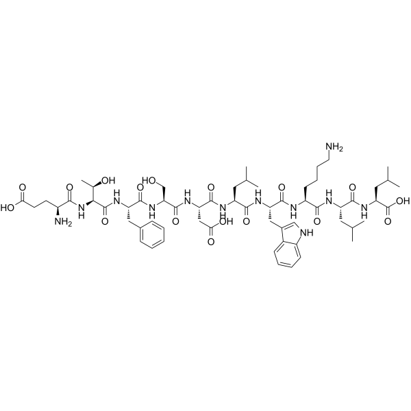
p53 (17-26) is a peptide derived from the P53 MDM2 binding domain, with a Kd of 50 nM for MDM2. p53 (17-26) causes cell lysis by damaging cancer cells and nuclear membranes, and induces cancer cell necrosis. p53 (17-26) exhibits antitumor activity and is applicable to research related to pancreatic cancer.
For research use only. We do not sell to patients.
- CAS No.: 488118-64-5
- Formula: C60H90N12O17
- Molecular Weight:1251.43
-
Storage:
Please store the product under the recommended conditions in the Certificate of Analysis.

To place orders, for customer services and technical support, please contact: MedChemExpress USA
Tel: 609-228-6898 E-mail: [email protected] [email protected]


-
 Biological Activity
-
Chemical Information
-
Solvent & Solubility
- Protocols
- Purity & Documentation
- References
-
 Help & FAQs




Biological Activity
Description
In Vitro
In Vivo
p53 (17-26) (1-20 mg; subcutaneous osmotic pump; 14 days) dose-dependently inhibits the growth of established subcutaneous xenograft pancreatic cancer in nude mice[2].
MedChemExpress (MCE) has not independently confirmed the accuracy of these methods. They are for reference only.
-
Animal Model:Hsd:Athymic Nu/Nu (7-8 weeks old, 22-24 g, i.p. xenotransplantation of BMRPA1.Tuc3 pancreatic carcinoma cells)[2]
-
Dosage:2 mg/mouse
-
Administration:subcutaneous osmotic pump; constant rate 0.25 μL/hr; 14 days
-
Result:Prevented ascites or liver metastasis on day 21.
Contained no viable tumor cells in peritoneal lavage fluid, only macrophages and lymphocytes that did not grow in culture.
-
Animal Model:Hsd:Athymic Nu/Nu (7-8 weeks old, 22-24 g, s.c. xenotransplanted BMRPA1.Tuc3 pancreatic carcinoma cells grown to 40-260 mg before treatment)[2]
-
Dosage:20 mg/mouse; 10 mg/mouse; 1 mg/mouse
-
Administration:subcutaneous osmotic pump; 14 days
-
Result:Reduced average tumor size.
Inhibited post-treatment tumor growth.
Caused no weight loss or toxicity in treated mice.


Chemical Information
-
CAS No. 488118-64-5
-
Molecular Weight 1251.43
-
Formula C60H90N12O17
-
Sequence
Glu-Thr-Phe-Ser-Asp-Leu-Trp-Lys-Leu-Leu
-
Sequence Shortening
ETFSDLWKLL
-
Shipping
Room temperature in continental US; may vary elsewhere.
-
Storage
Please store the product under the recommended conditions in the Certificate of Analysis.


Solvent & Solubility
In Vitro
H2O
Peptide Solubility and Storage Guidelines:
1. Calculate the length of the peptide.
2. Calculate the overall charge of the entire peptide according to the following table:
| Contents | Assign value | |
|---|---|---|
| Acidic amino acid | Asp (D), Glu (E), and the C-terminal -COOH. | -1 |
| Basic amino acid | Arg (R), Lys (K), His (H), and the N-terminal -NH2 | +1 |
| Neutral amino acid | Gly (G), Ala (A), Leu (L), Ile (I), Val (V), Cys (C), Met (M), Thr (T), Ser (S), Phe (F), Tyr (Y), Trp (W), Pro (P), Asn (N), Gln (Q) | 0 |
3. Recommended solution:
| Overall charge of peptide | Details |
|---|---|
| Negative (<0) |
1. Try to dissolve the peptide in water first. 2. If water fails, add NH4OH (<50 μL). 3. If the peptide still does not dissolve, add DMSO (50-100 μL) to solubilize the peptide. |
| Positive (>0) |
1. Try to dissolve the peptide in water first. 2. If water fails, try dissolving the peptide in a 10%-30% acetic acid solution. 3. If the peptide still does not dissolve, try dissolving the peptide in a small amount of DMSO. |
| Zero (=0) |
1. Try to dissolve the peptide in organic solvent (acetonitrile, methanol, etc.) first. 2. For very hydrophobic peptides, try dissolving the peptide in a small amount of DMSO, and then dilute the solution with water to the desired concentration. |


Purity & Documentation
References
[1]. Schon O, et al. Binding of p53-derived ligands to MDM2 induces a variety of long range conformational changes. J Mol Biol. 2004;336(1):197-202. [Content Brief]
[2]. Michl J, et al. PNC-28, a p53-derived peptide that is cytotoxic to cancer cells, blocks pancreatic cancer cell growth in vivo. Int J Cancer. 2006;119(7):1577-1585. [Content Brief]

Calculators
Concentration (start) × Volume (start) = Concentration (final) × Volume (final)


p53 (17-26) Related Classifications
HY-P1755 Related Classifications
 Powered by Bioz