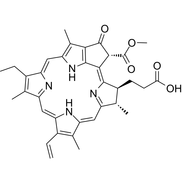
Pheophytin a
Based on 1 Customer Validation
Pheophytin a is a multi-target inhibitor, anticancer agent, antioxidant and antiviral agent. Pheophytin a directly binds to and inhibits HCV-NS3/4A protease (IC50=0.89 μM) to block viral replication. Pheophytin a also scavenges free radicals, reduces ferric ions, and exhibits cytotoxic activity against breast cancer cells. Pheophytin a effectively inhibits LPS-induced production of nitric oxide, prostaglandin E2, NOS2 and COX-2, as well as various pro-inflammatory cytokines, by downregulating the transcription levels of inflammatory mediators and blocking the ERK1/2 and STAT-1 pathways. In a low nerve growth factor environment, Pheophytin a also enhances ERK1/2 phosphorylation and synergistically promotes neurite outgrowth through MAPK pathway. Pheophytin a can be used to investigate the pathogenic mechanisms of diseases including chronic hepatitis C, sepsis, breast cancer and Alzheimer's disease.
For research use only. We do not sell to patients.
- Purity : 80.0%
- CAS No.: 603-17-8
- Formula: C55H74N4O5
- Molecular Weight:871.20
-
Storage:Powder -20°C, 3 years ; In solvent -80°C, 6 months , -20°C, 1 month
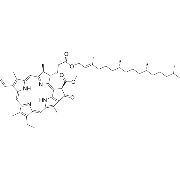
Biological Activity
Description
IC50 & Target
|
ERK1 |
ERK2 |
COX-2 |
STAT1 |
In Vitro
At concentrations of 5-25 μM with a 30-min pre-incubation followed by 24 h of LPS stimulation, Pheophytin a exhibits cytotoxicity toward LPS-stimulated RAW 264.7 murine macrophages at 25 μM, but shows no such effect at 5 μM or 10 μM[1].
Pheophytin a (1-10 μM; pre-incubated for 30 min followed by LPS stimulation for 6 h) reduces LPS-induced NOS2 protein expression in RAW 264.7 mouse macrophages in a dose-dependent manner; when cells are pre-incubated with concentrations of 1, 5, or 10 μM for 30 min, the inhibitory effect at 10 μM is significant, whereas administration after LPS stimulation shows no effect[1].
Pheophytin a (1-10 μM; pre-incubated for 30 min, followed by LPS stimulation for 3 h) dose-dependently inhibits LPS-induced NOS2 mRNA expression in RAW 264.7 mouse macrophages when pre-incubated at concentrations of 1, 5 or 10 μM for 30 min, and exhibits significant inhibitory effects at all tested concentrations[1].
Pheophytin a (10 μM; pre-incubated for 30 min, 10-30 min after LPS stimulation) enhances LPS-induced ERK1/2 phosphorylation in RAW 264.7 mouse macrophages when cells are pre-incubated with 10 μM Pheophytin a for 30 min and proteins are harvested at 10, 15 or 30 min after LPS stimulation; its inhibitory effect on LPS-induced NO production is partially reversed by the ERK1/2 inhibitor U0126 (HY-12031A); Pheophytin a has no effect on LPS-induced phosphorylation of JNK, p38 or Akt[1].
The IC50 values of Pheophytin a microparticles for scavenging superoxide anion radicals and nitric oxide radicals are 200.5 μg/mL and 132.7 μg/mL, respectively; the IC50 value for cytotoxicity against MCF-7 human breast cancer cells is 35.9 μg/mL[2].
Pheophytin a (0.9-31.2 μg/mL; 48 h) dose-dependently potentiates nerve growth factor-induced neurite outgrowth in PC12 cells; the effect at 3.9 μg/mL is comparable to that of high-concentration nerve growth factor, which enhances the phosphorylation level of ERK1/2 without affecting the total ERK level or the phosphorylation levels of p38 and JNK[3].
Pheophytin a (100 nM-10 μM; 60 min) inhibits FmLP-induced chemotaxis of human polymorphonuclear leukocytes in a dose-dependent manner[4].
Pheophytin a (0.1-6 μM; 48-96 h) inhibits the expression of HCV proteins and RNA in HCV subgenomic replicon cells, with an IC50 of 4.97 μM, and exhibits synergistic anti-HCV activity with IFNa-2a[5].
MedChemExpress (MCE) has not independently confirmed the accuracy of these methods. They are for reference only. Further protocols information, click here.
-
Cell Line:LPS-stimulated RAW 264.7 murine macrophages
-
Concentration:10 μM
-
Incubation Time:30 min pre-incubation; 10, 15, 30 min after LPS stimulation (protein harvest)
-
Result:Significantly increased LPS-induced ERK1/2 phosphorylation. Showed no effect on LPS-induced JNK, p38, or Akt phosphorylation. Had its suppressive effect on LPS-induced NO production partially reversed by the ERK1/2 inhibitor U0126.
-
Cell Line:PC12 rat phaeochromocytoma cells
-
Concentration:3.9 μg/ml (in presence of 10 ng/ml NGF)
-
Incubation Time:5 min (in presence of 10 ng/ml NGF)
-
Result:Did not increase total ERK protein levels. Significantly enhanced the phosphorylation of ERK1/2 in PC12 cells treated with 10 ng/ml NGF, with activation levels similar to those induced by 50 ng/ml NGF alone. Did not stimulate phosphorylation of p38 or c-Jun N-terminal kinase (JNK) in PC12 cells.
In Vivo
MedChemExpress (MCE) has not independently confirmed the accuracy of these methods. They are for reference only.
-
Animal Model:BALB/c (8 weeks old)[4]
-
Dosage:80 nmol/20 μl; 800 nmol/20 μl
-
Administration:topical; single application
-
Result:Reduced TPA-induced ear edema to a mean ear disk weight of 12.2 ± 1.4 mg (P < 0.05 vs TPA alone).\nReduced TPA-induced ear edema to a mean ear disk weight of 8.6 ± 1.1 mg (P < 0.005 vs TPA alone).
Chemical Information
-
CAS No. 603-17-8
-
Appearance Solid
-
Molecular Weight 871.20
-
Formula C55H74N4O5
-
Color Brown to black
-
SMILES
COC([C@@H]1/C2=C([C@H]([C@@H]3C)CCC(OC/C=C(C)/CCC[C@H](C)CCC[C@H](C)CCCC(C)C)=O)/N=C3/C=C(C(C)=C/4C=C)\NC4=C/C5=N/C(C(CC)=C5C)=C\C6=C(C)C(C1=O)=C2N6)=O
-
Structure Classification
-
Initial Source
-
Shipping
Room temperature in continental US; may vary elsewhere.
-
Storage
Powder -20°C 3 years In solvent -80°C 6 months -20°C 1 month
Protocols
-
Research Protocol for Infectious Diseases
Infectious-disease experiments test how pathogens interact with host barriers, innate immune receptors, inflammatory signaling, pathogen replication, and tissue injury; pattern-recognition receptors such as TLRs, RIG-I-like receptors, NOD-like receptors, and inflammasomes detect microbial molecules and activate NF-κB, interferon, and cytokine responses. The central hypothesis is that infection severity reflects the balance between pathogen burden and host response: protective inflammation restricts pathogen growth, whereas excessive or mislocalized inflammation contributes to tissue damage and disease phenotype. Unresolved questions include which host pathways are protective versus pathogenic, why some infection models fail to translate to human disease, and which combined readouts best predict clinically relevant infection outcomes.
-
Western Blot
Western blotting (WB) is a commonly used experimental method in molecular biology, biochemistry, and immunogenetics for identifying and quantifying target proteins. It combines gel electrophoresis with immunoassay, enabling researchers to analyze protein expression, post-translational modifications, and molecular weight.
-
Cell differentiation
Cell differentiation refers to the process in which cells of the same origin gradually produce cell groups with different morphological structure and functional characteristics.
-
Research Protocol for Inflammation-related Diseases
The NLRP3 inflammasome is a cytosolic innate immune signaling platform that integrates priming signals and danger-signal activation to promote caspase-1 activation, maturation of IL-1β and IL-18, and gasdermin D-mediated pyroptotic cell death. The core experimental logic is to determine whether inflammatory disease phenotypes are driven by increased NLRP3 expression, ASC-containing inflammasome assembly, caspase-1 cleavage, GSDMD cleavage, and extracellular release of IL-1β/IL-18 rather than by nonspecific cell injury alone. The pathway is strongly linked to inflammation-related disease phenotypes because monosodium urate crystals activate NALP3/NLRP3 inflammasome signaling in gout-like crystal inflammation, cholesterol crystals activate NLRP3 inflammasomes in atherogenesis models, and DSS-induced intestinal inflammation has been reported to involve NLRP3 inflammasome activity. However, experimental colitis studies also show context-dependent protective effects of NLRP3 inflammasome co
-
PC12 NGF-induced neuronal-like differentiation
PC12 cells are a rat adrenal pheochromocytoma-derived clonal cell line that responds to nerve growth factor by stopping proliferation and extending neurites, producing a sympathetic neuron-like phenotype used to study neuronal differentiation and neurite outgrowth. NGF acts through TrkA-dependent signaling, and neurite outgrowth is associated with ERK/Akt signaling, microtubule organization, neuronal-marker expression, and increased electrophysiological neuronal features such as sodium-channel density. The main assay readout is morphological differentiation, usually measured as the percentage of neurite-bearing cells, neurite length, neurite number, or total neurite length per cell. Additional readouts include GAP-43, tyrosine hydroxylase, βIII-tubulin, neurofilament, synapsin I, synaptophysin, ERK phosphorylation, Akt phosphorylation, and sodium-channel current density.
-
Breast Cancer Modeling
Breast cancer is a heterogeneous cancer, and it has been distinguished into four subtypes: luminal A, luminal B, HER2-positive and basal-like. Molecular mutations, epigenetic alterations, hormone exposure and immune microenvironment are related to the progression of breast cancer.
-
Cell Viability Determination by MTT Colorimetric Assay
The following protocol uses the MTT colorimetric assay as a classic literature-established method for assessing cell viability/metabolic activity in cultured mammalian cells. MTT[3-(4,5-dimethylthiazol-2-yl)-2,5-diphenyltetrazolium bromide] is reduced by metabolically active cells to a colored formazan product; the amount of formazan is quantified spectrophotometrically and provides an indirect measure of metabolically active viable cells. Importantly, MTT reduction reflects cellular oxidoreductase/metabolic activity rather than an absolute direct count of living cells, so changes in cellular metabolism can alter the signal independently of cell number.
-
Alzheimer’s Disease Modeling
Alzheimer’s Disease (AD) is a neurodegenerative disorder characterized by a progressive decline in cognitive functions and loss of specific types of neurons and synapses. Alzheimer's symptoms can be simulated in mice by injecting drugs (such as Aβ) or genetically modified.
-
Kinase activity and phosphorylation assays
Kinase activity assays measure the ability of kinases to transfer phosphate groups from ATP to specific substrates, while phosphorylation assays detect the presence and levels of phosphorylated proteins. Common methods include radiolabeled ATP incorporation (e. g. ,), ADP release detection via bioluminescence (e. g. ,[3]), enzyme-linked immunosorbent assays (ELISA) for phospho-specific epitopes (e. g. ,[6]), and microtiter-based formats for high-throughput screening (e. g. ,[8]). The ADP-Glo assay quantifies kinase activity by measuring ADP produced during phosphorylation using a luciferase-based system. Radiometric assays involve autoradiography or scintillation counting after incorporation of 32P-labeled ATP into substrate proteins. ELISA-based approaches rely on phospho-specific antibodies to detect activated kinases in cell lysates or purified samples.
-
PC12 NGF-Induced Neuronal Differentiation Culture
PC12 cells are a rat adrenal pheochromocytoma clonal line that responds to NGF by stopping proliferation and extending branching neurite-like processes; after longer NGF exposure, cells develop long processes and neuronal-like ultrastructural and functional features. NGF-induced differentiation is read out mainly by neurite outgrowth, reduced proliferation, microtubule assembly, and neuronal differentiation-associated proteins such as MAPs, tau, GAP-43, and synapsin-1.
-
Cell Cytotoxicity Assay
Cytotoxicity assays are usually based on the assessment of cell membrane damage, which can also be indirectly detected by measuring cell viability. Detection methods include MTT assay, CKK-8 assay, LDH assay and ATP assay, etc.
-
LPS-Induced Endotoxemia/Systemic Inflammation
Lipopolysaccharide (LPS)-induced endotoxemia is a widely used in vivo model of acute systemic inflammation in which LPS, a Gram-negative bacterial endotoxin, activates innate immune signaling primarily through TLR4, leading to rapid and transient induction of pro-inflammatory cytokines such as TNF-α, IL-6, and IL-1β in circulation and tissues. This cytokine surge is commonly used as a measurable readout of systemic inflammatory activation and immune dysregulation, and is typically assessed within hours after intraperitoneal LPS administration in mouse models of endotoxemia. The model captures key features of systemic inflammatory response syndrome, including cytokine release, immune cell activation, and downstream tissue responses, and has been used to evaluate anti-inflammatory interventions such as cytokine modulation, lipid mediators, and immune cell-targeting therapies.
-
SH-SY5Y Neuronal Differentiation Culture
SH-SY5Y neuronal differentiation culture uses sequential exposure to retinoic acid and neurotrophic factors to reduce proliferative neuroblastoma-like behavior and induce neuron-like morphology, including neurite extension, neuronal marker expression, and, in RA/BDNF protocols, greater synaptic-marker expression than undifferentiated culture. Retinoic acid is commonly used as the initiating differentiation cue, while BDNF in serum-reduced or serum-free medium supports later maturation and neurotrophic-factor-dependent neuron-like survival.
-
SH-SY5Y neuronal-like differentiation
SH-SY5Y neuronal-like differentiation uses defined culture conditions to shift proliferative human neuroblastoma cells toward a neuron-like state, mainly assessed by reduced proliferation, neurite extension, neuronal-marker expression, and, in some protocols, increased dependence on neurotrophic support. Retinoic acid (RA) is commonly used for the first differentiation phase, and sequential RA followed by brain-derived neurotrophic factor (BDNF) in serum-free medium is a well-characterized approach for generating neuron-like SH-SY5Y cultures with extensive neurite outgrowth. The primary readouts are morphology-based neurite outgrowth and marker-based confirmation using proteins such as βIII-tubulin, MAP2, GAP43, synaptophysin, NeuN, NSE, TH, or related neuronal/synaptic markers, depending on the study endpoint.
-
Protocol for Kinase activity and phosphorylation assays
Kinase activity assays measure transfer of phosphate from ATP to a protein or peptide substrate, generating phosphorylated substrate, ADP, or incorporated radiolabeled phosphate as the readout; phosphorylation assays measure site-specific phosphorylation in cells or tissues as a proxy for kinase-pathway activation, inhibition, or substrate regulation. Phosphorylation can be detected by phospho-specific Western blot, immunoprecipitation kinase assay, phospho-immunofluorescence, phospho-flow cytometry, luminescent ADP detection, radiolabeled ATP incorporation, or reporter-based pathway assays, and these readouts can be applied to cancer cells, primary neurons, mouse tumors, organoids, inflammatory macrophages, ferroptosis studies, and mitophagy studies when the kinase target is biologically relevant.
Purity & Documentation
-
Data Sheet (279 KB)
-
SDS (251 KB)
- English - EN (251 KB)
- Français - FR (251 KB)
- Deutsch - DE (251 KB)
- Norwegian - NO (251 KB)
- Español - ES (251 KB)
- Swedish - SV (251 KB)
- Italian - IT (251 KB)
- Korean - KR (251 KB)
- Portuguese - PT (251 KB)
-
Handling Instructions (2659 KB)
References
[1]. Lin CY, et al. Pheophytin a inhibits inflammation via suppression of LPS-induced nitric oxide synthase-2, prostaglandin E2, and interleukin-1β of macrophages. Int J Mol Sci. 2014;15(12):22819-22834. Published 2014 Dec 9. [Content Brief]
[2]. Mohammed HA, et al. Formulation of Ethyl Cellulose Microparticles Incorporated Pheophytin A Isolated from Suaeda vermiculata for Antioxidant and Cytotoxic Activities. Molecules. 2019 Apr 17;24(8):1501. [Content Brief]
[3]. Ina A, et al. Pheophytin a, a low molecular weight compound found in the marine brown alga Sargassum fulvellum, promotes the differentiation of PC12 cells. Int J Dev Neurosci. 2007;25(1):63-68. [Content Brief]
[4]. Okai Y, et al. Potent anti-inflammatory activity of pheophytin a derived from edible green alga, Enteromorpha prolifera (Sujiao-nori). Int J Immunopharmacol. 1997;19(6):355-358. [Content Brief]
[5]. Wang SY, et al. Bioactivity-guided screening identifies pheophytin a as a potent anti-hepatitis C virus compound from Lonicera hypoglauca Miq. Biochem Biophys Res Commun. 2009;385(2):230-235. [Content Brief]
Calculators
Concentration (start) × Volume (start) = Concentration (final) × Volume (final)