



Pleurocidin
Based on 2 publication(s) in Google Scholar
Pleurocidin is an Antimicrobial peptide. Pleurocidin is derived from the skin mucosa or intestinal secretions of Pseudopleuronectes americanus. Pleurocidin inhibits the expression of key proteins in the MAPK and NF-κB inflammatory signaling pathways. Pleurocidin alters serum inflammatory and immune cytokine levels, regulates the down-regulation of tight junction proteins, and modulates the intestinal flora. Pleurocidin exerts antibacterial activity by inducing bacterial membrane damage, hydroxyl radical formation, and NADH depletion, and also produces a synergistic effect with Antibiotics. Pleurocidin alleviates DSS-induced ulcerative colitis. Pleurocidin can be used for research on ulcerative colitis, bacterial infections, and bacterial biofilm-related infections.
For research use only. We do not sell to patients.
- Purity : 98.91%
- CAS No.: 190324-47-1
- Formula: C129H192N36O29
- Molecular Weight:2711.13
-
Storage:
Sealed storage, away from moisture.
Powder -80°C, 2 years , -20°C, 1 year* In solvent : -80°C, 6 months; -20°C, 1 month (sealed storage, away from moisture)

To place orders, for customer services and technical support, please contact: MedChemExpress USA
Tel: 609-228-6898 E-mail: [email protected] [email protected]


-
 Biological Activity
-
Chemical Information
-
Solvent & Solubility
- Protocols
- Purity & Documentation
- References
-
 Help & FAQs


Publications Citing Use of MedChemExpress (MCE) Pleurocidin
MoreAll Antibiotic Isoforms
More

Biological Activity
Description
IC50 & Target
[1]|
p38 MAPK |
In Vitro
Pleurocidin triggers the expression of upstream genes related to IL-1β and COX-2 in rainbow trout macrophages[1].
Pleurocidin exhibits antibacterial activity against Escherichia coli in bacterial lytic plate assays[2].
Pleurocidin (18 h) inhibits the growth of planktonic S. aureus ATCC 25923, E. faecium ATCC 19434, P. acnes ATCC 6919, E. coli ATCC 25922, E. coli O-157 ATCC 43895, and P. aeruginosa ATCC 27853, with an MIC of 1.9-3.8 μg/mL after incubation at 37°C for 18 h[3].
At its minimum inhibitory concentration (MIC), Pleurocidin (MIC; 30-120 min) transiently increases the NAD+/NADH ratio of planktonic S. aureus ATCC 25923 and P. aeruginosa ATCC 27853 to more than 4 times the original level at 30 min post-treatment, and this ratio returns to the baseline level at 60 min[3].
MedChemExpress (MCE) has not independently confirmed the accuracy of these methods. They are for reference only. Further protocols information, click here.
In Vivo
MedChemExpress (MCE) has not independently confirmed the accuracy of these methods. They are for reference only.
-
Animal Model:BALB/C6J (male, 7 weeks old, 16-20 g, DSS-induced colitis)[1]
-
Dosage:5 mg/kg
-
Administration:rectal injection; 4 doses (days 8, 10, 12, 14 of trial)
-
Result:Reduced body weight loss, lower disease activity index (DAI) score, longer colon length, and reduced rectal bleeding compared to DSS-only mice.
Significantly lowered histological scores, reduced crypt loss and intestinal villi destruction, increased goblet cell numbers, reduced epithelial cell apoptosis, and decreased colonic tissue infiltration of F4/80-positive macrophages and CD177-positive neutrophils compared to DSS-only mice.
Reduced levels of pro-inflammatory cytokines IL-1β, IL-6, TNF-α, and IFN-γ, and restored levels of anti-inflammatory cytokine IL-10 compared to DSS-only mice.
Restored colonic expression of tight junction proteins ZO-1, occludin, and claudin-1, and reduced serum levels of intestinal permeability markers LPS, D-LA, and DAO compared to DSS-only mice.
Reduced colonic tissue expression of phosphorylated NF-κB p65, p38 MAPK, JNK, and ERK proteins compared to DSS-only mice.
Increased α-diversity (Ace, Chao, and Sobs indices) compared to DSS-only mice, shifted microbiota composition closer to healthy controls, reduced relative abundance of harmful taxa (Gammaproteobacteria, Enterobacteriales, Escherichia), and increased relative abundance of beneficial taxa including Butyricicoccus and Saccharibacteria compared to DSS-only mice.


Chemical Information
-
CAS No. 190324-47-1
-
Appearance Solid
-
Molecular Weight 2711.13
-
Formula C129H192N36O29
-
Color White to off-white
-
Sequence
Gly-Trp-Gly-Ser-Phe-Phe-Lys-Lys-Ala-Ala-His-Val-Gly-Lys-His-Val-Gly-Lys-Ala-Ala-Leu-Thr-His-Tyr-Leu
-
Sequence Shortening
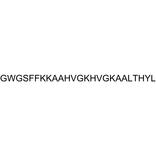
GWGSFFKKAAHVGKHVGKAALTHYL
-
Shipping
Room temperature in continental US; may vary elsewhere.
-
Storage
Sealed storage, away from moisture
Powder -80°C 2 years -20°C 1 year * In solvent : -80°C, 6 months; -20°C, 1 month (sealed storage, away from moisture)


Publications (2)
-
Journal Impact Factor
-
Most Recent
-
J Nanobiotechnology
A core-shell microneedle platform for the spatiotemporal codelivery of dual-agent therapeutics precisely orchestrates diabetic wound healing. [Abstract]2026 Mar 17;24(1):389. PMID: 41845345 -


Solvent & Solubility
In Vitro:
H2O : ≥ 50 mg/mL (18.44 mM)
* "≥" means soluble, but saturation unknown.
Please refer to the solubility information to select the appropriate solvent. Once prepared, please aliquot and store the solution to prevent product inactivation from repeated freeze-thaw cycles.
Storage method and period of stock solution: -80°C, 6 months; -20°C, 1 month (sealed storage, away from moisture). When stored at -80°C, please use it within 6 months. When stored at -20°C, please use it within 1 month.
* Note: If you choose water as the stock solution, please dilute it to the working solution, then filter and sterilize it with a 0.22 μm filter before use.
Please refer to the solubility information to select the appropriate solvent. Once prepared, please aliquot and store the solution to prevent product inactivation from repeated freeze-thaw cycles.
Storage method and period of stock solution: -80°C, 6 months; -20°C, 1 month (sealed storage, away from moisture). When stored at -80°C, please use it within 6 months. When stored at -20°C, please use it within 1 month.
* Note: If you choose water as the stock solution, please dilute it to the working solution, then filter and sterilize it with a 0.22 μm filter before use.
Concentration (start) × Volume (start) = Concentration (final) × Volume (final)

Protocols
-
Research Protocol for Infectious Diseases
Infectious-disease experiments test how pathogens interact with host barriers, innate immune receptors, inflammatory signaling, pathogen replication, and tissue injury; pattern-recognition receptors such as TLRs, RIG-I-like receptors, NOD-like receptors, and inflammasomes detect microbial molecules and activate NF-κB, interferon, and cytokine responses. The central hypothesis is that infection severity reflects the balance between pathogen burden and host response: protective inflammation restricts pathogen growth, whereas excessive or mislocalized inflammation contributes to tissue damage and disease phenotype. Unresolved questions include which host pathways are protective versus pathogenic, why some infection models fail to translate to human disease, and which combined readouts best predict clinically relevant infection outcomes.
-
RNA extraction experimental
By lysing cells, releasing RNA, and removing impurities such as proteins and DNA, high-purity RNA products are finally obtained. The commonly used traditional method is the guanidine isothiocyanate/phenol/chloroform method (Trizol), which is suitable for a variety of animal materials including animal tissues, microorganisms, cultured cells, etc., and most plant materials.
-
DSS-Induced Colitis
Dextran sulfate sodium (DSS)-induced colitis is generated by administering DSS in mouse drinking water, producing epithelial injury, barrier disruption, weight loss, diarrhea, fecal blood, colon shortening, histologic mucosal damage, and inflammatory mediator changes; the model is mainly used to study acute or chronic intestinal inflammation resembling selected features of ulcerative colitis. DSS injury is interpreted through clinical and tissue readouts rather than a single molecular endpoint: daily body weight, stool consistency, and bleeding are combined into a disease activity index, while colon length, histology, cytokines, myeloperoxidase activity, intestinal permeability, and tight-junction markers provide complementary measures of inflammation and barrier damage.
-
TNBS-Induced Colitis
TNBS-induced colitis is produced by intrarectal delivery of 2,4,6-trinitrobenzene sulfonic acid in ethanol, where ethanol disrupts the mucosal barrier and TNBS haptenates colonic proteins, generating immune-mediated colonic inflammation with weight loss, diarrhea, ulceration, transmural injury, inflammatory-cell infiltration, and cytokine responses. The model is used as an experimental intestinal inflammation model with Crohn’s disease–like features, especially when Th1-type responses, IL-12–dependent inflammation, chronic relapsing inflammation, or fibrosis-related endpoints are studied.
-
Research Protocol for Inflammation-related Diseases
The NLRP3 inflammasome is a cytosolic innate immune signaling platform that integrates priming signals and danger-signal activation to promote caspase-1 activation, maturation of IL-1β and IL-18, and gasdermin D-mediated pyroptotic cell death. The core experimental logic is to determine whether inflammatory disease phenotypes are driven by increased NLRP3 expression, ASC-containing inflammasome assembly, caspase-1 cleavage, GSDMD cleavage, and extracellular release of IL-1β/IL-18 rather than by nonspecific cell injury alone. The pathway is strongly linked to inflammation-related disease phenotypes because monosodium urate crystals activate NALP3/NLRP3 inflammasome signaling in gout-like crystal inflammation, cholesterol crystals activate NLRP3 inflammasomes in atherogenesis models, and DSS-induced intestinal inflammation has been reported to involve NLRP3 inflammasome activity. However, experimental colitis studies also show context-dependent protective effects of NLRP3 inflammasome co
-
Bacterial live/dead nucleic-acid viability staining
The LIVE/DEAD bacterial viability staining method is based on differential permeability of nucleic-acid-binding fluorescent dyes, most commonly SYTO 9 and propidium iodide (PI), which enables discrimination of bacterial populations with intact versus compromised cytoplasmic membranes. SYTO 9 penetrates both intact and damaged bacterial membranes and binds nucleic acids to produce green fluorescence, whereas propidium iodide penetrates only cells with compromised membranes and fluoresces red while also reducing SYTO 9 signal through competitive binding and fluorescence interactions. The resulting fluorescence pattern is interpreted as a proxy for membrane integrity, which is widely used as an indicator of bacterial viability in microscopy, flow cytometry, and spectroscopic platforms. However, mechanistic studies show that SYTO 9 and PI interactions involve displacement and fluorescence resonance energy transfer effects, which can influence signal interpretation depending on dye ratios a


Purity & Documentation
-
Data Sheet (276 KB)
-
SDS (252 KB)
- English - EN (252 KB)
- Français - FR (252 KB)
- Deutsch - DE (252 KB)
- Norwegian - NO (252 KB)
- Español - ES (252 KB)
- Swedish - SV (252 KB)
- Italian - IT (252 KB)
- Korean - KR (252 KB)
- Portuguese - PT (252 KB)
-
Handling Instructions (2659 KB)
References
[1]. Nong K, et al. Effect of the Pseudopleuronectes americanus-derived Pleurocidin on DSS-induced Ulcerative colitis in mice and its preliminary molecular mechanisms. Int Immunopharmacol. 2024;130:111757. [Content Brief]
[2]. Cole AM, et al. Isolation and characterization of pleurocidin, an antimicrobial peptide in the skin secretions of winter flounder. J Biol Chem. 1997;272(18):12008-12013. [Content Brief]
[3]. Choi H, et al. Antimicrobial peptide pleurocidin synergizes with antibiotics through hydroxyl radical formation and membrane damage, and exerts antibiofilm activity. Biochim Biophys Acta. 2012;1820(12):1831-1838. [Content Brief]


Complete Stock Solution Preparation Table
Please refer to the solubility information to select the appropriate solvent. Once prepared, please aliquot and store the solution to prevent product inactivation from repeated freeze-thaw cycles.
Storage method and period of stock solution: -80°C, 6 months; -20°C, 1 month (sealed storage, away from moisture). When stored at -80°C, please use it within 6 months. When stored at -20°C, please use it within 1 month.
| Optional Solvent | Concentration Solvent Mass | 1 mg | 5 mg | 10 mg | 25 mg |
|---|---|---|---|---|---|
| H2O | 1 mM | 0.3688 mL | 1.8442 mL | 3.6885 mL | 9.2212 mL |
| 5 mM | 0.0738 mL | 0.3688 mL | 0.7377 mL | 1.8442 mL | |
| 10 mM | 0.0369 mL | 0.1844 mL | 0.3688 mL | 0.9221 mL | |
| 15 mM | 0.0246 mL | 0.1229 mL | 0.2459 mL | 0.6147 mL |
* Note: If you choose water as the stock solution, please dilute it to the working solution, then filter and sterilize it with a 0.22 μm filter before use.


Pleurocidin Related Classifications
HY-P5641 Related Classifications
 Powered by Bioz