



Pteryxin
Based on 1 publication(s) in Google Scholar
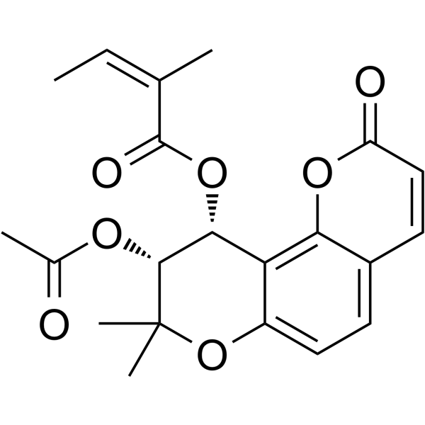
Pteryxin ((+)-Pteryxin) is an orally active multi-target inhibitor that targets NF-κB, MAPK, NLRP3 inflammasome, and Nrf2/ARE pathways. Pteryxin is also a BChE inhibitor (IC50=12.96 μg/mL) with a low inhibitory efficiency on AChE. Pteryxin inhibits the Ca2+-calcineurin-NFATc1 pathway by blocking NF-κB/MAPK signaling, inhibiting NLRP3 inflammasome activation, and reducing ROS generation, and activates Nrf2-mediated antioxidant enzyme expression. Pteryxin has anti-inflammatory, antioxidant, and osteoclastogenesis inhibitory activities. Pteryxin can be used in the study of inflammatory diseases, osteoporosis, diabetes, and Alzheimer's disease.
Nos produits utilisent uniquement pour la recherche. Nous ne vendons pas aux patients.
- Pureté : 99.55%
- CAS No.: 13161-75-6
- Formule: C21H22O7
- Masse moléculaire:386.40
-
Stockage:Powder -20°C, 3 years , 4°C, 2 years ; In solvent -80°C, 2 years , -20°C, 1 year

To place orders, for customer services and technical support, please contact: MedChemExpress USA
Tel: 609-228-6898 E-mail: [email protected] [email protected]


-
 Activité biologique
-
Chemical Information
-
Solvant et solubilité
- Protocole
- Pureté et documentation
- Références
-
 Help & FAQs


-
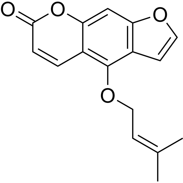
HY-N0286
IsoimperatorinCholinesterase (ChE) Inhibitor -
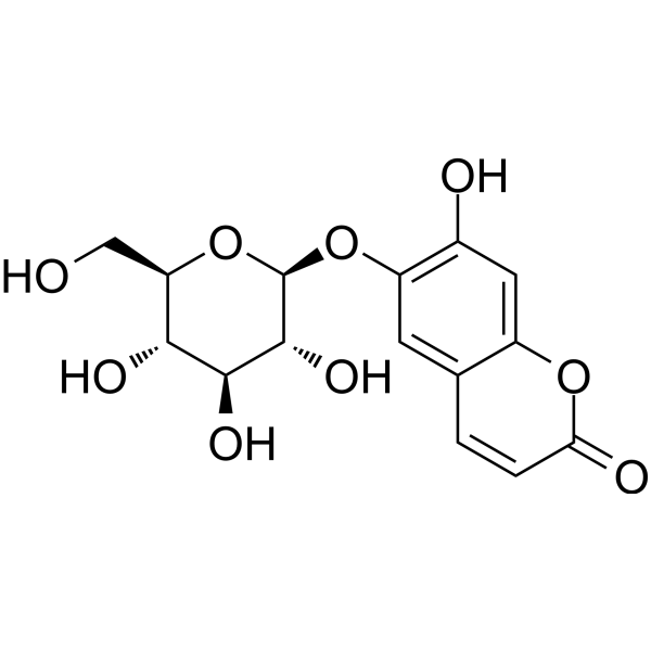
HY-N0188
Esculinp38 MAPK Inhibitor -
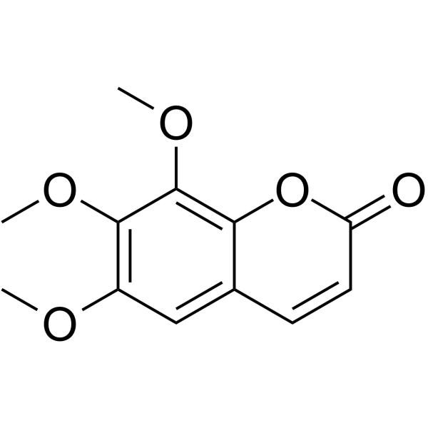
HY-N0085
DimethylfraxetinCarbonic Anhydrase Inhibitor -

HY-N0286R
Isoimperatorin (Standard)Cholinesterase (ChE) Inhibitor -
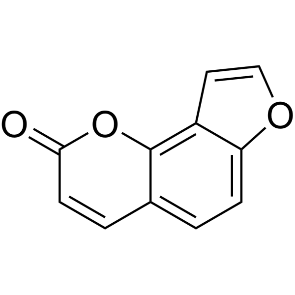
HY-N0763
AngelicinApoptosis Inducer -
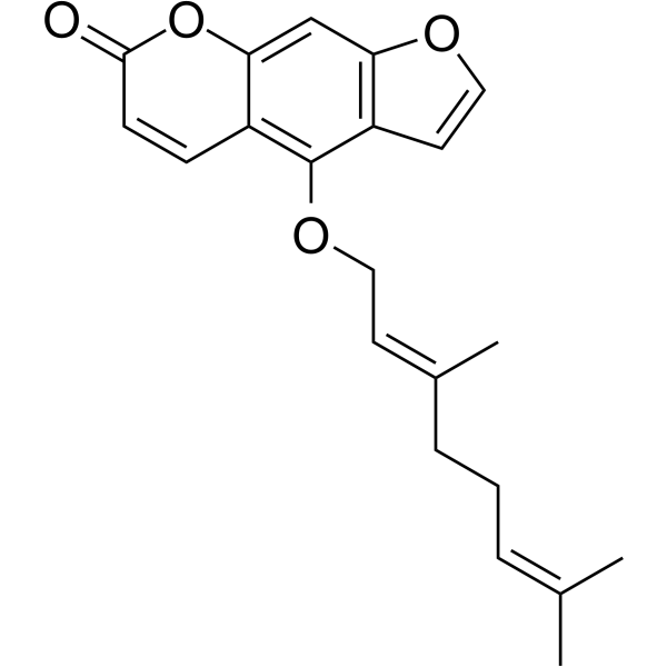
HY-N2194
BergamottinCytochrome P450 Inhibitor -
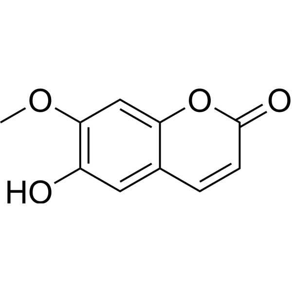
HY-N1365
IsoscopoletinHBV Inhibitor -

HY-N1365R
Isoscopoletin (Standard)HBV Inhibitor -
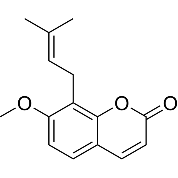
HY-N0054
OstholeApoptosis Inducer -
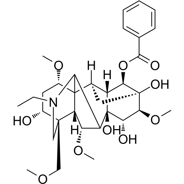
HY-N0217
BenzoylaconineAnti-inflammatory Agent -

HY-N0188R
Esculin (Standard)p38 MAPK Inhibitor -
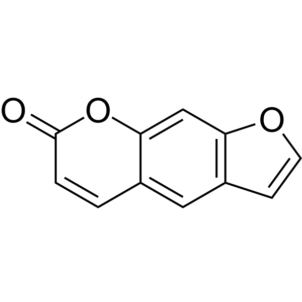
HY-N0053
PsoralenBioactive Natural Compound
Publications Citing Use of MedChemExpress (MCE) Pteryxin
More

Activité biologique
Description
IC50 & Target
[5]|
BChE 12.96 μg/mL (IC50) |
AChE |
NLRP3 |
NFATc1 |
Nrf2-ARE |
In Vitro
MTT cell viability assay:
Pteryxin (5-20 μM; 24 h) has no significant toxicity to RAW264.7 macrophages and BMMs osteoclast precursor cells[1][2].
ELISA inflammatory factor detection:
IL-6 and TNF-α in RAW264.7 cells[1].
WB protein analysis:
Pteryxin (5-20 μM; 24 h) concentration-dependently downregulates the expression of iNOS, COX-2, p-p38/p-ERK/p-JNK, p-p65 and NLRP3/ASC/Caspase-1 p20 proteins in RAW264.7 cells, and inhibits the expression of NFATc1/c-FOS/CTSK proteins in BMMs cells[1][2].
IF immunofluorescence:
Pteryxin (20 μM; 24 h) blocks NF-κB p65 nuclear translocation and NLRP3/ASC speck formation in RAW264.7 cells, and reduces F-actin ring area and NFATc1 nuclear localization in BMMs cells[1][2].
qPCR gene expression:
Pteryxin (5-20 μM; 24 h) upregulates antioxidant genes such as HO-1, GCLC, and Trxr1 in MIN6 islet cells, and downregulates Acp5/Mmp9/Dc-stamp[2][3] in BMMs cells[2][3].
MedChemExpress (MCE) has not independently confirmed the accuracy of these methods. They are for reference only.
-
Cell Line:BMMs (bone marrow-derived macrophages)
-
Concentration:5, 10, 15, 20, 25 μM
-
Incubation Time:48-96 h
-
Result:Resulted viability >85% at ≤20 μM, while reduced viability by 12% at 25 μM for 96 h, confirming safety for osteoclastogenesis assays.
In Vivo
Pteryxin (5/10 mg/kg; intraperitoneal injection; once a day; 3 days) significantly attenuates LPS (5 mg/kg, i.t.)-induced lung inflammation in C57BL/6 mice, reduces lung wet/dry weight ratio, myeloperoxidase (MPO) activity, inflammatory cell infiltration in bronchoalveolar lavage fluid (BALF), and IL-6/TNF-α levels[1].
Osteoporosis (OVX) model:
Pteryxin (10 mg/kg; oral; once a day; 8 weeks) significantly inhibits bone loss in ovariectomized C57BL/6 female mice, increases bone mineral density and trabecular number, reduces osteoclast number, and downregulates serum TRACP-5b and CTX-1 levels[2].
Diabetic nephropathy (DN) model:
Pteryxin (20 mg/kg; gavage; once daily; 12 weeks) improves STZ-induced renal function in db/db mice, reduced urine protein, serum creatinine and urea nitrogen, alleviates glomerular mesangial proliferation and fibrosis, and inhibits renal NF-κB activation and TGF-β1 expression[3].
MedChemExpress (MCE) has not independently confirmed the accuracy of these methods. They are for reference only.
-
Animal Model:C57BL/6 mice (male, 20-22 g, 8 weeks old), LPS (5 mg/kg, i.t.)-induced ALI[1]
-
Dosage:5 or 10 mg/kg
-
Administration:Intraperitoneal injection (i.p.), once daily for 3 days (starting 1 h prior to LPS).
-
Result:Decreased lung wet/dry ratio from 5.8±0.3 (LPS group) to 4.3±0.2 (10 mg/kg); reduced MPO activity from 2.5±0.2 U/mg to 1.4±0.1 U/mg, BALF total cells from 1.2×10⁶±0.1×10⁶ to 0.5×10⁶±0.05×10⁶, IL-6 from 850±60 pg/mL to 320±30 pg/mL, TNF-α from 520±40 pg/mL to 210±20 pg/mL.


Chemical Information
-
CAS No. 13161-75-6
-
Appearance Solid
-
Masse moléculaire 386.40
-
Formule C21H22O7
-
Color White to off-white
-
SMILES
C/C=C(C)\C(O[C@@H]1C2=C3C(C=CC(O3)=O)=CC=C2OC(C)(C)[C@@H]1OC(C)=O)=O
-
Synonyms
(+)-Pteryxin
-
Structure Classification
-
Initial Source
-
Livraison
Room temperature in continental US; may vary elsewhere.
-
Stockage
Powder -20°C 3 years 4°C 2 years In solvent -80°C 2 years -20°C 1 year


Publications (1)
-
Journal Impact Factor
-
Most Recent
-
Biomed Pharmacother
Pteryxin suppresses osteoclastogenesis and prevents bone loss via inhibiting the MAPK/Ca2+ signaling pathways mediated by ROS. [Abstract]2023 Sep:165:114898. PMID: 37352699


Solvant et solubilité
In Vitro:
DMSO : 100 mg/mL (258.80 mM; Need ultrasonic; Hygroscopic DMSO has a significant impact on the solubility of product, please use newly opened DMSO)
Please refer to the solubility information to select the appropriate solvent. Once prepared, please aliquot and store the solution to prevent product inactivation from repeated freeze-thaw cycles.
Storage method and period of stock solution: -80°C, 2 years; -20°C, 1 year. When stored at -80°C, please use it within 2 years. When stored at -20°C, please use it within 1 year.
Please refer to the solubility information to select the appropriate solvent. Once prepared, please aliquot and store the solution to prevent product inactivation from repeated freeze-thaw cycles.
Storage method and period of stock solution: -80°C, 2 years; -20°C, 1 year. When stored at -80°C, please use it within 2 years. When stored at -20°C, please use it within 1 year.
Concentration (start) × Volume (start) = Concentration (final) × Volume (final)
In Vivo:
Select the appropriate dissolution method based on your experimental animal and administration route.
- For the following dissolution methods, please ensure to first prepare a clear stock solution using an In Vitro approach and then sequentially add co-solvents:
- To ensure reliable experimental results, the clarified stock solution can be appropriately stored based on storage conditions. As for the working solution for In Vivo experiments, it is recommended to prepare freshly and use it on the same day.
- The percentages shown for the solvents indicate their volumetric ratio in the final prepared solution. If precipitation or phase separation occurs during preparation, heat and/or sonication can be used to aid dissolution.
Add each solvent one by one: 10% DMSO 40% PEG300 5% Tween-80 45% Saline
Solubility: ≥ 2.5 mg/mL (6.47 mM); Clear solution
This protocol yields a clear solution of ≥ 2.5 mg/mL (saturation unknown).
Taking 1 mL working solution as an example, add 100 μL DMSO stock solution (25.0 mg/mL) to 400 μL PEG300, and mix evenly; then add 50 μL Tween-80 and mix evenly; then add 450 μL Saline to adjust the volume to 1 mL.
Preparation of Saline: Dissolve 0.9 g sodium chloride in ddH₂O and dilute to 100 mL to obtain a clear Saline solution.
Add each solvent one by one: 10% DMSO 90% (20% SBE-β-CD in Saline)
Solubility: 2.5 mg/mL (6.47 mM); Suspended solution; Need ultrasonic
This protocol yields a suspended solution of 2.5 mg/mL. Suspended solution can be used for oral and intraperitoneal injection.
Taking 1 mL working solution as an example, add 100 μL DMSO stock solution (25.0 mg/mL) to 900 μL 20% SBE-β-CD in Saline, and mix evenly.
Preparation of 20% SBE-β-CD in Saline (4°C, storage for one week): 2 g SBE-β-CD powder is dissolved in 10 mL Saline, completely dissolve until clear.
In Vivo Dissolution Calculator
Please enter the basic information of animal experiments:
-
-
-
-
Recommended: Prepare an additional quantity of animals to account for potential losses during experiments.
Please enter your animal formula composition:
-
%DMSO +
Recommended: Keep the proportion of DMSO in working solution below 2% if your animal is weak.
-
%+
-
+%Tween-80 + +
-
%Saline +
The co-solvents required include: DMSO, . All of co-solvents are available by MedChemExpress (MCE). , Tween 80. All of co-solvents are available by MedChemExpress (MCE).
Working solution concentration: 0.22 mg/mL
Method for preparing stock solution: mg drug dissolved in μL DMSO. Stock solution concentration: mg/mL.
1. Take μL DMSO stock solution;
2. Add μL .
μL , mix evenly;
3. Then add μL Tween 80, mix evenly;
4. Then add μL
Please ensure that the stock solution in the first step is dissolved to a clear state, and add co-solvents in sequence. You can use ultrasonic heating (ultrasonic cleaner, recommended frequency 20-40 kHz), vortexing, etc. to assist dissolution.


Pureté et documentation
-
Fiche technique (288 KB)
-
SDS (393 KB)
- English - EN (393 KB)
- Français - FR (393 KB)
- Deutsch - DE (393 KB)
- Norwegian - NO (393 KB)
- Español - ES (393 KB)
- Swedish - SV (393 KB)
- Italian - IT (393 KB)
- Korean - KR (393 KB)
- Portuguese - PT (393 KB)
-
Instruction de manipulation (2659 KB)
Références
[1]. Zhen D, et al. Pteryxin attenuates LPS-induced inflammatory responses and inhibits NLRP3 inflammasome activation in RAW264.7 cells. J Ethnopharmacol. 2022 Feb 10;284:114753. [Content Brief]
[2]. Sun R, et al. Pteryxin suppresses osteoclastogenesis and prevents bone loss via inhibiting the MAPK/Ca2+ signaling pathways mediated by ROS. Biomed Pharmacother. 2023 Sep;165:114898. [Content Brief]
[3]. Taira J, et al. Cytoprotective Effect of Pteryxin on Insulinoma MIN6 Cells Due to Antioxidant Enzymes Expression via Nrf2/ARE Activation. Antioxidants (Basel). 2023 Mar 10;12(3):693. [Content Brief]
[4]. Kiris I, et al. Molecular Effects of Pteryxin and Scopoletin in the 5xFAD Alzheimer's Disease Mouse Model. Curr Med Chem. 2022;29(16):2937-2950. [Content Brief]
[5]. Orhan IE, et al. Pteryxin - A promising butyrylcholinesterase-inhibiting coumarin derivative from Mutellina purpurea. Food Chem Toxicol. 2017 Nov;109(Pt 2):970-974. [Content Brief]


Complete Stock Solution Preparation Table
Please refer to the solubility information to select the appropriate solvent. Once prepared, please aliquot and store the solution to prevent product inactivation from repeated freeze-thaw cycles.
Storage method and period of stock solution: -80°C, 2 years; -20°C, 1 year. When stored at -80°C, please use it within 2 years. When stored at -20°C, please use it within 1 year.
| Optional Solvent | Concentration Solvent Mass | 1 mg | 5 mg | 10 mg | 25 mg |
|---|---|---|---|---|---|
| DMSO | 1 mM | 2.5880 mL | 12.9400 mL | 25.8799 mL | 64.6998 mL |
| 5 mM | 0.5176 mL | 2.5880 mL | 5.1760 mL | 12.9400 mL | |
| 10 mM | 0.2588 mL | 1.2940 mL | 2.5880 mL | 6.4700 mL | |
| 15 mM | 0.1725 mL | 0.8627 mL | 1.7253 mL | 4.3133 mL | |
| 20 mM | 0.1294 mL | 0.6470 mL | 1.2940 mL | 3.2350 mL | |
| 25 mM | 0.1035 mL | 0.5176 mL | 1.0352 mL | 2.5880 mL | |
| 30 mM | 0.0863 mL | 0.4313 mL | 0.8627 mL | 2.1567 mL | |
| 40 mM | 0.0647 mL | 0.3235 mL | 0.6470 mL | 1.6175 mL | |
| 50 mM | 0.0518 mL | 0.2588 mL | 0.5176 mL | 1.2940 mL | |
| 60 mM | 0.0431 mL | 0.2157 mL | 0.4313 mL | 1.0783 mL | |
| 80 mM | 0.0323 mL | 0.1617 mL | 0.3235 mL | 0.8087 mL | |
| 100 mM | 0.0259 mL | 0.1294 mL | 0.2588 mL | 0.6470 mL |


Pteryxin Related Classifications
HY-N2157 Related Classifications
 Powered by Bioz