



CD3D-CD3E Heterodimer Protein, Rhesus Macaque (HEK293, hFc)

Based on 1 Customer Validation
The CD3D protein is an important component of the TCR-CD3 complex on T lymphocytes and is critical for adaptive immune responses. After APC activates TCR, CD3D, together with CD3E, CD3G and CD3Z, transmits signals through ITAM and activates downstream pathways. CD3D-CD3E Heterodimer Protein, Rhesus Macaque (HEK293, hFc) is a recombinant protein dimer complex containing Rhesus Macaque-derived CD3D-CD3E Heterodimer protein, expressed by HEK293 , with N-hFc.
- Species: Rhesus Macaque
- Source: HEK293
-
Storage:Stored at -20°C for 2 years from date of receipt. After reconstitution, it is stable at 4°C for 1 week or -20°C for longer (with carrier protein). It is recommended to freeze aliquots at -20°C or -80°C for extended storage.



To place orders, for customer services and technical support, please contact: MedChemExpress USA
Tel: 609-228-6898 E-mail: [email protected] [email protected]


-
 Biological Activity
-
Technical Parameters
-
Product Properties
- Documentation
- Help & FAQs




Biological Activity
Description
The CD3D protein is an important component of the TCR-CD3 complex on T lymphocytes and is critical for adaptive immune responses. After APC activates TCR, CD3D, together with CD3E, CD3G and CD3Z, transmits signals through ITAM and activates downstream pathways. CD3D-CD3E Heterodimer Protein, Rhesus Macaque (HEK293, hFc) is a recombinant protein dimer complex containing Rhesus Macaque-derived CD3D-CD3E Heterodimer protein, expressed by HEK293 , with N-hFc.
Background
The CD3D protein is a crucial component of the TCR-CD3 complex found on the surface of T-lymphocytes, playing a pivotal role in adaptive immune responses. Upon activation of the T-cell receptor (TCR) by antigen-presenting cells (APCs), CD3D, along with CD3E, CD3G, and CD3Z, transmits TCR-mediated signals across the cell membrane. These CD3 chains contain immunoreceptor tyrosine-based activation motifs (ITAMs) in their cytoplasmic domain, which, upon phosphorylation by LCK and FYN kinases, activate downstream signaling pathways. Beyond its role in signal transduction for T-cell activation, CD3D is indispensable in thymocyte differentiation, contributing to proper intracellular TCR-CD3 complex assembly and surface expression. Dysfunction in the TCR-CD3 complex leads to impaired thymocyte differentiation. CD3D further interacts with CD4 and CD8, establishing a functional link between the TCR and coreceptors CD4 and CD8, crucial for the activation and positive selection of CD4 or CD8 T-cells. The TCR-CD3 complex consists of CD3D/CD3E and CD3G/CD3E heterodimers, forming trimers that associate with TCRalpha and TCRbeta. Additionally, the hexamer interacts with CD3Z homodimer to complete the TCR-CD3 complex, wherein TCRalpha and TCRbeta can be replaced by TCRgamma and TCRdelta. This intricate interaction network highlights the multifaceted role of CD3D in orchestrating T-cell responses.
Verified Bioactivity
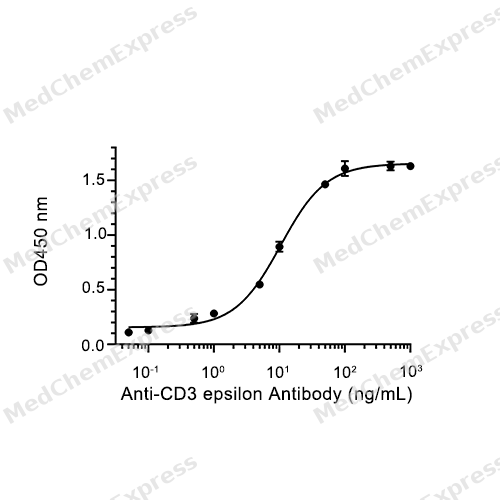
Immobilized Rhesus Macaque CD3D-CD3E at 2 μg/mL (100 μL/well) can bind CD3 epsilonAntibody. The ED50 for this effect is 10.45 ng/mL.
MCE Validation Data
-
Purity - SDS-PAGE
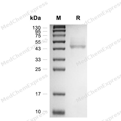
Purity - SDS-PAGE
-
Bioactivity - ELISA
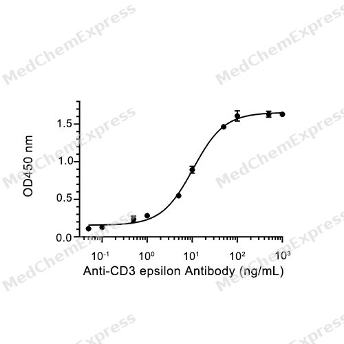
Bioactivity - ELISA


Technical Parameters
-
Species Rhesus Macaque
-
Source HEK293
-
Tag N-hFc
-
Molecular Construction
-
N-term
-
hFc
-
CD3D (F22-A105)
Accession # F6WI60 -
C-term
-
N-term
-
hFc
-
CD3E (Q22-D117)
Accession # G7NCB9 -
C-term
-
-
Synonyms
Mafa-AG; MHC class I antigen; CD3E; T3E; CD3 Epsilon Subunit Of T-Cell Receptor Complex; T-Cell Antigen Receptor Complex, Epsilon Subunit Of T3; T-Cell Surface Glycoprotein CD3 Epsilon Chain; CD3e Molecule; CD3-Epsilon; CD3e Antigen; CD3epsilon; CD3E Prot
-
Predicted Molecular Mass
37 kDa & 39 kDa
-
Molecular Weight
Approximately 43-45 kDa & 52-60 kDa, based on SDS-PAGE under reducing conditions, due to the glycosylation.
-
Purity
≥ 95%, as determined by reducing SDS-PAGE.


Product Properties
Lyophilized powder.
Lyophilized from a 0.22 μm filtered solution of PBS, pH 7.4.
<1 EU/μg, determined by LAL method.
It is not recommended to reconstitute to a concentration less than 100 μg/mL in ddH2O. For long term storage it is recommended to add a carrier protein (0.1% BSA, 5% HSA, 10% FBS or 5% Trehalose).
Stored at -20°C for 2 years from date of receipt. After reconstitution, it is stable at 4°C for 1 week or -20°C for longer (with carrier protein). It is recommended to freeze aliquots at -20°C or -80°C for extended storage.
Room temperature in continental US; may vary elsewhere.


Documentation
-
Data Sheet (235 KB)
-
SDS (251 KB)
- English - EN (251 KB)
- Français - FR (251 KB)
- Deutsch - DE (251 KB)
- Norwegian - NO (251 KB)
- Español - ES (251 KB)
- Swedish - SV (251 KB)
- Italian - IT (251 KB)
- Korean - KR (251 KB)
- Portuguese - PT (251 KB)
-
Handling Instructions (2659 KB)

Calculators
Concentration (start) × Volume (start) = Concentration (final) × Volume (final)


 Powered by Bioz

