



MGAT5 Protein, Human (HEK293, His)
Based on 1 Customer Validation
The MGAT5 protein regulates the cytoskeleton, adhesion, migration, and signaling. MGAT5 Protein, Human (HEK293, His) is the recombinant human-derived MGAT5 protein, expressed by HEK293 , with C-His labeled tag.
- Species: Human
- Source: HEK293
-
Storage:Stored at -20°C for 2 years from date of receipt. After reconstitution, it is stable at 4°C for 1 week or -20°C for longer (with carrier protein). It is recommended to freeze aliquots at -20°C or -80°C for extended storage.


To place orders, for customer services and technical support, please contact: MedChemExpress USA
Tel: 609-228-6898 E-mail: [email protected] [email protected]


-
 Biological Activity
-
Technical Parameters
-
Product Properties
- Documentation
- Help & FAQs




Biological Activity
Description
The MGAT5 protein regulates the cytoskeleton, adhesion, migration, and signaling. MGAT5 Protein, Human (HEK293, His) is the recombinant human-derived MGAT5 protein, expressed by HEK293 , with C-His labeled tag.
Background
MGAT5 protein is a key enzyme that catalyzes the addition of N-acetylglucosamine (GlcNAc) in beta 1-6 linkage to the alpha-linked mannose of biantennary N-linked oligosaccharides. This catalytic activity is crucial for the biosynthesis of branched, complex-type N-glycans found on various proteins, including EGFR, TGFR (TGF-beta receptor), and CDH2. Through its involvement in complex N-glycan biosynthesis, MGAT5 plays a pivotal role in cellular signaling pathways, actin cytoskeleton reorganization, cell-cell adhesion, and cell migration. Notably, MGAT5-dependent EGFR N-glycosylation prolongs EGFR signaling by preventing rapid endocytosis. MGAT5 is also essential for efficient interactions between TGFB1 and its receptor, enhancing intracellular signaling pathways activated by growth factors like FGF2, PDGF, IGF, TGFB1, and EGF. Additionally, MGAT5-mediated CDH2 N-glycosylation inhibits homotypic cell-cell adhesion, influencing downstream signaling pathways and promoting cell migration. In the immune response, MGAT5-dependent TCR N-glycosylation limits TCR clustering and dampens TCR-mediated responses to antigens, contributing to normal leukocyte evasion and accumulation at inflammatory sites. Moreover, MGAT5 inhibits monocyte attachment to vascular endothelium and subsequent diapedesis, while also promoting proliferation of endothelial cells and angiogenesis by facilitating the release of FGF2 from the extracellular matrix. In summary, MGAT5 emerges as a multifaceted regulator with significant implications in cellular processes, immune responses, and angiogenesis.
Verified Bioactivity
Measured in a cell proliferation assay using MDA-MB-231 cells. The ED50 for this effect is <0.1 μg/mL, corresponding to a activity is >1×10^4 units/mg.
MCE Validation Data
-
Purity - SDS-PAGE
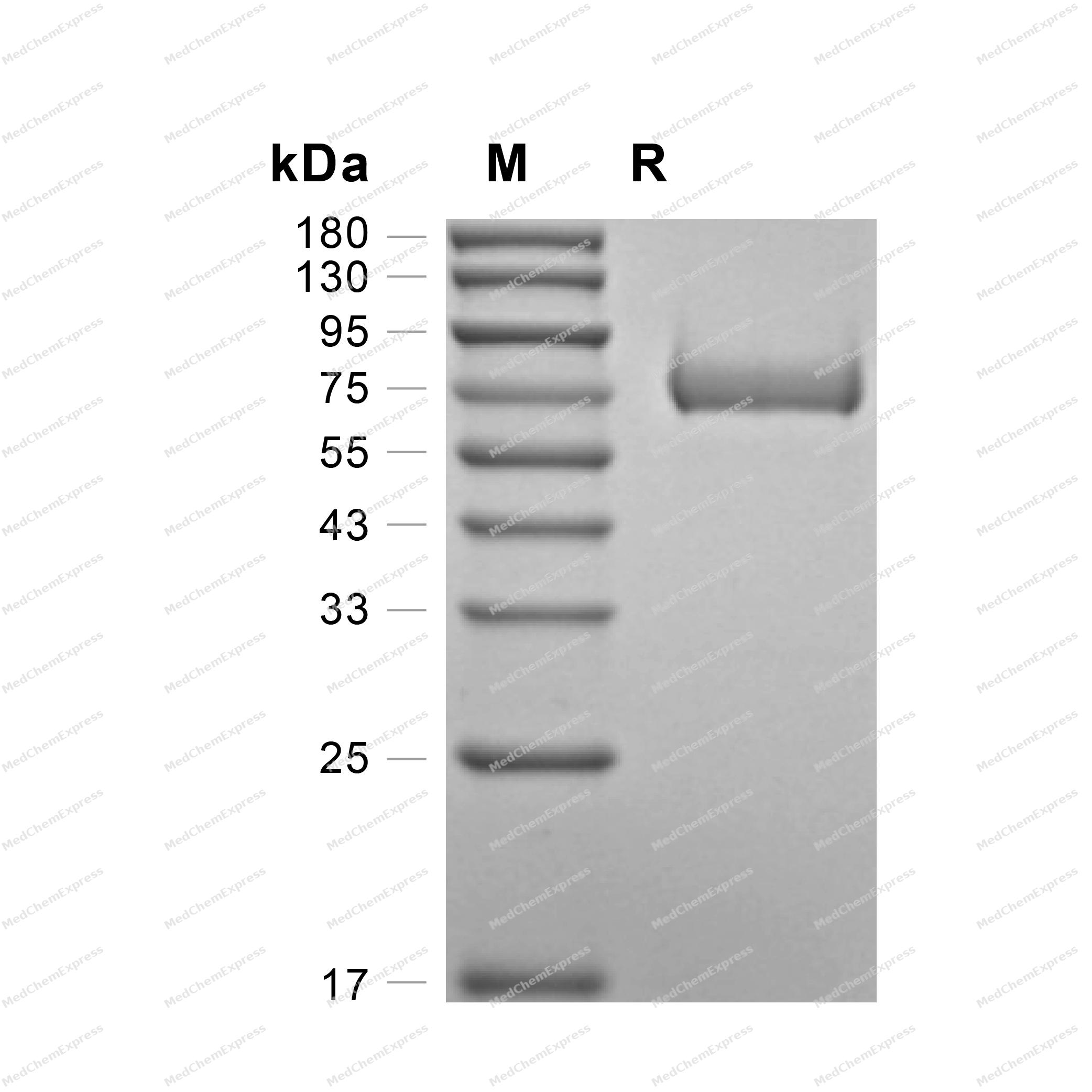
Purity - SDS-PAGE
-
Bioactivity - Cell-Based Assay
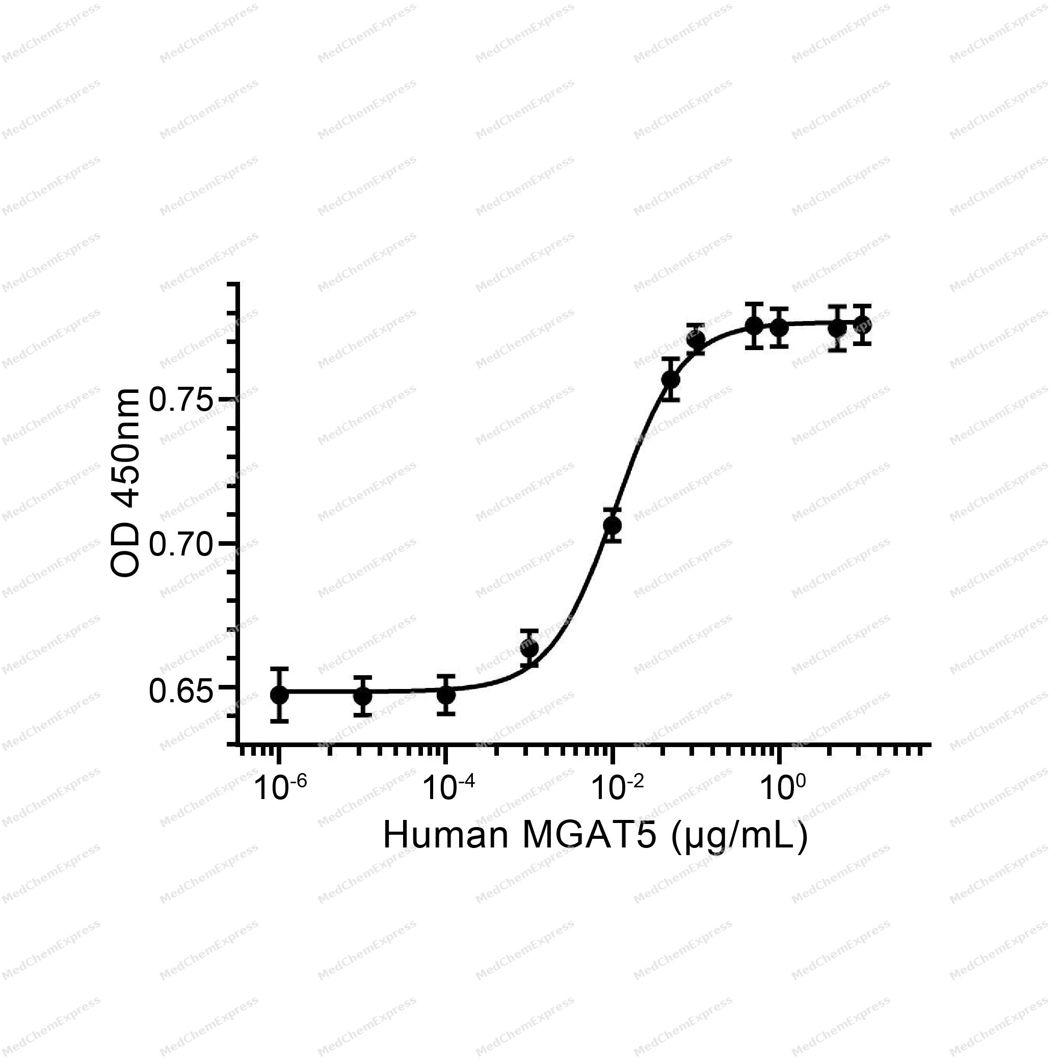
Bioactivity - Cell-Based Assay


Technical Parameters
-
Species Human
-
Source HEK293
-
Tag C-His
-
Accession
Q09328 (L189-L741)
-
Molecular Construction
-
N-term
-
MGAT5 (L189-L741)
Accession # Q09328 -
His
-
C-term
-
-
Protein Length
Partial
-
Synonyms
MGAT5; Mannoside Acetylglucosaminyltransferase 5; Alpha-1,6-Mannosylglycoprotein 6-Beta-N-Acetylglucosaminyltransferase; N-Acetylglucosaminyl-Transferase V; GNT-V; GlcNAc-T V; Alpha-1,6-Mannosylglycoprotein 6-Beta-N-Acetylglucosaminyltransferase A; Alpha-
-
AA Sequence
LSEVENWCPHLPWRAKNPYEEADHNSLAEIRTDFNILYSMMKKHEEFRWMRLRIRRMADAWIQAIKSLAEKQNLEKRKRKKVLVHLGLLTKESGFKIAETAFSGGPLGELVQWSDLITSLYLLGHDIRISASLAELKEIMKKVVGNRSGCPTVGDRIVELIYIDIVGLAQFKKTLGPSWVHYQCMLRVLDSFGTEPEFNHANYAQSKGHKTPWGKWNLNPQQFYTMFPHTPDNSFLGFVVEQHLNSSDIHHINEIKRQNQSLVYGKVDSFWKNKKIYLDIIHTYMEVHATVYGSSTKNIPSYVKNHGILSGRDLQFLLRETKLFVGLGFPYEGPAPLEAIANGCAFLNPKFNPPKSSKNTDFFIGKPTLRELTSQHPYAEVFIGRPHVWTVDLNNQEEVEDAVKAILNQKIEPYMPYEFTCEGMLQRINAFIEKQDFCHGQVMWPPLSALQVKLAEPGQSCKQVCQESQLICEPSFFQHLNKDKDMLKYKVTCQSSELAKDILVPSFDPKNKHCVFQGDLLLFSCAGAHPRHQRVCPCRDFIKGQVALCKDCL
-
Molecular Weight
Approximately 60-65 kDa, based on SDS-PAGE under reducing conditions.
-
Purity
≥ 95%, as determined by reducing SDS-PAGE.


Product Properties
Lyophilized powder
Lyophilized from a 0.22 μm filtered solution of PBS, 5% trehalose, mannitol, 0.01% Tween 80, pH 7.4.
<1 EU/μg, determined by LAL method.
It is not recommended to reconstitute to a concentration less than 100 μg/mL in ddH2O. For long term storage it is recommended to add a carrier protein (0.1% BSA, 5% HSA, 10% FBS or 5% Trehalose).
Stored at -20°C for 2 years from date of receipt. After reconstitution, it is stable at 4°C for 1 week or -20°C for longer (with carrier protein). It is recommended to freeze aliquots at -20°C or -80°C for extended storage.
Room temperature in continental US; may vary elsewhere.


Documentation
-
Data Sheet (240 KB)
-
SDS (251 KB)
- English - EN (251 KB)
- Français - FR (251 KB)
- Deutsch - DE (251 KB)
- Norwegian - NO (251 KB)
- Español - ES (251 KB)
- Swedish - SV (251 KB)
- Italian - IT (251 KB)
- Korean - KR (251 KB)
- Portuguese - PT (251 KB)
-
Handling Instructions (2659 KB)

Calculators
Concentration (start) × Volume (start) = Concentration (final) × Volume (final)


 Powered by Bioz

