



Ritlecitinib malonate
Based on 9 publication(s) in Google Scholar
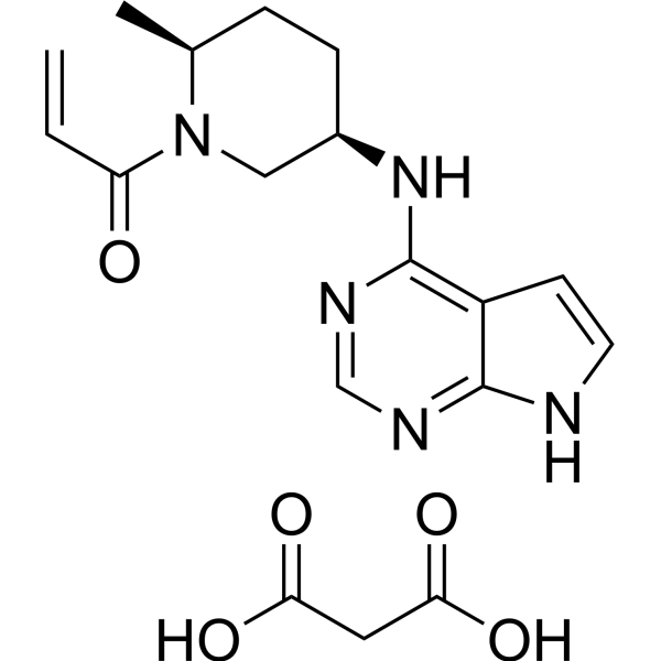
Ritlecitinib (PF-06651600) malonate is a highly selective, orally active, irreversible covalent JAK3 inhibitor (IC50=33 nM) without inhibitory activity towards JAK1, JAK2, and TYK2 (IC50 >10 μ M). Ritlecitinib malonate rapidly inactivates the JAK3 kinase, and blocks signaling and downstream STAT phosphorylation mediated by common gamma chain cytokines such as IL-2 and IL-15. Ritlecitinib malonate can inhibit Th1/Th17 cell differentiation and function, and effectively suppress preclinical animal models such as alopecia areata, adjuvant-induced arthritis (AIA), and experimental autoimmune encephalomyelitis (EAE).
For research use only. We do not sell to patients.
- CAS No.: 2140301-97-7
- Formula: C18H23N5O5
- Molecular Weight:389.41
-
Storage:
Please store the product under the recommended conditions in the Certificate of Analysis.

To place orders, for customer services and technical support, please contact: MedChemExpress USA
Tel: 609-228-6898 E-mail: [email protected] [email protected]


-
 Biological Activity
-
Chemical Information
- Protocols
- Purity & Documentation
- References
-
 Help & FAQs



Publications Citing Use of MedChemExpress (MCE) Ritlecitinib malonate
More- Ann Rheum Dis. 2025 Sep 25:S0003-4967(25)04383-3. [Abstract]
- Blood Adv. 2022 Jul 26;6(14):4283-4296. [Abstract]
- Int J Mol Sci. 2024 Dec 15;25(24):13452. [Abstract]
- Int J Mol Sci. 2023 May 25;24(11):9243. [Abstract]
- Expert Opin Investig Drugs. 2021 Dec;30(12):1169-1174. [Abstract]
- Clin Immunol. 2025 Apr:273:110445. [Abstract]
- Res Sq. 2026 Apr 13.
- bioRxiv. 2026 Mar 7.
- bioRxiv. 2023 Aug 17.
 Customer Validation & Images

-
RT-PCR
-
WB


Biological Activity
Description
IC50 & Target
|
JAK3 33.1 nM (IC50) |
IL-2 |
IL-4 |
IL7R |
IL-15 |
STAT3 |
STAT5 |
STAT6 |
In Vitro
Ritlecitinib malonate inhibits IL-15 in peripheral blood mononuclear cells (PBMCs) and heparin treated human whole blood (HWB) with IC50 of 51 nM and 197 nM, respectively[1].
Ritlecitinib malonate inhibits the phosphorylation of STAT proteins mediated by IL-2 (STAT5), IL-4 (STAT6), IL-7 (STAT5), IL-15 (STAT5), and IL-21 (STAT3) at concentrations of 244 nM, 340 nM, 407 nM, 266 nM, and 355 nM (IC50), respectively[2].
Ritlecitinib malonate (30 nM, 167 nM; 5 days, 6 days) inhibits cell differentiation in Th1 and Th17 cell differentiation inhibition experiments for 5 days under Th1 conditions and 6 days under Th17 conditions, respectively[2].
MedChemExpress (MCE) has not independently confirmed the accuracy of these methods. They are for reference only. Further protocols information, click here.
-
Cell Line:Mouse splenocytes, human whole blood lymphocytes (HWB)
-
Concentration:33.1 nM, 244 nM, 340 nM, 407 nM, 266 nM, 355 nM
-
Incubation Time:1 hour (pre-treatment with Ritlecitinib malonate) + 15-20 minutes (cytokine stimulation)
-
Result:In mouse splenocytes, 33.1 nM inhibited JAK3 kinase activity, while 244 nM, 340 nM, 407 nM, and 266 nM inhibited STAT5 phosphorylation induced by IL-2, IL-4, IL-7, and IL-15 respectively. In human whole blood lymphocytes, 355 nM inhibited IL-21-induced STAT3 phosphorylation.
In Vivo
Ritlecitinib (30, 100 mg/kg (reversal); 20, 60 mg/kg (prevention); oral; once daily) malonate reduces disease severity in the mouse experimental autoimmune encephalomyelitis (EAE) model[2].
Ritlecitinib (30 mg/kg; oral; once daily; 4 weeks) malonate can prevent the occurrence of alopecia areata in the C3H/HeJ mouse skin transplantation alopecia areata prevention model; Ritlecitinib (30 mg/kg; oral; once daily; 12 weeks) malonate can reverse the symptoms of alopecia areata, promote hair regeneration and reduce skin inflammation in the C3H/HeJ mouse alopecia areata model[3].
MedChemExpress (MCE) has not independently confirmed the accuracy of these methods. They are for reference only.
-
Animal Model:C3H/HeJ mice (8-week-old) with alopecia areata model[3]
-
Dosage:30 mg/kg Ritlecitinib
-
Administration:Oral via ALZET osmotic pump; once daily; 12 weeks
-
Result:Significantly induced hair regrowth, reduced skin-infiltrating CD45+ leukocytes, CD44+CD62L−CD8+ T cells, NKG2D+CD8+ T cells, and IFN-γ+CD8+ T cells, and decreased Alopecia areata (AA)-associated skin inflammation.


Chemical Information
-
CAS No. 2140301-97-7
-
Molecular Weight 389.41
-
Formula C18H23N5O5
-
SMILES
OC(CC(O)=O)=O.C=CC(N1[C@@H](C)CC[C@@H](NC2=C3C(NC=C3)=NC=N2)C1)=O
-
Synonyms
PF-06651600 malonate
-
Shipping
Room temperature in continental US; may vary elsewhere.
-
Storage
Please store the product under the recommended conditions in the Certificate of Analysis.


Publications (9)
-
Journal Impact Factor
-
Most Recent
-
Ann Rheum Dis
Augmentation of immunothrombosis as a key mechanism underlying JAK inhibition associated hypercoagulability in rheumatoid arthritis. [Abstract]2025 Sep 25:S0003-4967(25)04383-3. PMID: 41006174 -
Blood Adv
Latent membrane proteins from EBV differentially target cellular pathways to accelerate MYC-induced lymphomagenesis. [Abstract]2022 Jul 26;6(14):4283-4296. PMID: 35605249 -
Int J Mol Sci
Epstein-Barr Virus BRRF1 Induces Butyrophilin 2A1 in Nasopharyngeal Carcinoma NPC43 Cells via the IL-22/JAK3-STAT3 Pathway. [Abstract]2024 Dec 15;25(24):13452. PMID: 39769218
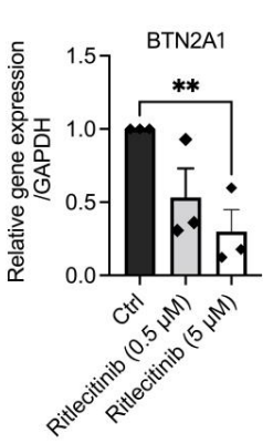
Ritlecitinib malonate purchased from MedChemExpress. Usage Cited in: Int J Mol Sci. 2024 Dec 15;25(24):13452. [Abstract]
Ritlecitinib (0.5-5 μM) significantly decreased the gene expression level of BTN2A1 in NPC43 cells.
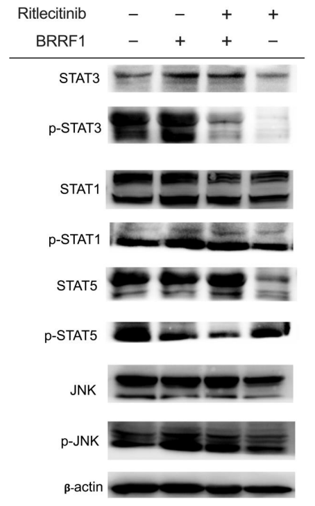
Ritlecitinib malonate purchased from MedChemExpress. Usage Cited in: Int J Mol Sci. 2024 Dec 15;25(24):13452. [Abstract]
STAT3, p-STAT3, STAT1, p-STAT1, STAT5, p-STAT5, JNK, and p-JNK protein expression of NPC43 cells by BRRF1 overexpression with or without Ritlecitinib (5 μM) treatment. Representative immunoblots are shown.
-
Int J Mol Sci
JAK Signaling Is Critically Important in Cytokine-Induced Viral Susceptibility of Keratinocytes. [Abstract]2023 May 25;24(11):9243. PMID: 37298195 -
Expert Opin Investig Drugs
Ritlecitinib: an investigational drug for the treatment of moderate to severe alopecia areata. [Abstract]2021 Dec;30(12):1169-1174. PMID: 34826225 -
Clin Immunol
Novel janus kinase 3 inhibitor ritlecitinib suppresses T and B cell responses to prevent acute cardiac allograft rejection in mice. [Abstract]2025 Apr:273:110445. PMID: 39954912 -
-
-

Protocols
-
Kinase activity and phosphorylation assays
Kinase activity assays measure the ability of kinases to transfer phosphate groups from ATP to specific substrates, while phosphorylation assays detect the presence and levels of phosphorylated proteins. Common methods include radiolabeled ATP incorporation (e. g. ,), ADP release detection via bioluminescence (e. g. ,[3]), enzyme-linked immunosorbent assays (ELISA) for phospho-specific epitopes (e. g. ,[6]), and microtiter-based formats for high-throughput screening (e. g. ,[8]). The ADP-Glo assay quantifies kinase activity by measuring ADP produced during phosphorylation using a luciferase-based system. Radiometric assays involve autoradiography or scintillation counting after incorporation of 32P-labeled ATP into substrate proteins. ELISA-based approaches rely on phospho-specific antibodies to detect activated kinases in cell lysates or purified samples.
-
Western Blot
Western blotting (WB) is a commonly used experimental method in molecular biology, biochemistry, and immunogenetics for identifying and quantifying target proteins. It combines gel electrophoresis with immunoassay, enabling researchers to analyze protein expression, post-translational modifications, and molecular weight.
-
Collagen-Induced Arthritis
Collagen-induced arthritis (CIA) is an autoimmune murine model of rheumatoid arthritis in which immunization with type II collagen (CII) emulsified in an adjuvant induces a T cell- and autoantibody-driven inflammatory arthritis characterized by synovial hyperplasia, immune cell infiltration, and joint destruction. The model typically relies on genetically susceptible mouse strains (e. g. , DBA/1) and reproduces key features of human rheumatoid arthritis, including anti-collagen immune responses and progressive joint inflammation. Disease onset generally occurs within ~3-4 weeks after immunization, depending on antigen/adjuvant combinations and protocol variation. The immunopathology is driven by adaptive immune activation against CII, leading to systemic and local joint inflammation mediated by pro-inflammatory cytokines and effector immune cells, making CIA a standard preclinical platform for evaluating immunomodulatory and anti-arthritic interventions.
-
Cell Cytotoxicity Assay
Cytotoxicity assays are usually based on the assessment of cell membrane damage, which can also be indirectly detected by measuring cell viability. Detection methods include MTT assay, CKK-8 assay, LDH assay and ATP assay, etc.
-
Naïve CD4+ T-cell subset differentiation/polarization
Naïve CD4+ T-cell subset differentiation/polarization is an in vitro assay in which purified naïve CD4+ T cells are activated through TCR and CD28 costimulation and cultured with defined cytokines and neutralizing antibodies to generate Th0, Th1, Th2, Th17, or induced Treg-like populations. Differentiation is detected by subset-associated cytokines and transcription factors: IFN-γ/T-bet for Th1, IL-4/GATA3 for Th2, IL-17A/RORγt for Th17, and Foxp3 for induced Treg cells. The assay readout is usually generated by intracellular cytokine staining after restimulation, transcription-factor staining by flow cytometry, ELISA of secreted cytokines, or gene-expression analysis. The result reflects cytokine-directed lineage commitment or polarization rather than antigen-specific immune protection by itself.
-
Protocol for Kinase activity and phosphorylation assays
Kinase activity assays measure transfer of phosphate from ATP to a protein or peptide substrate, generating phosphorylated substrate, ADP, or incorporated radiolabeled phosphate as the readout; phosphorylation assays measure site-specific phosphorylation in cells or tissues as a proxy for kinase-pathway activation, inhibition, or substrate regulation. Phosphorylation can be detected by phospho-specific Western blot, immunoprecipitation kinase assay, phospho-immunofluorescence, phospho-flow cytometry, luminescent ADP detection, radiolabeled ATP incorporation, or reporter-based pathway assays, and these readouts can be applied to cancer cells, primary neurons, mouse tumors, organoids, inflammatory macrophages, ferroptosis studies, and mitophagy studies when the kinase target is biologically relevant.
-
iPSC cell differentiation
Induced pluripotent stem cells (iPSCs) are a type of cell that has similar properties to embryonic stem cells through somatic cell reprogramming.


Purity & Documentation
References

Calculators
Concentration (start) × Volume (start) = Concentration (final) × Volume (final)


Ritlecitinib malonate Related Classifications
HY-100754A Related Classifications
 Powered by Bioz