





Scientific Reviews
Product Guides
Blogs
Topics
Targeted therapy! The Capetin Prize winning "Click Chemistry" can be used like this!Targeted therapy! The Nobel Prize winning "Click Chemistry" can be used like this!2025-02-06



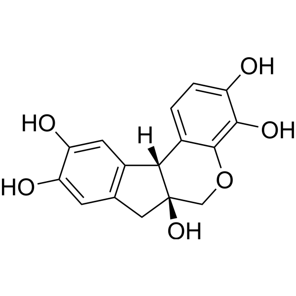
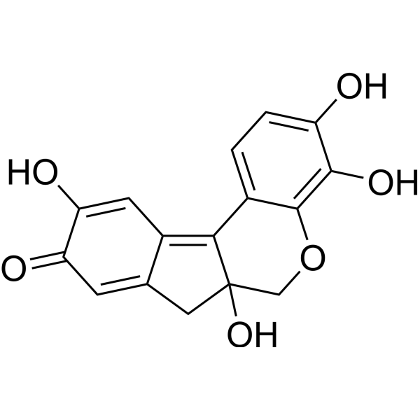
9 Results for "Hematoxylin" in MCE Product Catalog:
Leguminosae Caesalpinia sappan L. Other Diseases Phenols Polyphenols Plants Disease Research Fields
MCE Hematoxylin-Eosin Staining Kit (Including Differentiation and Bluing Solutions) contains staining solutions, differentiation solution, and bluing solution. Through optimized formulations, it helps to adjust the staining effect, ensuring proper staining intensity and further improving experimental accuracy.
MCE Sirius Red Staining Kit consists of hematoxylin staining solution and Sirius Red staining solution and is mainly used to observe abnormal collagen deposition or fibrosis in various pathological tissues. Under a conventional light microscope, collagen fibers in tissues such as the heart and blood vessels appear red. Under polarized light microscopy, this method is useful for the classification and evaluation of different types of fibrotic lesions.
MCE Modified Masson Staining Kit employs Celestine Blue hematoxylin for light nuclear staining, offering shorter differentiation time compared to conventional methods. It features low toxicity, environmental friendliness, simple operation, and stable performance. The staining results show clear coloration and high contrast. The stained sections can be stored for long periods with minimal fading, facilitating long-term preservation and image analysis. This kit is widely used in studies of connective tissue, muscle tissue, and collagen fibers, and is suitable for histological observation and related pathological analyses.
MCE Modified Van Gieson Staining Kit employs Celestine Blue and Mayer’s hematoxylin for nuclear staining, providing clearer and more stable nuclear visualization while facilitating longer preservation of stained sections. Ponceau S is used for collagen fiber staining, offering stable coloration and strong resistance to fading. This method enables effective differentiation between collagen fibers and muscle fibers, and can assist in distinguishing collagen fiber–derived tumors from myogenic tumors to a certain extent. It is also suitable for observing tissue or organ injury, repair processes, and the degree of fibrosis.
MCE Oil Red O Staining Kit for Cultured Cells consists of Oil Red O staining solution and hematoxylin. It is used to demonstrate fat degeneration and abnormal lipid deposition in cultured cells, particularly when multiple neutral fat droplets are present within the cells. This kit helps in identifying lipid changes and their nature in cultured cells. It is important to note that samples should not be fixed with ethanol-containing fixatives, as ethanol may interfere with lipid staining. The positive staining result for fat typically appears orange-yellow to red, with the exact color depending on the lipid concentration.

Scientific Reviews
Product Guides
Blogs
Topics
Targeted therapy! The Capetin Prize winning "Click Chemistry" can be used like this!Targeted therapy! The Nobel Prize winning "Click Chemistry" can be used like this!2025-02-06
Protocols
Targeted therapy! The Capetin Prize winning "Click Chemistry" can be used like this!Targeted therapy! The Nobel Prize winning "Click Chemistry" can be used like this!2025-02-06



