Search Result
Results for "
staining
" in MedChemExpress (MCE) Product Catalog:
33
Biochemical Assay Reagents
8
Isotope-Labeled Compounds
| Cat. No. |
Product Name |
Target |
Research Areas |
Chemical Structure |
-
- HY-D0815
-
|
|
Fluorescent Dye
DNA/RNA Synthesis
|
Others
|
|
Propidium Iodide (PI) is a nuclear staining agent that stains DNA. Propidium Iodide is an analogue of ethidine bromide that emits red fluorescence upon embedding in double-stranded DNA. Propidium Iodide cannot pass through living cell membranes, but it can pass through damaged cell membranes to stain the nucleus. Propidium Iodide has a fluorescence wavelength of 493/617 nm and a wavelength of 536/635 nm after Mosaic with DNA. Propidium Iodide is commonly used in the detection of apoptosis (apoptosis) or necrosis (necrosis), and is often used in flow cytometry analysis.
|
-
-
- HY-D0718
-
|
Nile Blue A oxazone; Phenoxazone 9
|
Fluorescent Dye
|
Others
|
|
Nile red (Nile blue oxazone) is a lipophilic stain. Nile red has environment-sensitive fluorescence. Nile red is intensely fluorescent in a lipid-rich environment while it has minimal fluorescence in aqueous media. Nile red is an excellent vital stain for the detection of intracellular lipid droplets by fluorescence microscopy and flow cytof uorometry. Nile red stains intracellular lipid droplets red. The fluorescence wavelength is 559/635 nm .
|
-
-
- HY-B0324A
-
|
Basic Violet 3; Gentian Violet; Methyl Violet 10B
|
Environmental Pollutants
Fluorescent Dye
Influenza Virus
Bacterial
|
Infection
|
|
Crystal Violet, also known as Gentian violet, methyl violet 10B, is a triphenyl-methane, an alkaline dye that binds to DNA in the nucleus of a cell, staining it a deep purple. It is often used for Gram staining to classify bacteria, or for cell or histological staining[1].
|
-
-
- HY-Y0016
-
|
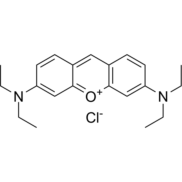
Basic Violet 10; Brilliant Pink B; Rhodamine O; Tetraethylrhodamine
|
Environmental Pollutants
Fluorescent Dye
|
Others
|
|
Rhodamine B is a staining fluorescent dye, commonly used for dyeing textiles, paper, soap, leather, and agents.
|
-
-
- HY-D1020
-
-
-
- HY-N0116
-
|
Natural Black 1; Haematoxylin
|
Amyloid-β
|
Others
|
|
Hematoxylin (Natural Black 1), a naturally occurring flavonoid compound derived from Caesalpinia sappan Linn.. Hematoxylin is a nuclear stain in histology and is also a potent Aβ42 fibrillogenesis inhibitor with an IC50 of 1.6 μM.
|
-
-
- HY-D0914
-
|
FD&C Green No. 3; Food green 3; C.I. 42053
|
Environmental Pollutants
α-synuclein
|
Neurological Disease
|
|
Fast Green FCF is a sea green triarylmethane food dye, with absorption maximum ranging from 622 to 626 nm. Fast Green FCF inhibits α-synuclein aggregation, as well as Aβ and P2X4 receptor, and TLR4/Myd88/NF-κB. Fast Green FCF is widely used as a staining agent like quantitative stain for histones at alkaline pH after acid extraction of DNA, and as a protein stain in electrophoresis. Fast Green FCF improves cognitive impairment, depression, relieves pain allergies, and promotes reproductive function .
|
-
-
- HY-D0213
-
-
-
- HY-13735
-
-
-
- HY-12489
-
|
Acid Red 112
|
Fluorescent Dye
|
Others
|
|
Ponceau S (Acid Red 112) is a non-specific protein dye commonly used as a stain for Western blot. Ponceau S is used in an acidic aqueous solution that is compatible with antibody-antigen binding and dyes the proteins on the membrane red .
|
-
-
- HY-120601
-
|
ARS sodium
|
Fluorescent Dye
|
Others
|
|
Alizarin Red S sodium is an anthraquinone derivative dye. When combined with cations such as calcium ions, the functional group of Alizarin Red S sodium can form a coordination bond with the cation through the oxygen atom to show orange-red fluorescence. Alizarin Red S sodium can be used for screening of calcium compounds in synovial fluid and detecting osteoblast differentiation, and can also be used for bone staining in mice. Excitation/emission wavelength: 500/570 nm .
|
-
-
- HY-D0970
-
|
Direct Blue 14; Trypan Blue
|
Fluorescent Dye
|
Others
|
|
Diphenyl Blue (Trypan Blue) is a cell active dye, the most commonly used dye for the identification of dead cells, of en used to test cell membrane integrity and cell viability. Diphenyl Blue staining is one of the methods for tissue and cell culture. When cells are deactivated or have incomplete cell membranes, Diphenyl Blue can stain them Blue. Normal living cells with intact cell membranes reject Diphenyl blue and do not stain them blue. However, macrophages are capable of phagocytosis of Diphenyl Blue, so it can be used as a living stain for macrophages .
|
-
-
- HY-D0987
-
|
|
Calmodulin
|
Others
|
|
Stains-All, a cationic carbocyanine dye, is a convenient probe to study the structural features of the individual calcium-binding sites of calmodulin (CaM) and related calcium-binding proteins (CaBP) .
|
-
-
- HY-D0952
-
|
|
Parasite
|
Others
|
|
Acridine Orange base is a cell-permeable fluorescent dye that stains organisms (bacteria, parasites, viruses, etc.) bright orange and, when used under appropriate conditions (pH=3.5, Ex=460 nm), distinguishes human cells in green for detection by fluorescence microscopy. Acridine Orange base fluoresces green when bound to dsDNA (Ex=488, Em=520-524) and red when bound to ssDNA (Ex=457, Em=630-644) or ssRNA (Ex=457, Em=630-644), also can be used in cell cycle assays .
|
-
-
- HY-D0367
-
|
Calcofluor White M2R
|
Fluorescent Dye
|
Others
|
|
Fluorescent brightener 28 is a fluorescent whitening agent commonly used in the padding process of the textile industry. Fluorescent brightener 28 is capable of staining polysaccharides such as cellulose, and when the plasma membrane ruptures, it also weakly stains the cytoplasm and strongly stains the cell nucleus. Additionally, Fluorescent brightener 28 can be utilized to detect intracellular chitin in living cells. Fluorescent Brightener 28 also is a visible light emitting diode (LED)-light sensitive photoinitiator for free radical photopolymerizations .
|
-
-
- HY-D0944
-
|
|
Fluorescent Dye
|
Others
|
|
Giemsa stain is a composite dye composed of methylene azure, methylene blue, eosin and other components. Giemsa stain stains chromatin, nuclear membranes, specific cytoplasmic components and microorganisms. Giemsa stain is used in research on cytological and parasitological staining .
|
-
-
- HY-D0955
-
|
Thionine acetate
|
Biochemical Assay Reagents
Bacterial
|
Infection
|
|
Thionin acetate (Thionine acetate) is a compound present in the seeds, stems, roots, and leaves of many plant species with antibacterial activity. Thionin acetate is a metachromatic cationic histological dye widely used in biological staining .
|
-
-
- HY-D0004
-
|
Azure B chloride
|
Monoamine Oxidase
|
Neurological Disease
|
|
Azure B is a cationic dye and the major metabolite of Methylene blue. Azure B is used in making Azure eosin stains for blood smear staining. Azure B is a high-potency, selective and reversible inhibitor of monoamine oxidases (MAO)-A, with IC50s of 11 and 968 nM for recombinant human MAO-A and MAO-B, respectively. Azure B possesses significant antidepressant-like effects .
|
-
-
- HY-D0232
-
-
-
- HY-D0093
-
|
EthD-1
|
DNA Stain
|
Others
|
|
Ethidium homodimer (EthD-1) is a high-affinity fluorescent nucleic acid dye commonly used to stain mammals, bacteria, yeast, and fungi. Ethidium homodimer binds to DNA or RNA, enhancing fluorescence more than 30 times. The Ethidium homodimer has a strong positive charge, so it cannot cross cell membranes and stain living cells; But the Ethidium homodimer can cross the disordered region of the dead cell membrane to reach the nucleus and embed the DNA double strand to produce red fluorescence. Therefore, Ethidium homodimer is a relatively sensitive nucleic acid stain that can accurately detect nucleic acids in solution or in decomposing cells. Ethidium homodimer binds DNA, Ex/Em=528/617 nm .
|
-
-
- HY-D0333
-
|
Sirius Red
|
Amyloid-β
|
Others
|
|
Direct Red 80 (Sirius Red) is a polyazo dye used principally in staining methods for collagen and amyloid. Direct Red 80 does not release benzidine upon degradation and is safer than many traditional direct dyes .
|
-
-
- HY-W094758A
-
|
|
Fluorescent Dye
|
Cancer
|
|
4-Di-1-ASP is a styryl dye used to stain glioma cells in living brain tissue for analysis of cell structure, viability, proliferation and endocytosis, cytokinesis and phagocytosis, as well as for observation of mitochondrial structures in living cells. 4-Di-1-ASP fluoresces green when imaged microscopically (λex /λem = 475/606 nm) .
|
-
-
- HY-N0335
-
Indigo
1 Publications Verification
|
Fluorescent Dye
|
Others
|
|
Indigo is a blue dye. Indigo stains cellulose fibers such as cotton. Indigo stains food or biological samples to visualize and quantify the uptake or distribution of substances by organisms .
|
-
-
- HY-101888
-
-
-
- HY-D0003
-
|
|
Environmental Pollutants
Bacterial
|
Infection
|
|
Methyl blue belongs to the group of triaminotriphenylmethane dyes. Methyl blue is widely used as antiseptic dye in polychrome staining method and has applications in histological and microbiological staining solutions. Methyl blue has been used as a model to study the effect of various catalysts on photodegradation of dyes .
|
-
-
- HY-B1539A
-
|
Magenta base monohydrochloride; Basic Fuchsin monohydrochloride; Rosaniline Base monohydrochloride
|
Fluorescent Dye
Bacterial
Fungal
|
Infection
|
|
Fuchsine base (Magenta base; Basic Fuchsin) monohydrochloride is a triaminotriphenylmethane dye. Fuchsine base monohydrochloride has anesthetic, bactericidal and fungicidal properties. Fuchsine base monohydrochloride can be used for the staining of collagen, muscle, mitochondria and tuberculosis. Fuchsine base monohydrochloride is commonly used as a counterstain in Gram staining .
|
-
-
- HY-D0996
-
|
|
DNA Stain
|
Others
|
|
Lds-751 is a nucleic acid stain that mainly detects DNA. Lds-751 is a nucleic acid stain that mainly detects DNA. Lds-751 has a high affinity for DNA and fluorescence is enhanced after binding, but the maximum emission wavelength is 670nm. Lds-751 and Thiazole orange can be used for the differentiation of red blood cells, platelets, reticulocytes, and nucleated cells and can be stimulated at 488nm. Studies have shown that LDS-751 binds almost exclusively to mitochondria when incubated with nucleated living cells. After nucleated Acridine Orange (HY-101879) staining and LDS-751 treatment of cells, confocal microscopy revealed almost no co-location of the cells. Staining with Rhodamine 123 (HY-D0816), a dye known to bind polarized mitochondria, was almost identical to the pattern observed with LDS-751 .
|
-
-
- HY-120601A
-
|
ARS
|
Fluorescent Dye
|
Others
|
|
Alizarin Red S (ARS) is an anthraquinone derivative dye. When combined with cations such as calcium ions, the functional group of Alizarin Red S can form a coordination bond with the cation through the oxygen atom to show orange-red fluorescence. Alizarin Red S can be used for screening of calcium compounds in synovial fluid and detecting osteoblast differentiation, and can also be used for bone staining in mice. Excitation/emission wavelength: 500/570 nm .
|
-

-
- HY-DY1008
-
|
|
Fluorescent Dye
|
Others
|
Nile Red (solution) is a lipophilic stain. Nile red has environment-sensitive fluorescence. Nile red is intensely fluorescent in a lipid-rich environment while it has minimal fluorescence in aqueous media. Nile red is an excellent vital stain for the detection of intracellular lipid droplets by fluorescence microscopy and flow cytof uorometry. Nile red stains intracellular lipid droplets red. The fluorescence wavelength is 559/635 nm .
Solvent and concentration: DMSO: 1 mM
|
-

-
- HY-D1122
-
|
|
Fluorescent Dye
|
Others
|
|
Janus green B is a supravital stain. Janus green B staining reaction is oxygen dependent, and is reversibly inhibited by cyanide. Janus green B has been used for staining peripheral nerves in live insects, lymphatic vessels of rabbits and mitochondria .
|
-
-
- HY-W127770
-
|
Basic red 9
|
Fluorescent Dye
|
Infection
|
|
Pararosaniline hydrochloride (Basic red 9) is a pH-responsive basic dye, as a biological stain to track certain proteins. The pH of the acidified Pararosaniline hydrochloride reagent has a significant effect on the color and the maximum absorption wavelength (λmax) of the reaction system, with its optimum pH 0.48 and a λmax at 549 nm. Pararosaniline hydrochloride is also a strong modifier of RNA splicing. Pararosaniline hydrochloride has been used in the analysis of SO2 and formaldehyde and staining of bacteria or other organisms. Pararosaniline hydrochloride is extensively used in industries like textile, printing, paper, cosmetic, and leather .
|
-
-
- HY-W040198
-
|
|
Fluorescent Dye
|
Others
|
|
Phenosafranine is a phenazine dye. Phenosafranine exhibits higher binding affinity for triple-stranded RNA than for double-stranded RNA, and binds to both types of RNA via intercalation. Phenosafranine interacts with hemoglobin. Phenosafranine can be used for plant cell staining, as well as the detection of hemoglobin, dopamine, serotonin, etc.
|
-
-
- HY-D0163
-
|
|
DNA Stain
|
Others
|
|
Methyl Green is a non-intercalating fluorescent labeling agent that selectively binds to the major groove of DNA. Methyl Green electrostatically interacts with the major groove of DNA through positively charged groups, exhibiting key activities such as high affinity, resistance to photobleaching, and stable fluorescence emission. Methyl Green can be directly measured by microscopy and flow cytometry, with peaks at 633 and 677 nm. Methyl Green can be used for fluorescent labeling of the nuclei of embryonic tissues or cells, or DNA staining and cell activity detection in gel electrophoresis[1][2][3].
|
-
-
- HY-126367
-
|
Acid Green 5
|
Fluorescent Dye
|
Cancer
|
|
Light green SF yellowish (Acid Green 5) is a water-soluble triarylmethane dye. Light green SF yellowish serves as a histological stain that selectively labels mitochondria, collagen, and cartilage, while being an essential component of Papanicolaou staining. Light green SF yellowish is commonly used as a cytoplasmic counterstain for nuclear stains, and is applied in Masson's trichrome staining for collagen fibers, Pap staining, and cytological polychromatic staining in histopathology. Light green SF yellowish also induces growth inhibition and local fibrosarcomas in rats and exerts mild pulmonary tumorigenicity in mice .
|
-
-
- HY-DY2001
-
|
|
Fluorescent Dye
Influenza Virus
Bacterial
|
Infection
|
Crystal Violet (solution) , also known as Gentian violet, methyl violet 10B, is a triphenyl-methane, an alkaline dye that binds to DNA in the nucleus of a cell, staining it a deep purple. It is often used for Gram staining to classify bacteria, or for cell or histological staining .
Solvent and Concentration: Sterile water: 5 mg/mL (0.5%)
|
-

-
- HY-W088068
-
|
|
Fluorescent Dye
|
Others
|
|
Wright's stain is a composite cell stain that mainly binds to intracellular nucleic acids, proteins and other components through thiazine dyes (such as methylene blue) and eosin. Wright's stain is pH-dependent (optimal pH 6.4-6.7) and achieves cell morphology resolution by differentially staining the cytoplasm and nucleus. Under alkaline conditions, thiazine dyes bind to nucleic acids to form purple, and acidic eosin binds to cytoplasmic proteins to form red, which can form contrasting cell morphological features. Wright's stain can clearly display the fine structures of blood cells and bone marrow cells (such as nuclear chromatin and granules) and quickly evaluate cell morphological abnormalities .
|
-

-
- HY-W247131
-
DASPEI
2 Publications Verification
|
Fluorescent Dye
|
Neurological Disease
|
|
DASPEI is a cationic styrenyl mitochondrial dye with large Stokes shift. DASPEI has excitation and emission wavelength at 550/573 nm, which has good light chromogenic property. DASPEI can stain mitochondria in living cells with good labeling property. And DASPEI can also be used to stain presynaptic nerve endings independently of neuronal activity .
|
-
-
- HY-D0220A
-
|
Toluidine Blue O (purity 36%)
|
Fluorescent Dye
|
Others
Cancer
|
|
Toluidine Blue (Toluidine Blue O) purity 36% is an alkaline quinonimine dye (vivo dye) with high affinity for acidic tissue components, staining nuclei blue and polysaccharides purple. Toluidine Blue purity 36% shows heterostaining properties for mast cells, mucins and chondrocytes. Toluidine Blue purity 36% can stain different components of plant tissues and cells in different colours. Toluidine Blue purity 36% is also used as a diagnostic aid to identify malignant lesions, such as cancer .
|
-
-
- HY-D0950A
-
|
|
DNA Stain
|
Others
|
|
Methyl Green zinc chloride is a potent fluorescent dye. Methyl Green zinc chloride is a DNA stains of cells and electrophoretic gels. Methyl Green zinc chloride can be used as direct measuring of viability by both microscopy and flow cytometry, with peaks at 633 and 677 nm .
|
-
-
- HY-D1491A
-
|
|
Fluorescent Dye
|
Others
|
|
Fast Red Violet LB Zinc chloride is a stain that stains tartrate-resistant acid phosphatase (TRAP) and Fast Red Violet LB Zinc chloride can be used to stain alkaline phosphatase (ALP) activity .
|
-
-
- HY-126395
-
|
|
Fluorescent Dye
|
Others
|
|
Patent Blue V calcium salt is a triarylmethane dye used for tissue staining and lymphatic tracing, mainly applied by topical injection or eye drops. Patent Blue V calcium salt has affinity for specific tissues (such as corneal endothelium, lymphatic system), and stains the target structure by adsorption or binding, assisting in precise operation during surgery. Patent Blue V calcium salt is mainly used in ophthalmic surgery (such as graft staining for Descemet's membrane endothelial keratoplasty) and lymphatic drainage localization for sentinel lymph node biopsy of tumors .
|
-
-
- HY-137896
-
|
|
Fluorescent Dye
|
Others
|
|
4-Acetamido-4'-isothiocyanatostilbene-2,2'-disulfonic acid disodium is a fluorescent dye. 4-Acetamido-4'-isothiocyanatostilbene-2,2'-disulfonic acid disodium can be used to demonstrate retrograde axonal transport to label secondary antibodies and as a fluorescent whole cell stain .
|
-
-
- HY-D0954
-
|
|
Fluorescent Dye
|
Others
|
|
Jenner's Stain is a dye that is used in microscopy for staining blood smears. Jenner's Stain can be used for the chromosome stain by C-banding technique. Jenner's Stain can be used for the stain for routine blood examinations and malarial staining .
|
-

-
- HY-145612
-
-

-
- HY-D0983
-
-
-
- HY-W750459
-
|
|
Fluorescent Dye
|
Others
|
|
Fluorescent brightener 28 (Technical Grade) is a fluorescent whitening agent commonly used in the padding process of the textile industry. Fluorescent brightener 28 is capable of staining polysaccharides such as cellulose, and when the plasma membrane ruptures, it also weakly stains the cytoplasm and strongly stains the cell nucleus. Additionally, Fluorescent brightener 28 can be utilized to detect intracellular chitin in living cells. Fluorescent Brightener 28 also is a visible light emitting diode (LED)-light sensitive photoinitiator for free radical photopolymerizations .
|
-

-
- HY-W250151
-
|
|
Fluorescent Dye
|
Others
|
|
Leishman's stain is an essential staining tool for for staining of the peripheral blood and bone marrow smears (displayed pale bluish-grey to deep blue under oil-immersion lens) .
|
-

-
- HY-D1174
-
|
|
Fluorescent Dye
|
Others
|
|
Rhodamine 700, a Lambdachrome laser dye, is one of the few rhodamine dyes with near infrared fluorescence. Rhodamine 700 can be used for mitochondrial staining .
|
-
-
- HY-B2241A
-
|
Aluminum potassium sulfate dodecahydrate, for cell culture
|
Biochemical Assay Reagents
|
Inflammation/Immunology
|
|
Potassiumalum, for cell culture (Aluminum potassium sulfate dodecahydrate, for cell culture) is an egg white adjuvant. Potassiumalum, for cell culture acts as a mordant to help dyes bind to tissue components, enhancing the staining effect and stability during staining. Potassiumalum, for cell culture can induce allergic reactions in mice. Potassium alum, for cell culture can be used in research for bacterial and tissue staining .
|
-

-
- HY-D1544
-
|
|
Fluorescent Dye
|
Others
|
|
Uniblue A sodium is a reactive protein stain that can be used in the covalent pre-gel staining of the protein (Ex=594 nM) .
|
-
- HY-D1480
-
|
|
Fluorescent Dye
|
Others
|
|
Crystal Ponceau 6R is a red azo dye. Crystal Ponceau 6R used in histology, for staining fibrin with the martius, scarlet and blue (MSB) Trichrome stain .
|
-
- HY-118320
-
|
Mordant orange 1
|
Fluorescent Dye
|
Others
|
|
Alizarine Yellow R (Mordant orange 1), Salicylic acid derivative (HY-B0167), is an azo dye. Alizarine Yellow R is used as a pH indicator and a biological stain in chemical examinations and dyeing industries .
|
-
- HY-W250143
-
|
|
Biochemical Assay Reagents
|
Others
|
|
Toluidine blue (ZnCl2) is a basic thiazine dye commonly used as a biological stain for microscopy. It has a deep bluish-purple color and is commonly used to stain nucleic acids such as DNA and RNA, as well as to stain mast cells, cartilage, and other connective tissues. Toluidine blue (ZnCl2) stains the acidic components of these tissues, such as sulfated or carboxylated mucopolysaccharides. It is frequently used in histology, cytology, and pathology applications to aid in the diagnosis of various diseases and conditions. The dye is usually applied to tissue sections prior to microscopic examination and can be differentiated using an acidic alcohol solution. Toluidine blue (ZnCl2) is a relatively simple and inexpensive stain with good reproducibility, making it a popular choice for many laboratories.
|
-
- HY-D1220
-
|
Indanthrene
|
Fluorescent Dye
|
|
|
Pigment blue 60 is an excellent paint and ceramic stain.
|
-
- HY-D2582
-
|
|
Fluorescent Dye
|
Infection
|
|
DMAO is a membrane-permeable DNA fluorescent dye stains live and dead bacteria. (Ex/Em = 490/540 nm) .
|
-

- HY-D0895
-
-
- HY-N8365
-
|
|
Others
|
Others
|
|
Alpha-D-Glucose 1,6-bisphosphate (tetrapotassium) is a negative stain. Alpha-D-Glucose 1,6-bisphosphate (tetrapotassium) as an accuracy of 86???and has a somewhat higher image contrast .
|
-
- HY-W099576
-
|
EHDA bromide
|
Environmental Pollutants
Biochemical Assay Reagents
|
Others
|
|
Ethylhexadecyldimethylammonium (EHDA) bromide, a surfactant, has been used in a number of adsorptive separational methods, such as the removal of nickel, zinc and chromium ions. Ethylhexadecyldimethylammonium (EHDA) bromide also can be used to prepare dye of staining intracellular ions .
|
-
- HY-D0404
-
|
|
Fluorescent Dye
|
Neurological Disease
|
|
Direct Red 254 is a bisazo dye for textile applications. Direct Red 254 can be used for amyloid tissue staining in basic research on neurodegenerative diseases (e.g., Alzheimer's disease) .
|
-
- HY-101889
-
-
- HY-W250306
-
|
|
Biochemical Assay Reagents
|
Others
|
|
Carbol fuchsin is a histological stain used in microbiology to distinguish acid-fast bacteria from non-acid-fast bacteria. It is a mixture of basic fuchsin, phenol, and water and is commonly used in the Ziehl-Neelsen staining technique for the detection of tuberculosis and other mycobacterial infections. Carbol fuchsin stains the cell walls of acid-fast bacteria bright red, while other cells are unstained or slightly stained. This makes it easier to see and identify these microbes under a microscope. Carbol fuchsin is also used in veterinary medicine and phytopathology for similar purposes.
|
-
- HY-D1491
-
|
|
Fluorescent Dye
|
Others
|
|
Fast Red Violet LB is a dye for staining tartrate resistant acid phosphatase (TRAP). Fast Red Violet LB can be used for alkaline phosphatase (ALP) activity staining .
|
-
- HY-B0324AS
-
|
Basic Violet 3-d6; Gentian Violet-d6; Methyl Violet 10B-d6
|
Isotope-Labeled Compounds
|
Others
|
|
Crystal Violet-d6 is the deuterium labeled Crystal Violet (HY-B0324A). Crystal Violet, also known as Gentian violet, methyl violet 10B, is a triphenyl-methane, an alkaline dye that binds to DNA in the nucleus of a cell, staining it a deep purple. It is often used for Gram staining to classify bacteria, or for cell or histological staining .
|
-
- HY-114227
-
|
|
DNA Stain
|
Inflammation/Immunology
|
|
Hexidium iodide, a fluorescent nucleic binding acid stain (excitation/emission ~ 518/600 nm), permeants to mammalian cells and selectively stains almost all gram-positive bacteria. Hexidium iodide can bind to the DNA of all bacteria after permeabilization by EDTA .
|
-
- HY-D1674
-
|
|
Fluorescent Dye
|
Others
|
|
Sulforhodamine G is a fluorescent stain with broad dynamic ranges. Sulforhodamine G can be used for the research of protein stains .
|
-
- HY-D0946
-
-

- HY-W053871
-
-
- HY-W342930
-
|
Acid Red 17
|
Biochemical Assay Reagents
|
Others
|
|
Bordeaux Red (Acid Red 17) is a redox indicator that can be used for cytoplasm staining, such as spleen, testis, and liver slice staining. Bordeaux Red is a kind of biological materials or organic compounds that are widely used in life science research .
|
-

- HY-D0004R
-
|
Azure B chloride (Standard)
|
Reference Standards
Monoamine Oxidase
|
Neurological Disease
|
|
Azure B (Standard) is the analytical standard of Azure B. This product is intended for research and analytical applications. Azure B is a cationic dye and the major metabolite of Methylene blue. Azure B is used in making Azure eosin stains for blood smear staining. Azure B is a high-potency, selective and reversible inhibitor of monoamine oxidases (MAO)-A, with IC50s of 11 and 968 nM for recombinant human MAO-A and MAO-B, respectively. Azure B possesses significant antidepressant-like effects .
|
-

- HY-D1386
-
|
|
Fluorescent Dye
|
Others
|
|
JF526-Taxol (TFA) is a versatile scaffold for fluorogenic probes including ligands for self-labeling tags, stains for endogenous structures, and spontaneously blinking labels for super-resolution immunofluorescence .
|
-
- HY-13735R
-
|
Acriquine (Standard)
|
Parasite
Sodium Channel
DNA Stain
Apoptosis
Reference Standards
|
Infection
Neurological Disease
Cancer
|
|
Quinacrine (Standard) is the analytical standard of Quinacrine. This product is intended for research and analytical applications. Quinacrine (Acriquine) is an antimalarial and anti-cancer agent. Quinacrine also inhibits human aldehyde oxidase (IC50: 3.3 μM). Quinacrine has affinity for nucleic acids, and stains DNA and RNA in fixed cells (Ex/Em: 436/525 nm) .
|
-

- HY-D1246
-
|
|
DNA Stain
|
Others
|
|
Ethidium monoazide bromide is a DNA intercalating fluorescent dye that enters bacteria with damaged membranes. Ethidium monoazide bromide can be covalently linked to DNA by photoactivation. Ethidium monoazide bromide stains only dead cells . Ethidium monoazide (bromide) is a click chemistry reagent, it contains an Azide group and can undergo copper-catalyzed azide-alkyne cycloaddition reaction (CuAAc) with molecules containing Alkyne groups. It can also undergo strain-promoted alkyne-azide cycloaddition (SPAAC) reactions with molecules containing DBCO or BCN groups.
|
-
- HY-D1102
-
|
|
Fluorescent Dye
|
|
|
DNA intercalator 3 is a DNA intercalating agent that can be used for DNA staining.
|
-
- HY-N3290
-
|
Methyl p-methoxyphenylpropionate
|
Others
|
Others
|
|
Methyl p-methoxyhydrocinnamate is extracted from in liquid culture by the blue stain fungus Ophiostoma crassioaginata .
|
-
- HY-D1173
-
|
|
Fluorescent Dye
|
Others
|
|
Acid blue 113 is an azo dye whose staining effect is effectively removed by nanoscale zero-valent iron (NZVI) particles.
|
-
- HY-D0914A
-
|
|
α-synuclein
|
Neurological Disease
|
|
Fast green FCF free acid is a dye that is acid-resistant. Fast Green FCF free acid inhibits α-synuclein aggregation, as well as Aβ, P2X4 receptor and TLR4/Myd88/NF-κB. Fast Green FCF free acid is widely used as a staining agent like quantitative stain for histones at alkaline pH after acid extraction of DNA, and as a protein stain in electrophoresis. Fast Green FCF free acid improves cognitive impairment, depression, relieves pain allergies, and promotes reproductive function .
|
-
- HY-W441766
-
-
- HY-D1385
-
|
|
Fluorescent Dye
|
Others
|
|
JF526–Pepstatin A TFA is a fluorescent dye that can be used for lysosomal staining in live cells. The excitation maximum is 530 nm and the emission maximum is 549 nm .
|
-
- HY-W341583
-
|
Pontacyl green B
|
Biochemical Assay Reagents
|
Others
|
|
Acid green 3 (Pontacyl green B) is an organic compound that can be used as a biological stain. Acid green 3 is a kind of biological materials or organic compounds that are widely used in life science research .
|
-

- HY-W020211
-
-

- HY-D1493
-
|
|
PKC
|
Others
|
|
FIM-1 is a fluorescent PKC (protein kinase C) probe that can be used for mitochondrial staining. FIM-1 inhibits PKC and acts as ATP-competitive catalytic site inhibitor .
|
-
- HY-D1536
-
|
|
Fluorescent Dye
|
Neurological Disease
|
|
Glycine cresol red is a complexometric indicator. Glycine cresol red forms coloured complexes with Al 3+, Ga 3+ and In 3+ ions in aqueous solutions. Glycine cresol red can been used for the spectrophotometric determination of inorganic ions. Glycine cresol red can be used as a stain in neurohistology .
|
-
- HY-W414447
-
|
DB1
|
Biochemical Assay Reagents
|
Others
|
|
Disperse blue 1 is an organic dye. Disperse blue 1 can be used in staining experiments in biology to stain cells and tissues .
|
-

- HY-D1771
-
-
- HY-145612S
-
|
|
Isotope-Labeled Compounds
|
Others
|
|
Sudan Red 7B-d5 is a deuterated labeled Sudan red 7B . Sudan red 7B is a red non-fluorescent stain that can be used to stain fat bodies .
|
-

- HY-D0371
-
|
|
Fluorescent Dye
|
|
|
Acid blue 62 is an acid dye whose staining effect can be removed by sepiolite
|
-
- HY-D1101
-
|
|
Fluorescent Dye
|
|
|
DNA intercalator 2 is a DNA intercalating agent that can be used for DNA staining.
|
-
- HY-D1100
-
|
|
Fluorescent Dye
|
|
|
DNA intercalator 1 is a DNA intercalating agent that can be used for DNA staining.
|
-
- HY-D1203
-
|
|
Fluorescent Dye
|
|
|
Acid Brown 4 is a brown dye whose staining effects can be removed by the electrocoagulation (EC) process.
|
-
- HY-D0389
-
|
Echt Brown M; AB14
|
Fluorescent Dye
|
|
|
Acid Brown 14 is a brown dye whose staining effects can be removed by the electrocoagulation (EC) process.
|
-
- HY-D0659
-
|
|
Fluorescent Dye
|
|
|
Acid brown 58 is a brown dye whose staining effects can be removed by the electrocoagulation (EC) process.
|
-
- HY-D0388
-
|
|
Fluorescent Dye
|
|
|
Acid Brown 5 is a brown dye whose staining effects can be removed by the electrocoagulation (EC) process.
|
-
- HY-D1511
-
|
|
Fluorescent Dye
|
Others
|
|
Oxonol Blue is a staining dye. Oxonol Blue can be used as a monitor of membrane potential .
|
-
- HY-W019997
-
|
1-Pyrenyldiazomethane
|
Fluorescent Dye
Endogenous Metabolite
|
|
|
PDAM (1-Pyrenyldiazomethane) is a compound 1-Pyrenyldiazomethane with strong biomarker and fluorescence activity. PDAM is used as a stain for cell imaging in histological studies, which can clearly mark specific cell structures. PDAM is also used as a hematological stain to improve the visualization of cellular components and help make more accurate diagnoses.
|
-

- HY-DY1099
-
|
7-AAD (solution)
|
DNA/RNA Synthesis
DNA Stain
Bacterial
Antibiotic
|
Cancer
|
7-Aminoactinomycin D (solution) (7-AAD (solution)) a cell-impermeant fluorescent DNA stain, is a potent RNA polymerase inhibitor. 7-Aminoactinomycin D selectively binds to GC regions of the DNA. 7-Aminoactinomycin D also has antibacterial effects .
Solvent and concentration: DMSO: 1 mg/mL
|
-

- HY-W713888
-
|
|
Fluorescent Dye
|
Others
|
|
Aniline blue diammonium is a component of commonly used polychrome stains. Aniline blue diammonium is used to stain collagen fibers in tissue sections using Masson′s trichrome protocol for staining multiple components. Collagen is stained blue by this method. The dye is suitable for selective staining of callose in plant specimens and staining histones for assessing nuclear maturity. Aniline blue diammonium is used in Gomori′s one-step trichrome stain and Mallory′s connective tissue stain for tissue including kidney and intestine.
|
-

- HY-203233
-
|
|
Fluorescent Dye
|
Others
|
|
Rhodamine-DHPE is a fluorescently labeled phosphatidylethanolamine lipid that labels phospholipid bilayers. Rhodamine-DHPE serves as a fluorescence quenching substrate and membrane stain. The fluorescence lifetime of Rhodamine-DHPE decreases significantly in the presence of Cu 2+-PS complexes. Rhodamine-DHPE effectively stains the membranes of human red blood cells and mouse fibroblasts, and supports lifetime-resolved imaging via pump-probe fluorescence microscopy .
|
-

- HY-B1750
-
|
Quinaldine blue
|
Fluorescent Dye
|
Others
|
|
Pinacyanol chloride (Quinaldine blue) is a bright blue cyanine dye. Pinacyanol chloride is used for staining chromosomes in histology and for staining bacteria in biochemistry .
|
-

- HY-172754
-
|
|
Fluorescent Dye
|
Cancer
|
|
ICAAc is a solvatochromic fluorophore with reduced basicity. ICAAc demonstrates significant solvatochromic behavior across solvents of varying polarity, with a large dipole moment difference and low quantum yield in water, making it a tunable solvatochromic fluorophore. ICAAc enables pH sensing via UV-vis/fluorescence detection and in microenvironments including sodium lauryl sulfate micelle Stern layers. ICAAc acts as a supravital cell stain for epifluorescence imaging of live cancer cells .
|
-

- HY-D2733
-
|
|
Fluorescent Dye
|
Infection
|
|
N14G is a fluorescent dye that can be used to stain bacteria such as Mycobacterium tuberculosis .
|
-

- HY-D1075
-
|
HIDC
|
Fluorescent Dye
|
Others
|
|
1,1',3,3,3',3'-Hexamethylindodicarbocyanine iodide is a carbocyanine dye that stains mitochondria of live cells.
|
-
- HY-D0164
-
-
- HY-D1168
-
|
|
Environmental Pollutants
Fluorescent Dye
|
Metabolic Disease
|
|
Oil Red O is a fat-soluble diazol dye, with a maximum absorption at 518 nm. Oil Red O stains neutral lipids and cholesteryl esters but not biological membranes. Oil Red O can be used for detecting and quantifying hepatic steatosis in mouse liver biopsies. Oil Red O staining efficiently helps to visualize the radical changes that occur in tissues as metabolic disease occurs and progresses .
|
-
- HY-D0021
-
|
EtBr; Homidium bromide
|
DNA Stain
|
Others
|
|
EthD bromide is an intercalating agent commonly used as a fluorescent tag (nucleic acid stain) in molecular biology laboratories for techniques such as agarose gel electrophoresis.
|
-
- HY-D0166
-
|
|
Fluorescent Dye
|
Others
|
|
Neutral Red, a nitrogenous pH-indicator with a pKi of 6.8, is an indicator for the internal acidification of thylakoids. Neutral Red stains lysosomes red .
|
-
- HY-DY1006
-
|
|
Fluorescent Dye
DNA/RNA Synthesis
|
Others
|
Propidium Iodide (PI) (solution) is a nuclear staining agent that stains DNA. Propidium Iodide is an analogue of ethidine bromide that emits red fluorescence upon embedding in double-stranded DNA. Propidium Iodide cannot pass through living cell membranes, but it can pass through damaged cell membranes to stain the nucleus. Propidium Iodide has a fluorescence wavelength of 493/617 nm and a wavelength of 536/635 nm after Mosaic with DNA. Propidium Iodide is commonly used in the detection of apoptosis (apoptosis) or necrosis (necrosis) , and is often used in flow cytometry analysis.
Solvent and Concentration: Sterile water: 1 mg/mL
The 1 mL volume is defined as the base specification. All larger sizes correspond to incremental volumes of this base.
|
-

- HY-D0957
-
|
|
Fluorescent Dye
Bacterial
|
Infection
|
|
Ethyl Violet is a triphenylmethane cationic dye with antibacterial activity. Ethyl Violet is applicable to research related to antibacterial therapy and histological staining .
|
-
- HY-DY1077
-
|
|
Fluorescent Dye
|
Others
|
Rhodamine B (solution) is a staining fluorescent dye, commonly used for dyeing textiles, paper, soap, leather, and agents.
Solvent and concentration: DMSO: 10 mM
|
-

- HY-101900
-
|
Nile blue sulfate
|
Fluorescent Dye
|
Others
|
|
Nile Blue A (Nile blue sulfate) is used to differentiate melanins and lipofuscins. It is also useful for staining fats and preparation of an amperometric glucose sensor .
|
-
- HY-116234
-
-

- HY-D0220
-
|
Toluidine Blue O
|
Fluorescent Dye
|
Cancer
|
|
Toluidine Blue (Toluidine Blue O) is an alkaline quinonimine dye (vivo dye) with high affinity for acidic tissue components, staining nuclei blue and polysaccharides purple. Toluidine Blue shows heterostaining properties for mast cells, mucins and chondrocytes. Toluidine Blue can stain different components of plant tissues and cells in different colours. Toluidine Blue is also used as a diagnostic aid to identify malignant lesions, such as cancer .
|
-
- HY-W154341
-
|
|
Biochemical Assay Reagents
|
Others
|
|
3-Indolyl-β-D-glucuronide cyclohexanamine is the glucose component of X-Gluc staining buffer. 3-Indolyl-β-D-glucuronide cyclohexanamine can be used to detect gene expression. The active ingredient of the stain, β-Glucuronidase (GUS), reacts with the enzyme, causing the target gene to appear blue-purple in tissues or cells, so that the expression level and tissue distribution of the gene can be visually observed .
|
-
- HY-NP163C
-
|
WGA-AF555
|
Fluorescent Dye
|
Others
|
|
Wheat germ agglutinin-AF555 (WGA-AF555) is a membrane-staining lectin conjugate that combines wheat germ agglutinin with the Alexa Fluor 555 fluorescent dye. Wheat germ agglutinin-AF555 is used for precise staining and contour delineation of cell membranes. Wheat germ agglutinin-AF555 also effectively distinguishes between surface vimentin and intracellular vimentin in cells .
|
-

- HY-W356116
-
|
Nbd-ceramide
|
Fluorescent Dye
|
Others
|
|
C6 NBD Ceramide is a Golgi apparatus fluorescent probe with cell membrane permeability. C6 NBD Ceramide can be used for fast and convenient green fluorescent labeling of Golgi in living and fixed cells, and can be used to observe changes in Golgi morphology in living cells (Ex=466 nm, Em=536 nm). C6-NBD-ceramide is metabolized to fluorescent sphingomyelin and glucosylceramide, can be used for the study of sphingolipid transport and metabolic mechanism .
|
-
- HY-D2170
-
|
|
Fluorescent Dye
|
Others
|
|
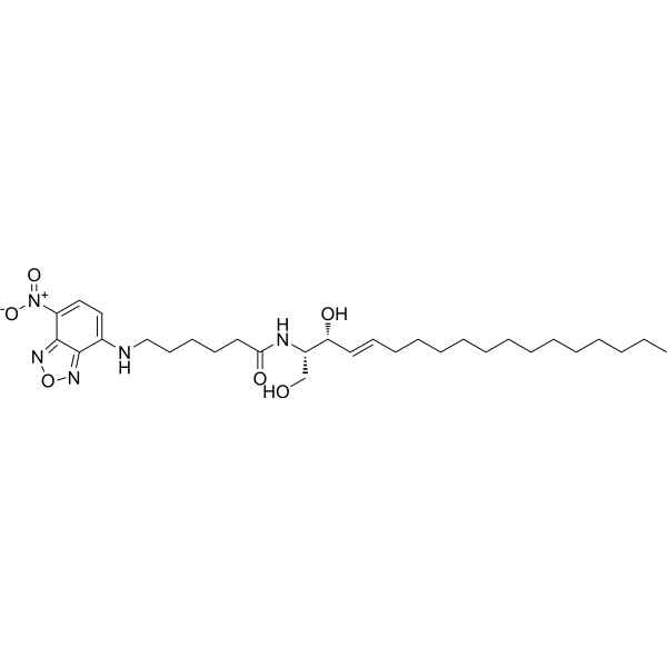
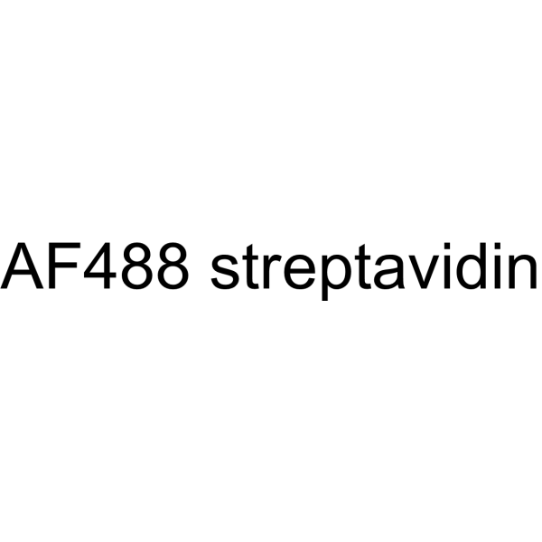
AF488 streptavidin is a fluorescently labeled streptavidin. AF488 streptavidin is a streptavidin conjugated to Alexa Fluor 488, with 4 fluorophores per protein molecule, enabling stoichiometric fluorescent labeling of cell surface targets. AF488 streptavidin can form a complex with biotinylated E07 aptamer to stain cells expressing EGFR, and the staining is reversible after treatment with mA9 detoxifying oligonucleotides (Ex/Em = 470/520 nm) .
|
-
- HY-123429
-
|
|
Antibiotic
|
Infection
|
|
CK0683A is a novel biaminourea antibiotic with antiplaque effects and demonstrated efficacy in experimental mouse dental disease models. A 12-week study in beagles compared its efficacy in terms of plaque and gingivitis reduction, staining potential, and safety. Dogs were selected as the study subjects because the onset and progression of canine periodontal disease is similar to that of humans. As a positive control for plaque and gingivitis reduction and staining, chlorhexidine acetate was selected because it is effective in both humans and dogs.
|
-

- HY-W011831
-
|
3-(Benzyldimethylammonio)propane-1-sulfonate; PDA
|
Biochemical Assay Reagents
|
Others
|
|
NDSB-256 is a non-stain remover sulfabetaine. NDSB-256 prevents protein aggregation and promotes denaturation of chemically and thermally denatured proteins .
|
-
- HY-D0166A
-
|
|
Biochemical Assay Reagents
|
Others
|
|
Neutral Red (IND) is an organic dye commonly used in biology and cytology laboratories. It can be used to stain living cells, secreted proteins and other molecular structures, etc., and has a wide range of applications in cell imaging and staining. In addition, Neutral Red (IND) is widely used in industrial fields such as water treatment, food processing and paper manufacturing, for example as an indicator or colorant. Although the compound has no direct medical application, it has important application value in the fields of biology, chemistry and industry.
|
-
- HY-D3172
-
|
|
Fluorescent Dye
|
Metabolic Disease
|
|
BD-105 is a glucagon-binding fluorescent probe with a Ka value of 13.3 μM. BD-105 exhibits changes in fluorescence intensity upon interaction with glucagon, and colocalizes with glucagon in cells and tissues. BD-105 is a selective cell stain that labels glucagon-secreting cells without staining insulin-secreting cells or non-endocrine control cells. BD-105 serves as an imaging reagent for glucagon in live cells and tissues .
|
-

- HY-123479
-
|
|
Antibiotic
|
Infection
|
|
CK0492B is a novel biaminourea antibiotic that has antiplaque properties in vitro and has shown efficacy in an experimental mouse dental disease model. A 12-week study in beagles compared its efficacy in reducing plaque and gingivitis, staining potential, and safety. Dogs were selected as the study subjects because the onset and progression of canine periodontal disease are similar to humans. Chlorhexidine acetate was selected as a positive control for plaque and gingivitis reduction and staining because it is effective in both humans and dogs.
|
-

- HY-D1615
-
|
|
Fluorescent Dye
|
Others
|
|
BODIPY 530/550 NHS ester can be used for the stain of protein. BODIPY 530/550 NHS ester can be used for fluorescence OIM (oblique illumination microscopic) image .
|
-
- HY-139440
-
|
|
Biochemical Assay Reagents
|
Others
|
|
DL-Leucyl-DL-phenylalanine is a dipeptide and can be used as a substrate to detecte two regions of dipeptidase staining on a gel in Drosophila simulans as well as in Drosophila melanogaster .
|
-

- HY-107629
-
|
|
Fluorescent Dye
|
Cancer
|
|
Flutax 1 is a fluorescent Paclitaxel (HY-B0015) derivative, which can be used for microtubule staining (Ex/Em = 495 nm/520 nm) .
|
-
- HY-108166
-
|
|
Fluorescent Dye
|
Inflammation/Immunology
|
|
Hydroxystilbamidine, a dye capable of binding to both DNA and RNA, is a powerful inhibitor of cellular ribonucleases. Hydroxystilbamidine is a retrograde fluorescent tracer and a histochemical stain [1]
|
-
- HY-A0216
-
|
|
Fluorescent Dye
DNA Stain
|
Others
|
|
Hydroxystilbamidine diisethionate, a dye capable of binding to both DNA and RNA, is a powerful inhibitor of cellular ribonucleases. Hydroxystilbamidine diisethionate is a retrograde fluorescent tracer and histochemical stain .
|
-

- HY-D0127
-
|
|
Fluorescent Dye
|
Others
|
|
Merocyanin 540 is a fluorescent membrane probe that selectively stains the membranes of a wide variety of electrically excitable cells, but not those of nonexcitable cells (Ex/Em: 540/580 nm) .
|
-
- HY-P990810
-
|
|
Arenavirus
|
Infection
|
|
Anti-LCMV Nucleoprotein Antibody (VL-4) is rat-derived IgG2a κ type antibody inhibitor, targeting to LCMV nucleoprotein. Anti-LCMV Nucleoprotein Antibody (VL-4) reacts with lymphocytic choriomeningitis virus (LCMV) nucleoprotein. Anti-LCMV Nucleoprotein Antibody (VL-4) stain LCMV-infected cell internally with no surface staining. Anti-LCMV Nucleoprotein Antibody (VL-4) can be used for the detections of immunohistochemistry and flow cytometry in LCMV infection .
|
-

- HY-101888R
-
|
|
Fluorescent Dye
Reference Standards
|
Neurological Disease
|
|
Cresyl Violet acetate (Standard) is the analytical standard of Cresyl Violet acetate (HY-101888). This product is intended for research and analytical applications. Cresyl Violet acetate is a dye, which can be used to stain neurons.
|
-

- HY-D1543
-
|
|
DNA Stain
|
Infection
|
|
Pyronin B is an organic cationic dye used for the staining of bacteria, mycobacteria and ribonucleic acids. Pyronin B is also used as a small hydrophobic (SH) protein channel inhibitor .
|
-
- HY-W133997
-
|
|
Fluorescent Dye
|
Others
|
|
Chromotrope 2R can be used as a chromogenic analytical probe for the quantification of proteins. Basic proteins stained red and the peak wavelength red shifts from 501.6 nm to 567 nm .
|
-
- HY-117070
-
|
|
Fluorescent Dye
|
Others
|
|
TO-PRO-3 iodide is a cell-impermeant nucleic acid stain. TO-PRO-3 iodide is useful as a nuclear counterstain and dead cell indicator (Ex/Em = 642/661 nm).
|
-
- HY-DY1050
-
|
|
DNA Stain
|
Others
|
Ethidium bromide (solution) is an intercalating agent commonly used as a fluorescent tag (nucleic acid stain) in molecular biology laboratories for techniques such as agarose gel electrophoresis.
Solvent and Concentration: Sterile water: 10 mg/Ml
|
-

- HY-N12515
-
|
|
Others
|
Metabolic Disease
|
|
Pinuseldarone (compound 1), extracted from Pinus eldarica needles regulats brown adipogenesis and thermogenesis .
|
-
- HY-D1585
-
|
|
Fluorescent Dye
|
Others
|
|
BODIPY TR methyl ester is a lipophilic GFP Counterstain. BODIPY TR methyl ester dye readily permeates cell membranes and localizes in endomembranous organelles but not localize strongly in plasma membranes. BODIPY TR methyl ester is an excellent red fluorescent vital dye (Ex=568 nm, Em=625 nm), can be used to reveal the location and shapes of cell nuclei, the shapes of cells within embryonic tissues, as well as the bound aries of organ-forming tissues within the whole embryo .
|
-
- HY-D0721
-
|
6-Carboxyfluorescein diacetate
|
Fluorescent Dye
|
Others
|
|
6-CFDA is a common aliphatic luciferin-line organism. CFDA conducts free diffusion into cells, and then it is hydrolyzed into carboxyl fluorescein (CF) by intracellular non-specific lipase. CF containing portion contains an additional negative charge so that it is better retained in cells, compared to fluorescein dyes .
|
-
- HY-D1121
-
|
|
Fluorescent Dye
|
|
|
Acid black 24 is a black agent whose staining effect is effectively removed by nanoscale zero-valent iron (NZVI) particles. The maximum unit removal capacity is 609.4 mg of dye per gram of NZVI.
|
-
- HY-W088190
-
|
Cyclohexyl ketone
|
Drug Intermediate
Biochemical Assay Reagents
|
Others
|
|
Dicyclohexyl ketone (Cyclohexyl ketone) is a toxic compound that causes toxic effects in rats at high doses, including death, decreased activity, staining of the fur on the lower body, reddening of the tears, and reproductive and developmental effects.
|
-

- HY-Y0016R
-
|
Basic Violet 10 (Standard); Brilliant Pink B (Standard); Rhodamine O (Standard); Tetraethylrhodamine (Standard)
|
Fluorescent Dye
Reference Standards
|
Others
|
|
Rhodamine B (Standard) is the analytical standard of Rhodamine B. This product is intended for research and analytical applications. Rhodamine B is a staining fluorescent dye, commonly used for dyeing textiles, paper, soap, leather, and agents.
|
-

- HY-15922A
-
|
|
Fluorescent Dye
|
Others
|
|
Luminol sodium salt is a chemical that exhibits chemiluminescence with pKa values of 6.74 and 15.1. Luminol sodium salt exhibits chemiluminescence (CL) at 425 nm λmax. Luminol sodium salt is commonly used in forensics as a diagnostic tool for the detection of blood stains .
|
-
- HY-D0021S
-
|
EtBr-d5; Homidium-d5 bromide
|
Isotope-Labeled Compounds
DNA Stain
|
Others
|
|
EthD-d5 bromide is the deuterium labeled Ethidium bromide. EthD bromide is an intercalating agent commonly used as a fluorescent tag (nucleic acid stain) in molecular biology laboratories for techniques such as agarose gel electrophoresis.
|
-

- HY-15922
-
Luminol
2 Publications Verification
Diogenes reagent
|
Fluorescent Dye
|
Others
|
|
Luminol is a chemical that exhibits chemiluminescence with pKa values of 6.74 and 15.1. Luminol exhibits chemiluminescence (CL) at 425 nm λmax. Luminol is commonly used in forensics as a diagnostic tool for the detection of blood stains .
|
-
- HY-W075283
-
|
1-(4-Aminophenyl)-1,2,2-triphenylethene
|
|
Others
|
|
4-(1,2,2-Triphenylvinyl)aniline (1-(4-Aminophenyl)-1,2,2-triphenylethene) is a tetraphenylethene-based aggregation-induced luminescent molecule. 4-(1,2,2-Triphenylvinyl)aniline exhibits weak or no fluorescence in dilute solution/dispersed state, and strong fluorescence with increased quantum yield when aggregated into nanoparticles .
|
-

- HY-Y0700
-
|
|
Fluorescent Dye
|
Others
|
|
Calconcarboxylic acid, an azo dye, acts as a silver-ion sensitizer to stain protein in SDS-PAGE gels. Calconcarboxylic acid increases silver binding on protein bands or spots by the formation of a silver-dye complex and also increases the reducing power o
|
-
- HY-15925
-
|
NBT
|
Fluorescent Dye
|
Others
|
|
Nitro blue tetrazolium chloride (NBT) is a substrate for dehydrogenases; is used with the alkaline phosphatase substrate 5-Bromo-4-Chloro-3-Indolyl Phosphate (BCIP) in western blotting and immunohistological staining procedures .
|
-
- HY-P3430
-
|
|
Biochemical Assay Reagents
|
Cancer
|
|
JM3A is a highly specific peptoid reagent that targets newly appears cell surface vimentin (CSV) on tumor-transformed early lung cancer cells. JM3A can detect and stain CSV by coupling with fluorophores .
|
-
- HY-D1353
-
|
|
Fluorescent Dye
|
Others
|
|
LipidGreen 2 is a second generation small molecule probe for lipid imaging. LipidGreen 2 has a better fluorescence signal compared with the previous LipidGreen, and selectively stains neutral lipids in cells and fat deposits in live zebrafish .
|
-
- HY-D1144
-
|
|
Fluorescent Dye
|
|
|
Acid blue 260 is an azo dye whose staining effect is effectively removed by multi-walled carbon nanotubes (MWCNTs). At 298 K, the adsorption capacity of MWCNT is 233.34 mg/g; and increases with the increase of dye concentration and temperature.
|
-
- HY-W110798
-
|
|
Environmental Pollutants
Biochemical Assay Reagents
|
Others
|
|
Bromophenol blue indicator (3.0-4.6) is a synthetic dye commonly used as an acid-base indicator with a transition range of pH 3.0-4.6. Bromophenol blue indicator (3.0-4.6) is water soluble and changes color from yellow to blue as the pH of the solution changes from acidic to basic. Its unique chemical properties make it an important ingredient in a variety of scientific applications, especially in biochemistry and molecular biology. In addition, it can be used as a stain in microbiology and histology. However, Bromophenol blue indicator (3.0-4.6) has potential irritating and staining properties.
|
-
- HY-W007967R
-
-

- HY-W007967
-
|
|
Biochemical Assay Reagents
|
Others
|
|
4-Chloronaphthalen-1-ol is a substrate of horseradish peroxidase. 4-Chloronaphthalen-1-ol can be used to visualize protein bands in western blotting and immunohistochemical staining .
|
-
- HY-Y0695
-
|
Naphthol Blue Black
|
Environmental Pollutants
Fluorescent Dye
|
Others
|
|
Amido Black 10B (Naphthol Blue Black) is a highly toxic azo dye. Amido Black 10B is genotoxic and mutagenic. Amido Black 10B can cause respiratory problems. Amido Black 10B is used for amino acid dyeing .
|
-
- HY-D1630
-
|
|
Fluorescent Dye
|
Others
|
|
4-Di-10-ASP is a fluorescent lipophilic tracer (Excitation 485 nm; Emission 620 nm). 4-Di-10-ASP can be used to stain phospholipid membranes in a specific manner .
|
-
- HY-D0714
-
|
2,3,5-Triphenyltetrazolium chloride; TPTZ; TTC
|
Biochemical Assay Reagents
|
Cardiovascular Disease
|
|
Tetrazolium Red (2,3,5-Triphenyltetrazolium chloride; TTC) is a not brain-penetrant, colorless, water-soluble dye that is reduced by mitochondrial enzymes to a deep red, water-insoluble compound (formazan) mainly in the mitochondria of living cells. Tetrazolium Red is used to observe the activity of dehydrogenase, and it turns colorless to red when exposed to hydrogen. Tetrazolium Red distinguishes between surviving and infarcted brain tissue after stroke. Tetrazolium Red has been used to stain heart tissue to measure the extent of acute lesions and also used to stain brain tissue to detect the size of the infarcted area. The absorption wavelength of Tetrazolium Red is 570 nm .
|
-
- HY-D3334
-
|
|
Fluorescent Dye
CD74
|
Others
|
|
PE-CF594 is a labeled monoclonal antibody conjugate that specifically binds to HLA-DR on the surface of monocytes and B cells, while acting as a signal attenuator. Through steric hindrance and a possible fluorescence resonance energy transfer mechanism, PE-CF594 specifically reduces the fluorescence intensity of PE-CD124 staining, but does not interfere with the staining of other PE-labeled antibodies such as CD40, CD4 or CD14. PE-CF594 can also be used to detect the emission signal of mt-Keima after excitation with a 561-nm laser, thereby effectively evaluating mitophagy activity .
|
-

- HY-D3410
-
|
|
Fluorescent Dye
GLUT
|
Others
|
|
CDr17 is a GLUT1 substrate and selective fluorescent dye staining M1 microphages. CDr17 utilizes the Gating-Oriented Live-cell Distinction (GOLD) mechanism to enter M1 macrophages (Ex/Em = 646/662 nm) .
|
-

- HY-W761989
-
|
|
Fluorescent Dye
|
Others
|
|
Direct Brown 95, a synthetic dye, finds extensive application in the textile industry for imparting color to fabrics. It serves as a stain in histology and cytology, aiding in the differentiation of diverse cell types. Additionally, it serves as a marker in molecular biology, facilitating the detection of DNA and RNA.
|
-

- HY-W440917
-
|
|
Liposome
|
Others
|
|
DSPE-PEG5000-FITC is a fluorescein attached PEG lipid. It can be used to prepare liposomes as drug carrier in targeted drug delivery. The polymer is modified with fluorescein (green) dye which can be used for staining cells, tissues, biomarkers, or nanoparticles.
|
-

- HY-W440934
-
|
|
Liposome
|
Others
|
|
Stearic acid-PEG2000-Rhodamine is an amphiphilic PEG polymer which can form micelles in water. The rhodamine can be used for staining sample and easily traced by fluorescence microscopy. Rhodamine has maximum absorption at 570 nm and emission around 595 nm.
|
-
- HY-W110781
-
|
|
DNA Stain
|
Others
|
|
Basic Blue 20 is a very convenient red-emitting DNA stains. Basic Blue 20 has relatively narrow excitation and emission spectra, with peaks at 633 and 677 nm, respectively. Basic Blue 20 also has a very high resistance to photobleaching .
|
-
- HY-W440916
-
|
|
Liposome
|
Others
|
|
DSPE-PEG3400-FITC is a fluorescein attached PEG lipid. It can be used to prepare liposomes as drug carrier in targeted drug delivery. The polymer is modified with fluorescein (green) dye which can be used for staining cells, tissues, biomarkers, or nanoparticles.
|
-

- HY-D1426
-
|
|
Fluorescent Dye
|
Neurological Disease
|
|
Di-12-ANEPPQ is a fast-responding membrane potential dye. Di-12-ANEPPQ, the lipophilic dye, shows cell-specific loading and Golgi-like staining patterns with minimal background fluorescence in the slices of neocortex and hippocampus .
|
-
- HY-D0001
-
|
|
Fluorescent Dye
|
Others
|
|
Alcian Blue 8GX is a commonly used phthalocyanine dye that binds to glycoproteins and glycosaminoglycans. Alcian Blue 8GX has a wide range of applications in biological staining, including proteins in brain tumors and DNA in cells and tissues .
|
-

- HY-D1074
-
|
3,3'-Dipropyloxacarbocyanine iodide
|
Fluorescent Dye
|
Others
|
|
DiOC3(3) (3,3'-Dipropyloxacarbocyanine iodide) is a green fluorescent lipophilic dye with cell membrane permeability. DiOC3(3) can be used to stain cell membranes and other lipid-soluble biological structures .
|
-
- HY-DY1074
-
|
|
Fluorescent Dye
|
Others
|
ER-Tracker dye is a derivative of BODIPY series dyes coupled with Glibenclamide (HY-15206) , highly selective binding to the endoplasmic reticulum, non-toxic to cells at low concentrations, this type of dye is an environmentally sensitive probe, and formaldehyde treatment can still retain part of the fluorescence, with high fluorescence life, good extinction coefficient and other characteristics. Glibenclamide is an atp-dependent K + channel blocker (Kir6, KATP) and CFTR Cl-channel blocker that binds in the endoplasmic reticulum. ER-Tracker is not suitable for staining fixed cells. Staining followed by fixation is possible, but cells fixed with aldehydes will only retain partial fluorescence .
Solvent and concentration: DMSO: 1 mM
|
-

- HY-D1429
-
|
|
Fluorescent Dye
|
Others
|
|
ER-Tracker dye is a derivative of BODIPY series dyes coupled with Glibenclamide (HY-15206), highly selective binding to the endoplasmic reticulum, non-toxic to cells at low concentrations, this type of dye is an environmentally sensitive probe, and formaldehyde treatment can still retain part of the fluorescence, with high fluorescence life, good extinction coefficient and other characteristics. Glibenclamide is an atp-dependent K + channel blocker (Kir6, KATP) and CFTR Cl-channel blocker that binds in the endoplasmic reticulum. ER-Tracker is not suitable for staining fixed cells. Staining followed by fixation is possible, but cells fixed with aldehydes will only retain partial fluorescence (Ex/Em = 374/ 430-640 nm) .
|
-
- HY-NP078A
-
|
PSA (FITC)
|
Biochemical Assay Reagents
CD3
|
Inflammation/Immunology
Endocrinology
|
|
Pisum sativum Agglutinin (PSA) FITC is a plant lectin conjugated with FITC (HY-66019). Pisum sativum Agglutinin FITC serves as an acrosome stain for detecting the acrosomal status of sperm. Pisum sativum Agglutinin exhibits lymphocyte mitogenic and immunomodulatory activities .
|
-

- HY-D0953
-
|
Solvent Blue 38
|
Environmental Pollutants
Fluorescent Dye
|
Neurological Disease
|
|
Direct Blue 86 (Solvent Blue 38) is a myelin-sheath stain, commonly utilized in microscopy to detect demyelination in the central nervous system. Direct Blue 86 also is a dye with various applications including as a commercial dye in the printing of cotton and mucilage glue fabrics .
|
-
- HY-D1775
-
|
|
Fluorescent Dye
|
Neurological Disease
Cancer
|
|
LysoTracker blue DND-22 is a blue fluorescent dye (Ex/Em: 373/422 nm). LysoTracker blue DND-22 stains acidic regions in living cells. LysoTracker blue DND-22 is used in the researches of neurodegenerative diseases and leukemia .
|
-
- HY-D2972
-
|
|
Fluorescent Dye
Apoptosis
Tim3
|
Cancer
|
|
Apotracker Red is a fluorogenic peptide (excitation/emission: 561/610 nm). Apotracker Red binds to PtdSer on the surface of cells. Apotracker Red rapidly and selectively stains Apoptotic cells but not viable cells. Apotracker Red can be used to detect cancer cell death in real time .
|
-

- HY-D1249
-
|
|
Fluorescent Dye
|
Others
|
|
Calcein (mixture of isomers), Calcein (HY-D0040) Derivative, is a fluorescent dye (Ex/Em = 495/515 nm). Calcein (mixture of isomers) can be used for the researches for live cell staining, calcium ion detection, bone fluorescence labeling and membrane permeability leakage detection.
|
-
- HY-D0933
-
-
- HY-103305
-
|
|
Fluorescent Dye
Calcium Channel
|
Others
|
|
cis-Ned19 is an irreversible nicotinic acid adenine dinucleotide phosphate (NAADP) antagonist. cis-Ned19 localizes to lysosomes and endolysosomal vesicles, stains two-pore calcium channels and fluorescently labels NAADP receptors (Ex/Em = 365/410 nm) .
|
-
- HY-DY1017
-
|
|
Antibiotic
Fungal
|
Infection
|
Filipin complex (solution) is a potent polyene macrolide antifungal antibiotic. Filipin complex inserts into membranes and sequester cholesterol into complexes and inhibits PRRSV entry. The Filipin complex consists of about 75.8% Filipin III (HY-N6718) , 10.8% Filipin IV, 9.1% Filipin II, and 1.2% Filipin I (Ex/Em = 380/430 nm) .
Solvent and concentration: DMSO: 5 mg/mL
|
-

- HY-N6716
-
|
|
Fungal
Antibiotic
|
Infection
|
|
Filipin complex is a potent polyene macrolide antifungal antibiotic. Filipin complex inserts into membranes and sequester cholesterol into complexes and inhibits PRRSV entry. The Filipin complex consists of about 75.8% Filipin III (HY-N6718), 10.8% Filipin IV, 9.1% Filipin II, and 1.2% Filipin I (Ex/Em = 380/430 nm) .
|
-
- HY-NP0147
-
|
WGA (Fluorescein)
|
Fluorescent Dye
|
Infection
Neurological Disease
|
|
Wheat Germ Agglutinin (WGA) Fluorescein is a classic fluorescent label that specifically binds to sugar residues such as N-acetylglucosamine, N-acetylneuraminic acid and sialic acid. Wheat Germ Agglutinin Fluorescein performs regionally differential fluorescent staining of the ocular surface epithelial glycocalyx to assess its integrity, and causes no damage to the eye at safe concentrations. Wheat Germ Agglutinin Fluorescein is also used for staining structures including red blood cells, cultured cells, bacteria and pine wood nematodes, and facilitates the isolation of wheat-associated plant-growth-promoting rhizobacterial strains. Wheat Germ Agglutinin Fluorescein can be applied to the detection of ocular glycocalyx integrity and the research of related diseases such as pine wilt disease .
|
-

- HY-N3290R
-
|
Methyl p-methoxyphenylpropionate (Standard)
|
Others
Reference Standards
|
Others
|
|
Methyl p-methoxyhydrocinnamate (Standard) (Methyl p-methoxyphenylpropionate (Standard)) is the analytical standard of Methyl p-methoxyhydrocinnamate (HY-N3290). This product is intended for research and analytical applications. Methyl p-methoxyhydrocinnamate is extracted from in liquid culture by the blue stain fungus Ophiostoma crassioaginata .
|
-

- HY-W133919
-
|
|
Environmental Pollutants
Biochemical Assay Reagents
|
Others
|
|
Aniline Blue sodium is a water-soluble dye commonly used as a biological stain for the detection of nucleic acids and proteins in various laboratory procedures such as electrophoresis and microscopy. Aniline Blue sodium has unique chemical properties that allow it to bind to specific cellular components, producing a color change that facilitates their visualization and analysis.
|
-
- HY-N15299
-
|
|
Endogenous Metabolite
|
Others
|
|
iso-C15:0 3-OH is a fatty acid that can be isolated from strain PLR T. PLR T is a rod shaped, Gram-stain-negative, chemo-organotrophic, heterotrophic, strictly aerobic bacterium isolated from faeces of the mollusc Aplysia punctata .
|
-

- HY-135056
-
|
|
Fluorescent Dye
|
Others
|
|
Mito-Tracker Green is a green fluorescent dye that selectively accumulates in the mitochondrial matrix. MitoTracker Green FM covalently binds mitochondrial proteins by reacting with free mercaptan of cysteine residues, allowing staining of mitochondrial membrane potential independent of membrane potential. Excitation/emission wavelength 490/523 nm.
|
-
- HY-101900R
-
|
Nile blue sulfate (Standard)
|
Fluorescent Dye
Reference Standards
|
Others
|
|
Nile Blue A sulfate (Standard) is the analytical standard of Nile Blue A sulfate (HY-101900). This product is intended for research and analytical applications. Nile Blue A (Nile blue sulfate) is used to differentiate melanins and lipofuscins. It is also useful for staining fats and preparation of an amperometric glucose sensor .
|
-

- HY-D0023
-
|
HPTS; Solvent Green 7
|
Environmental Pollutants
Fluorescent Dye
|
Others
|
|
Pyranine (HPTS; Solvent Green 7) is a cell-impermeant pH-sensitive fluorescent indicator. Pyranine acts as a class of fluorescent chemosensor for the Cu + ion. Pyranine is used as a coloring agent, biological stain, optical detecting reagent, and pH indicator (Ex/Em = 450/510 nm) .
|
-
- HY-D1723
-
|
|
DNA Stain
|
Others
|
|
EthD-III is a nucleic acid probe. EthD-III is a red fluorescent stain that can be used to detect dead cells. EthD-III enters cells with damaged membranes and binds to nucleic acids, resulting in bright red fluorescence in dead cells (Ex/Em=530/645 nm) .
|
-
- HY-137889
-
|
|
CMV
|
Infection
|
|
MLS8969 is a selective human cytomegalovirus (HCMV) entry inhibitor with an EC50 of 0.12 μM (HFFs). MLS8969 reduces viral protein levels and inhibits nuclear staining of HCMV pp65. MLS8969 can be used in studies related to human cytomegalovirus infection .
|
-

- HY-D3276
-
|
|
Fluorescent Dye
|
Others
|
|
PE-Cy7 is a tandem fluorescent dye composed of R-phycoerythrin (PE) coupled with the near-infrared dye Cy7. It is primarily excited by a 488 nm laser, with a maximum emission wavelength of approximately 780 nm. PE-Cy7 is widely used in flow cytometry and immunofluorescence staining.
|
-

- HY-D0023A
-
|
HPTS hydrate; Solvent Green 7 hydrate
|
Fluorescent Dye
|
Others
|
|
Pyranine (HPTS; Solvent Green 7) hydrate is a cell-impermeant pH-sensitive fluorescent indicator. Pyranine hydrate acts as a class of fluorescent chemosensor for the Cu + ion. Pyranine hydrate is used as a coloring agent, biological stain, optical detecting reagent, and pH indicator (Ex/Em = 450/510 nm) .
|
-
- HY-D1359
-
|
|
Fluorescent Dye
|
Others
|
|
Mito Red is a vital dye and mitochondrial stain that can be used to detect and evaluate mitochondrial function and status. Mito Red accumulates in mitochondria, and its fluorescence intensity is positively correlated with mitochondrial membrane potential. When the mitochondrial membrane potential increases, the fluorescence signal of Mito Red increases .
|
-
- HY-183274
-
|
|
Nuclear Hormone Receptor 4A/NR4A
Apoptosis
PARP
|
Cancer
|
|
Nur77 modulator 5 is a Nur77 modulator. Nur77 modulator 5 induces lysosomal dysfunction, impaired autophagic flux, and apoptosis with increased PARP cleavage, TUNEL positivity, and Annexin V/PI staining. Nur77 modulator 5 can be used for the research of gastric cancer .
|
-

- HY-D0116
-
|
HPTS free acid; Solvent Green 7 free acid
|
Fluorescent Dye
|
Others
|
|
Pyranine (HPTS; Solvent Green 7) free acid is a pH-sensitive fluorescent indicator. Pyranine free acid acts as a class of fluorescent chemosensor for the Cu + ion. Pyranine free acid is used as a coloring agent, biological stain, optical detecting reagent, and pH indicator (Ex/Em = 450/510 nm) .
|
-
- HY-D0286
-
|
2,8-Dimethylnaphtho[3,2,1-kl]xanthene
|
Fluorescent Dye
|
Others
|
|
Fluorol Yellow 088 is a fluorescent dye that can be used as a biological material or an organic compound for life science research. Fluorol Yellow 088 is suitable for lipid staining in plant tissues, particularly for the fluorescence imaging of suberized cell walls and hydrophobic structures, with an excitation wavelength of 365 nm and an emission wavelength of ≥420 nm .
|
-
- HY-D2222
-
|
|
Fluorescent Dye
|
Others
|
|
5-HMSiR-Hoechst is a DNA probe. 5-HMSiR-Hoechst comprises Hoechst 33258 and spontaneously blinking far-red hydroxymethyl silicon-rhodamine (HMSiR). 5-HMSiR-Hoechst can stain living cells. Ex/Em=640 nm/675 nm .
|
-
- HY-P11714
-
|
|
EBV
MHC
|
Infection
Cancer
|
|
EBNA3 246-254 (RYSIFFDYM) is an EBV peptide. EBNA3 246-254 is submitted by HLA-A24. EBNA3 246-254 is amplified in vitro, followed by HLA multimer staining to measure EBV-specific CTLs. EBNA3 246-254 can be used for the research of lung cancer .
|
-

- HY-D3173
-
|
TPG-456
|
Fluorescent Dye
GCGR
|
Metabolic Disease
|
|
TP-α is a glucagon-selective two-photon fluorescent probe with a Kd of 65 μM. TP-α directly interacts with glucagon to produce significant fluorescence enhancement. TP-α selectively stains viable glucagon-secreting α cells in pancreatic islets, enabling visualization of their localization, distribution and availability. TP-α is applicable to diabetes research .
|
-

- HY-NP0158
-
|
UEA I (Rhodamine)
|
Fluorescent Dye
|
Others
|
|
Ulex Europaeus Agglutinin I Rhodamine (UEA I Rhodamine) is a fluorescently labeled lectin. Ulex Europaeus Agglutinin I Rhodamine exhibits specificity for cell surface glycoconjugates containing α-L-fucosyl groups. Ulex Europaeus Agglutinin I Rhodamine can be used in combination with other lectins for double staining .
|
-

- HY-DY1032
-
|
|
Fluorescent Dye
|
Others
|
Mito-Tracker Green (solution) is a green fluorescent dye that selectively accumulates in the mitochondrial matrix. MitoTracker Green FM covalently binds mitochondrial proteins by reacting with free mercaptan of cysteine residues, allowing staining of mitochondrial membrane potential independent of membrane potential. Excitation/emission wavelength 490/523 nm.
Solvent and concentration: DMSO: 1 mM
|
-

- HY-135009
-
|
DASPI
|
G-quadruplex
|
Others
|
|
2-Di-1-ASP (DASPI; Compound 18a) is a mono-stryryl dye, and widely used as mitochondrial stain and groove-binding fluorescent probes for double-stranded DNA. 2-Di-1-ASP is selective for G-quadruplex (G4) and double-stranded DNA .
|
-
- HY-D3419
-
|
|
Fluorescent Dye
|
Neurological Disease
|
|
Neuro-DiO 4-chlorobenzenesulfonate is a hydrophobic C18 alkyl chain carbocyanine dye with green fluorescence, commonly used as a vascular marker, cell internalizer and deposition agent. Neuro-DiO 4-chlorobenzenesulfonate inserts its alkyl chain into the endothelial plasma membrane via liposome-mediated perfusion to achieve vascular labeling. Neuro-DiO chlorobenzenesulfonate can also stain the cell membrane and cytoplasm of cancer cells to assist in confocal microscopy observations. Neuro-DiO chlorobenzenesulfonate can be released from nanosponges and accumulate on the surface of mouse retina, then internalize into retinal ganglion cells, which is applicable to researches related to glaucoma and other diseases. It should be noted that during liposome-mediated vascular staining in mice, Neuro-DiO 4-chlorobenzenesulfonate may cause leakage of airway lavage fluid .
|
-

- HY-125962
-
|
|
Fluorescent Dye
Amyloid-β
|
Neurological Disease
|
|
X-34 is a lipophilic and bright yellow-green fluorescent derivative of Congo red (HY-D0236). X-34 can be used to stain neuritic and diffuse plaques, neurofibrillary tangles (NFTs), neuropil threads, and cerebrovascular amyloid in the brain. X-34 can be used for research of Alzheimer’s disease .
|
-
- HY-W440938
-
|
|
Liposome
|
Others
|
|
Stearic acid-PEG2000-FITC is an amphiphatic polyPEG which can self assemble to form micelles in water. The polymer can be used to encapsulate therapeutic agent. FITC is a green dye with peak absorption at 494 nm and maximum emission at 520 nm and can be used for staining biological samples or nanoparticles. FITC can be easily traced by fluorescence microscopy.
|
-

- HY-118907
-
|
|
Fluorescent Dye
|
Others
|
|
Methyl Orange is a soluble azo dye commonly used as an acid-base indicator and for staining cells and tissue sections, as well as for dyeing textiles. Methyl Orange appears red at a pH of 3.1 and changes to bright yellow as the pH increases to 4.4. Methyl Orange (500 mg/L) exhibits cytotoxicity and can cause DNA damage .
|
-
- HY-158725
-
|
|
Endogenous Metabolite
|
Others
|
|
N-Octadecenoyl-(cis-9) -Sulfatide is a glycolipid found in the brains of mice. N-Octadecenoyl-(cis-9) -Sulfatide can be used as a standard for the determination of C18:13 '-mergogalactosyllactose in dry blood stains of patients with chromatic leukodystrophy (MLD) by ultra-high performance liquid chromatography-mass spectrometry (UPLC-MS) .
|
-

- HY-122266
-
|
|
Biochemical Assay Reagents
|
Metabolic Disease
|
|
Orcein is an irreversible stain that specifically targets elastic fibers and can interact hydrophobically with the protein components in elastic fibers. Orcein makes elastic fibers in tissues appear purple or purple-red. Orcein can be used for morphological studies of Drosophila polytene chromosomes and for qualitative and quantitative analysis of elastic fibers, collagen fibers and other components in atherosclerotic plaques .
|
-

- HY-W440939
-
|
|
Liposome
|
Others
|
|
Stearic acid-PEG3400-FITC is a PEG lipid which forms micelles in water and can be used for drug delivery applications. The FITC fluorescent can be easily traced by miscroscopy. FITC is a green dye with peak absorption at 494 nm and maximum emission at 520 nm and can be used for staining biological samples or nanoparticles. FITC can be easily traced by fluorescence microscopy.
|
-
- HY-W440940
-
|
|
Liposome
|
Others
|
|
Stearic acid-PEG5000-FITC is a PEG lipid which forms micelles in water and can be used for drug delivery applications. The FITC fluorescent can be easily traced by miscroscopy. FITC is a green dye with peak absorption at 494 nm and maximum emission at 520 nm and can be used for staining biological samples or nanoparticles. FITC can be easily traced by fluorescence microscopy.
|
-
- HY-113383
-
-

- HY-D2208
-
|
|
Fluorescent Dye
|
Others
|
|
SYTM Green is a cell impermeant DNA dye that fluoresces green by binding to dsDNA (Ex/Em=503/530 nm). SYTM Green can also be used to stain bacteria, including Gram-positive and Gram-negative bacteria. Note: SYTM Green cannot penetrate living cells, and this product is equivalent to (Ex/Em=488/530 nm) .
|
-

- HY-D2346
-
|
|
Fluorescent Dye
|
Others
|
|
HBmito Crimson is a deep red fluorescent probe (λex: 658 nm, λem: 678 nm) for the inner mitochondrial membrane. HBmito Crimson is a cell membrane-permeable probe with high selectivity for the mitochondrial inner membrane, suitable for specific fluorescence staining of the inner mitochondrial membrane in living cells. HBmito Crimson has high photostability and brightness, suitable for long-term dynamic fluorescence imaging.
|
-

- HY-D0128
-
|
7-Methoxy-4-methylcoumarin
|
Bacterial
|
Infection
|
|
4-Methylherniarin (7-Methoxy-4-methylcoumarin) is a coumarin derivative and fluorescent label, has an antimicrobial activitiy against both gram positive and gram negative bacterial stains. 4-Methylherniarin displays good activity against B. subtilis and S.sonnei with IC50 values of 11.76 μg/ml and 13.47 μg/ml .
|
-
- HY-15625
-
|
|
DNA Stain
|
Others
|
|
Hoechst 33258 analog 3 is a marker dye in Hoechst series. Hoechst is A live nuclear marker dye. Hoechst binds to the grooves in the DNA double strand, which tends to be A/ T-rich DNA strand. Although it binds to all nucleic acids, the A/ T-rich double strand DNA significantly enhances fluorescence intensity Therefore,Hoechst dye can be used for living cell labeling. The fluorescence intensity of Hoechst dye increases with the increase of pH of solution .
|
-
- HY-15559
-
|
bisBenzimide H 33342; HOE 33342
|
Autophagy
|
Others
|
|
Hoechst 33342 is a marker dye in Hoechst series. Hoechst is A live nuclear marker dye. Hoechst binds to the grooves in the DNA double strand, which tends to be A/ T-rich DNA strand. Although it binds to all nucleic acids, the A/ T-rich double strand DNA significantly enhances fluorescence intensity Therefore,Hoechst dye can be used for living cell labeling. The fluorescence intensity of Hoechst dye increases with the increase of pH of solution .
|
-
- HY-15422
-
|
|
Fluorescent Dye
|
Others
|
|
Dye 937, substituted unsymmetrical cyanine dyes with selected permeability, useful in the detection of DNA in electrophoretic gels.
|
-
- HY-15562
-
|
|
Fluorescent Dye
|
Others
|
|
HOE 32021 is a marker dye in Hoechst series. Hoechst is A live nuclear marker dye. Hoechst binds to the grooves in the DNA double strand, which tends to be A/ T-rich DNA strand. Although it binds to all nucleic acids, the A/ T-rich double strand DNA significantly enhances fluorescence intensity Therefore,Hoechst dye can be used for living cell labeling. The fluorescence intensity of Hoechst dye increases with the increase of pH of solution .
|
-
- HY-15561
-
|
meta-Hoechst
|
Fluorescent Dye
|
Cancer
|
|
HOE-S 785026 is a marker dye in Hoechst series. Hoechst is A live nuclear marker dye. Hoechst binds to the grooves in the DNA double strand, which tends to be A/ T-rich DNA strand. Although it binds to all nucleic acids, the A/ T-rich double strand DNA significantly enhances fluorescence intensity Therefore,Hoechst dye can be used for living cell labeling. The fluorescence intensity of Hoechst dye increases with the increase of pH of solution .
|
-
- HY-15559A
-
|
bisBenzimide H 33342 trihydrochloride; HOE 33342 trihydrochloride
|
Autophagy
|
Others
|
|
Hoechst 33342 trihydrochloride is a marker dye in Hoechst series. Hoechst is A live nuclear marker dye. Hoechst binds to the grooves in the DNA double strand, which tends to be A/ T-rich DNA strand. Although it binds to all nucleic acids, the A/ T-rich double strand DNA significantly enhances fluorescence intensity Therefore,Hoechst dye can be used for living cell labeling. The fluorescence intensity of Hoechst dye increases with the increase of pH of solution .
|
-
- HY-15619
-
|
Nuclear yellow
|
Fluorescent Dye
|
Others
|
|
Hoechst S 769121 is a marker dye in Hoechst series. Hoechst is A live nuclear marker dye. Hoechst binds to the grooves in the DNA double strand, which tends to be A/ T-rich DNA strand. Although it binds to all nucleic acids, the A/ T-rich double strand DNA significantly enhances fluorescence intensity Therefore,Hoechst dye can be used for living cell labeling. The fluorescence intensity of Hoechst dye increases with the increase of pH of solution .
|
-
- HY-15560
-
|
HOE 34580
|
Amyloid-β
|
Neurological Disease
|
|
Hoechst 34580 is a marker dye in Hoechst series. Hoechst is A live nuclear marker dye. Hoechst binds to the grooves in the DNA double strand, which tends to be A/ T-rich DNA strand. Although it binds to all nucleic acids, the A/ T-rich double strand DNA significantly enhances fluorescence intensity Therefore,Hoechst dye can be used for living cell labeling. The fluorescence intensity of Hoechst dye increases with the increase of pH of solution .
|
-
- HY-D0917
-
|
|
DNA Stain
|
Cancer
|
|
NIR-Red Dead Cell-1 Dye is a DNA-binding fluorescent dye for non-living cells (Ex/Em=515 nm/531 nm). NIR-Red Dead Cell-1 Dye can intercalate into base pairs of double-stranded DNA and produce stronger fluorescence. NIR-Red Dead Cell-1 Dye is suitable for necrotic cells or late apoptotic cells with damaged cell membranes, showing green fluorescence under fluorescence microscopy or flow cytometry. NIR-Red Dead Cell-1 Dye can be used to distinguish live cells from dead cells and distinguish cell membrane integrity. NIR-Red Dead Cell-1 Dye can be attached to the surface of Feraheme (FH) nanoparticles (NPs) to obtain fluorescent dye-functionalized NPs for drug delivery studies .
|
-
- HY-15630A
-
|
|
Fluorescent Dye
|
Others
|
|
Hoechst 33342 analog 2 trihydrochloride is a marker dye in Hoechst series. Hoechst is A live nuclear marker dye. Hoechst binds to the grooves in the DNA double strand, which tends to be A/ T-rich DNA strand. Although it binds to all nucleic acids, the A/ T-rich double strand DNA significantly enhances fluorescence intensity Therefore,Hoechst dye can be used for living cell labeling. The fluorescence intensity of Hoechst dye increases with the increase of pH of solution .
|
-
- HY-108166A
-
|
|
Fluorescent Dye
|
Infection
|
|
Hydroxystilbamidine bis(methanesulfonate) is a dye that can bind to DNA and RNA; it's a fluorescent cationic dye, often used as a retrograde neuronal tracer and has also been found to be a potent inhibitor of cellular ribonucleases.
|
-
- HY-15630
-
|
|
Fluorescent Dye
|
Cancer
|
|
Hoechst 33342 analog 2 is a marker dye in Hoechst series. Hoechst is A live nuclear marker dye. Hoechst binds to the grooves in the DNA double strand, which tends to be A/ T-rich DNA strand. Although it binds to all nucleic acids, the A/ T-rich double strand DNA significantly enhances fluorescence intensity Therefore,Hoechst dye can be used for living cell labeling. The fluorescence intensity of Hoechst dye increases with the increase of pH of solution .
|
-
- HY-15563
-
|
|
Fluorescent Dye
|
Others
|
|
HOE 33187 is a marker dye in Hoechst series. Hoechst is A live nuclear marker dye. Hoechst binds to the grooves in the DNA double strand, which tends to be A/ T-rich DNA strand. Although it binds to all nucleic acids, the A/ T-rich double strand DNA significantly enhances fluorescence intensity Therefore,Hoechst dye can be used for living cell labeling. The fluorescence intensity of Hoechst dye increases with the increase of pH of solution .
|
-
- HY-15631
-
|
|
Fluorescent Dye
|
Others
|
|
Hoechst 33258 analog 6 is a marker dye in Hoechst series. Hoechst is A live nuclear marker dye. Hoechst binds to the grooves in the DNA double strand, which tends to be A/ T-rich DNA strand. Although it binds to all nucleic acids, the A/ T-rich double strand DNA significantly enhances fluorescence intensity Therefore,Hoechst dye can be used for living cell labeling. The fluorescence intensity of Hoechst dye increases with the increase of pH of solution (Ex/Em = 356/451 nm) .
|
-
- HY-15558A
-
|
bisBenzimide H 33258 trihydrochloride; H 33258 trihydrochloride
|
Fluorescent Dye
|
Cancer
|
|
Hoechst 33258 trihydrochloride is a marker dye in Hoechst series. Hoechst is A live nuclear marker dye. Hoechst binds to the grooves in the DNA double strand, which tends to be A/ T-rich DNA strand. Although it binds to all nucleic acids, the A/ T-rich double strand DNA significantly enhances fluorescence intensity Therefore,Hoechst dye can be used for living cell labeling. The fluorescence intensity of Hoechst dye increases with the increase of pH of solution .
|
-

- HY-15561B
-
|
meta-Hoechst trihydrochloride
|
Fluorescent Dye
|
Others
|
|
HOE-S 785026 trihydrochloride is a marker dye in Hoechst series. Hoechst is A live nuclear marker dye. Hoechst binds to the grooves in the DNA double strand, which tends to be A/ T-rich DNA strand. Although it binds to all nucleic acids, the A/ T-rich double strand DNA significantly enhances fluorescence intensity Therefore,Hoechst dye can be used for living cell labeling. The fluorescence intensity of Hoechst dye increases with the increase of pH of solution .
|
-
- HY-15560B
-
|
HOE 34580 tetrahydrochloride
|
Amyloid-β
|
Neurological Disease
|
|
Hoechst 34580 tetrahydrochloride is a marker dye in Hoechst series. Hoechst is A live nuclear marker dye. Hoechst binds to the grooves in the DNA double strand, which tends to be A/ T-rich DNA strand. Although it binds to all nucleic acids, the A/ T-rich double strand DNA significantly enhances fluorescence intensity Therefore,Hoechst dye can be used for living cell labeling. The fluorescence intensity of Hoechst dye increases with the increase of pH of solution .
|
-
- HY-15626
-
|
|
Fluorescent Dye
|
Others
|
|
ortho-iodoHoechst 33258 is a marker dye in Hoechst series. Hoechst is A live nuclear marker dye. Hoechst binds to the grooves in the DNA double strand, which tends to be A/ T-rich DNA strand. Although it binds to all nucleic acids, the A/ T-rich double strand DNA significantly enhances fluorescence intensity Therefore,Hoechst dye can be used for living cell labeling. The fluorescence intensity of Hoechst dye increases with the increase of pH of solution .
|
-
- HY-15627
-
|
|
Fluorescent Dye
|
Others
|
|
Hoechst 33342 analog is a marker dye in Hoechst series. Hoechst is A live nuclear marker dye. Hoechst binds to the grooves in the DNA double strand, which tends to be A/ T-rich DNA strand. Although it binds to all nucleic acids, the A/ T-rich double strand DNA significantly enhances fluorescence intensity Therefore,Hoechst dye can be used for living cell labeling. The fluorescence intensity of Hoechst dye increases with the increase of pH of solution .
|
-
- HY-D1539
-
|
CY 5.5 azide; Lumiprobe CY 5.5 azide
|
Fluorescent Dye
|
Others
|
|
Cyanine 5.5 azide (CY 5.5 azide) is a potent fluorescent dye. Cyanine 5.5 azide can label DNA. Cyanine 5.5 azide can be used for NIR live organism imaging. (λex=684 nm, λem=710 nm) . Cyanine 5.5 azide is a click chemistry reagent, it contains an Azide group and can undergo copper-catalyzed azide-alkyne cycloaddition reaction (CuAAc) with molecules containing Alkyne groups. It can also undergo strain-promoted alkyne-azide cycloaddition (SPAAC) reactions with molecules containing DBCO or BCN groups.
|
-
- HY-D1444
-
|
|
Fluorescent Dye
Bacterial
|
Infection
|
|
Propidium monoazide is a cell-impermeant photoreactive DNA-binding dye that preferentially binds to dsDNA. Propidium monoazide (PMA) prevents DNA from dead bacteria from being amplified during the PCR. PMA-PCR enhanced both the specificity and the sensitivity of PCR . Propidium monoazide is a click chemistry reagent, it contains an Azide group and can undergo copper-catalyzed azide-alkyne cycloaddition reaction (CuAAc) with molecules containing Alkyne groups. It can also undergo strain-promoted alkyne-azide cycloaddition (SPAAC) reactions with molecules containing DBCO or BCN groups.
|
-
- HY-D0942
-
|
Euchrysine 3RX
|
Parasite
Fluorescent Dye
DNA Stain
|
Others
|
|
Acridine Orange (Euchrysine 3RX) zinc chloride salt is a cell-penetrable nucleic acid-selective fluorescent dye. Acridine Orange zinc chloride salt produces orange fluorescence when it binds to ssDNA or RNA, and green fluorescence when it binds to dsDNA (Ex: 488 nM; Em: green fluorescence at 530 nm, orange fluorescence at 640 nm) .
|
-
- HY-15629
-
|
|
DNA Stain
|
Others
|
|
HOE 32020 is a marker dye in Hoechst series. Hoechst is A live nuclear marker dye. Hoechst binds to the grooves in the DNA double strand, which tends to be A/ T-rich DNA strand. Although it binds to all nucleic acids, the A/ T-rich double strand DNA significantly enhances fluorescence intensity Therefore,Hoechst dye can be used for living cell labeling. The fluorescence intensity of Hoechst dye increases with the increase of pH of solution .
|
-
- HY-15623
-
|
|
DNA Stain
|
Others
|
|
Hoechst 33258 analog is a marker dye in Hoechst series. Hoechst is A live nuclear marker dye. Hoechst binds to the grooves in the DNA double strand, which tends to be A/ T-rich DNA strand. Although it binds to all nucleic acids, the A/ T-rich double strand DNA significantly enhances fluorescence intensity Therefore,Hoechst dye can be used for living cell labeling. The fluorescence intensity of Hoechst dye increases with the increase of pH of solution .
|
-
- HY-15622
-
|
|
DNA Stain
|
Cancer
|
|
meta-iodoHoechst 33258 is a marker dye in Hoechst series. Hoechst is A live nuclear marker dye. Hoechst binds to the grooves in the DNA double strand, which tends to be A/ T-rich DNA strand. Although it binds to all nucleic acids, the A/ T-rich double strand DNA significantly enhances fluorescence intensity Therefore,Hoechst dye can be used for living cell labeling. The fluorescence intensity of Hoechst dye increases with the increase of pH of solution .
|
-
- HY-15621A
-
-
- HY-15628
-
|
|
DNA Stain
|
Others
|
|
Hoechst 33258 analog 5 is a marker dye in Hoechst series. Hoechst is A live nuclear marker dye. Hoechst binds to the grooves in the DNA double strand, which tends to be A/ T-rich DNA strand. Although it binds to all nucleic acids, the A/ T-rich double strand DNA significantly enhances fluorescence intensity Therefore,Hoechst dye can be used for living cell labeling. The fluorescence intensity of Hoechst dye increases with the increase of pH of solution .
|
-
- HY-15624
-
|
|
DNA Stain
|
Cancer
|
|
Hoechst 33258 analog 2 is a marker dye in Hoechst series. Hoechst is A live nuclear marker dye. Hoechst binds to the grooves in the DNA double strand, which tends to be A/ T-rich DNA strand. Although it binds to all nucleic acids, the A/ T-rich double strand DNA significantly enhances fluorescence intensity Therefore,Hoechst dye can be used for living cell labeling. The fluorescence intensity of Hoechst dye increases with the increase of pH of solution .
|
-
- HY-15632
-
|
|
Fluorescent Dye
|
Others
|
|
para-iodoHoechst 33258 is a marker dye in Hoechst series. Hoechst is A live nuclear marker dye. Hoechst binds to the grooves in the DNA double strand, which tends to be A/ T-rich DNA strand. Although it binds to all nucleic acids, the A/ T-rich double strand DNA significantly enhances fluorescence intensity Therefore,Hoechst dye can be used for living cell labeling. The fluorescence intensity of Hoechst dye increases with the increase of pH of solution .
|
-
- HY-15621
-
-
- HY-15558
-
|
bisBenzimide H 33258; H 33258
|
Fluorescent Dye
DNA Stain
DNA Alkylator/Crosslinker
|
Others
|
|
Hoechst 33258 is a blue to blue-green fluorescent live cell dye that can label DNA. Hoechst 33258 can specifically bind to the minor groove of DNA (and tends to bind to A/T-rich DNA), resulting in a significant increase in fluorescence intensity. Hoechst 33258 can cross the cell membrane and cause changes in DNA structure, such as G2/M phase arrest. Hoechst 33258 can bind to live or fixed cells, and the fluorescence intensity increases with increasing solution pH. As a DNA-specific probe, Hoechst 33258 can be used to detect DNA content, analyze cell cycle, etc. The excitation wavelength of Hoechst 33258 is 350-365 nm, and the emission wavelength is 460-490 nm .
|
-
- HY-D1691
-
|
|
Fluorescent Dye
|
Others
|
|
BODIPY-581/591 NHS ester is a bright, red fluorescent dye (excitation: 581 nm; emission: 591 nm). BODIPY-581/591 NHS ester can be used for the labeling of amine containing biomolecules .
|
-
- HY-D1421
-
|
|
Fluorescent Dye
|
Others
|
|
PKH67 is a fluorescent cell binding dye with green fluorescence. PKH67 can stain the cell membrane and the Ex/Em is 490/502 nm. PKH67 is often used in combination with the non-specific red fluorescent dye PKH26 (Ex/Em=551/567 nm) to label cells, detect cell proliferation in vitro, and trace cells in vitro and in vivo .
|
-

- HY-W998679
-
|
N-(Fluorescein-5-carbamidoethyl)biotinamide
|
Fluorescent Dye
Endogenous Metabolite
|
|
|
Biotin (5-fluorescein) conjugate (N-(Fluorescein-5-carbamidoethyl)biotinamide) is a fluorescently labeled biotin derivative with high cell labeling activity. Biotin (5-fluorescein) conjugate can be used to detect and locate target molecules in biomedical research. Biotin (5-fluorescein) conjugate is widely used in immunofluorescence staining experiments to improve the sensitivity and specificity of the experiment.
|
-

- HY-D0945
-
|
|
Fluorescent Dye
|
Others
|
|
Xylene Cyanol FF is an acid triphenylmethane dye. Xylene Cyanol FF can be used for histochemical staining of hemoglobin peroxidase or as a tracking dye for DNA sequencing in electrophoresis. Xylene Cyanol FF will be catalyzed by Fe and Al to accelerate oxidation under the addition of double oxidant hydrogen peroxide and potassium periodate. Xylene Cyanol FF thus enables the spectrophotometric determination of Fe and Al in the solution to be tested .
|
-
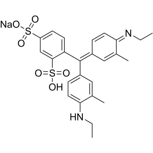
- HY-W440915
-
|
|
Liposome
|
Inflammation/Immunology
|
|
DSPE-PEG2000-FITC is a PEG lipid conjugated with fluorescein. FITC is a green dye with a peak absorption at 494 nm and a maximum emission at 520 nm, which is used for staining biological samples or nanoparticles. DSPE-PEG2000-FITC spontaneously forms lipid bilayers or micelles in water and can be used to prepare liposomes for delivering substances such as mRNA vaccines .
|
-
- HY-DY1105
-
|
MTDR FM (solution)
|
Fluorescent Dye
|
Others
|
MitoTracker Deep Red FM (solution) (MTDR FM (solution)) selectively accumulates in the mitochondrial matrix. MitoTracker Deep Red FM covalently binds mitochondrial proteins by reacting with free mercaptan of cysteine residues, allowing staining of mitochondrial membrane potential independent of membrane potential. The Ex/Em of MitoTracker Deep Red (MTDR) FM is 644/665 nm.
Solvent and concentration: DMSO: 1 mM
|
-

- HY-161283
-
|
|
MAP3K
|
Others
|
|
JT21-25 (compound 9h) is a potent and selective inhibitor of apoptosis signal-regulating kinase 1 (ASK1), with IC50 of 5.1 nM .
|
-
- HY-D3255
-
|
|
Fluorescent Dye
|
Others
|
|
CTAP-1 is a monovalent copper ion probe. CTAP-1 binds to monovalent copper Cu (I) to inhibit photoinduced electron transfer, thereby enhancing fluorescence emission. CTAP-1 produces copper-dependent perinuclear staining that colocalizes with mitochondria and the Golgi apparatus. CTAP-1 can be used for imaging Cu (I) pools, excitation/emission Wavelength: 365/480 nm .
|
-

- HY-D1346
-
|
|
Fluorescent Dye
|
Others
|
|
610CP is a new type of actin labeling dye. It dissolves in organic solvents. In DMSO the 610CP excitation/emission wavelength is between 609 and 634 nm. 610CP is a fluorescent dye that penetrates living cells. Upon cell entry, 610CP binds to Bromo-des-methyl-Jasplakinolide Therefore, 610CP dye can be used to stain actin fluorescence images with low background and high resolution.
|
-
- HY-D3375
-
|
|
Fluorescent Dye
|
Neurological Disease
|
|
RAPID DiI is a cyanine dye with orange-red fluorescenc (EX≈550 nm; Em≈565 nm). RAPID DiI is a lipophilic dye that labels cell membranes by inserting its two long hydrocarbon (C18 carbon) chains into the lipid bilayer. RAPID DiI is weakly fluorescent until incorporated into membranes. RAPID DiI diffuses laterally to stain the entire cell, allowing it to be used as an anterograde and retrograde tracer of neurons.
|
-

- HY-W591060
-
|
|
Biochemical Assay Reagents
Endogenous Metabolite
|
|
|
Bis(bipyridine)-(5-aminophenanthroline) is an organic catalyst with photocatalytic activity. The use of Bis(bipyridine)-(5-aminophenanthroline) in hematology involves its use as a stain for tissue diagnosis. Bis(bipyridine)-(5-aminophenanthroline) can be used as a transition metal catalyst in catalytic reactions. Bis(bipyridine)-(5-aminophenanthroline) has also been used in the study of ruthenium catalysts.
|
-

- HY-D3372
-
|
Dilinoleyl DiO
|
Fluorescent Dye
|
Neurological Disease
|
|
RAPID DiO (Dilinoleyl DiO) is a cyanine dye with green fluorescence (EX≈490 nm; Em≈505 nm). RAPID DiO is a lipophilic dye that labels cell membranes by inserting its two long hydrocarbon (C18 carbon) chains into the lipid bilayer. RAPID DiO is weakly fluorescent until incorporated into membranes. RAPID DiO diffuses laterally to stain the entire cell, allowing it to be used as an anterograde and retrograde tracer of neurons.
|
-

- HY-D0992
-
|
Peridinin-chlorophyll-protein complex
|
Fluorescent Dye
|
Cancer
|
|
PerCP is a peridinin-chlorophyll protein complex derived from the dinoflagellate *Glenodinium*, suitable for immunofluorescence staining of formalin-fixed, paraffin-embedded human tumor sections. As a red fluorescent dye, PerCP enables clear differentiation between antibody-bound tumor regions and yellow-green autofluorescence of tissues. PerCP allows simultaneous observation of tissue morphology without counterstaining. PerCP can be used in cancer research (Ex/Em = 482/677 nm) .
|
-

- HY-D2769
-
|
Difluorocarboxyfluorescein azide, 6-isomer
|
Fluorescent Dye
|
Others
|
|
Oregon green 488 azide (Difluorocarboxyfluorescein azide, 6-isomer) is a bright green fluorescent azide-activated probe that reacts with terminal alkynes via copper-catalyzed azide-alkyne cycloaddition (CuAAC). Oregon green 488 azide can label goat anti-mouse IgG and exhibits excellent luminescence efficiency. Oregon green 488 azide, as a streptavidin conjugate, is used for flow cytometry staining of macrophages .
|
-

- HY-12489A
-
|
Acid Red 112, BS
|
Biochemical Assay Reagents
|
Others
|
|
Both Ponceau S and Ponceau BS are synthetic dyes commonly used in biological research. They are commonly used as protein stains to visualize proteins in western blots and other protein detection analyses. Ponceau S is a red dye, while Ponceau BS is a blue shade of the same dye. Both dyes bind to positively charged amino acid residues in proteins for easy visualization. However, Ponceau S is more commonly used due to its higher sensitivity.
|
-

- HY-W110793
-
|
Diphenylthiocarbazone
|
Biochemical Assay Reagents
|
Metabolic Disease
|
|
Dithizone can be toxic to pancreatic islet cells and cause experimental diabetes. Dithizone can be used for in vitro in vivo staining of transplanted islets and cytoplasmic granules of bone marrow cells. Dithizone can be used as a chelating agent and colorimetric indicator for the detection and extraction of various heavy metals, including lead and mercury, in analytical and biochemical applications. Dithizone forms stable colored complexes with metal ions and can be quantitatively analyzed by methods such as spectrophotometry .
|
-
- HY-103240
-
|
|
Amyloid-β
|
Others
|
|
Methoxy-X04 is a fluorescent dye that crosses the blood-brain barrier and selectively binds to beta-pleated sheets found in dense core amyloid Aβ plaques. Methoxy-X04 retains in vitro binding affinity for amyloid b (Ab) fibrils (Ki= 26.8 nM). Methoxy-X04 is fluorescent and stains plaques, tangles, and cerebrovascular amyloid in postmortem sections of AD brain with good specificity .
|
-
- HY-D0014
-
|
|
Environmental Pollutants
Biochemical Assay Reagents
|
Others
|
|
Brilliant Blue G-250 is a dye commonly used for the visualization of proteins separated by SDS-PAGE, offering a simple staining procedure and high quantitation. In the Bradford protein assay, protein concentrations are determined by the absorbance at 595 nm due to the binding of Brilliant Blue G-250 to proteins. Brilliant Blue G-250 is a safe highly selective P2×7R antagonist with promising consequent inactivation of NLRP3 inflammasome .
|
-
- HY-118907R
-
|
|
Fluorescent Dye
Reference Standards
|
Others
|
|
Methyl Orange (Standard) is the analytical standard of Methyl Orange. This product is intended for research and analytical applications. Methyl Orange is a soluble azo dye commonly used as an acid-base indicator and for staining cells and tissue sections, as well as for dyeing textiles. Methyl Orange appears red at a pH of 3.1 and changes to bright yellow as the pH increases to 4.4. Methyl Orange (500 mg/L) exhibits cytotoxicity and can cause DNA damage .
|
-

- HY-D2639
-
|
|
Fluorescent Dye
|
Metabolic Disease
|
|
TPE-PY is a fluorescent dye targeting mitochondria, which has cationic and lipophilic characteristics, enabling specific targeting of mitochondria. TPE-PY functions through the aggregation-induced emission (AIE) mechanism. TPE-PY mechanism of action is based on the restriction of intramolecular rotation (RIR), which enhances fluorescence in the aggregated state. TPE-PY is mainly used in the field of biological imaging, especially for the specific staining of mitochondria in living cells, facilitating the observation of the location, morphology, and quantity of mitochondria .
|
-

- HY-118647
-
|
Etisul
|
Bacterial
|
Infection
|
|
Ditophal is an orally active thiol ester anti-tuberculosis and anti-leprosy agent. Ditophal combats Mycobacterium tuberculosis infections in subcutaneous and intracerebral sites in guinea pigs, including isoniazid (HY-B0329)-resistant strains. Ditophal alters the staining characteristics of Mycobacterium leprae and reduces the bacterial index of leprosy. Ditophal causes ulcers at the injection site when administered subcutaneously to guinea pigs at high doses. Ditophal can be used in the research of tuberculosis and leprosy .
|
-

- HY-158742
-
|
GL13; SBB-A-B
|
Tyrosinase
|
Others
|
|
SBB-Analogue (GL13) Biotin (GL13; SBB-A-B) consists of a Sudan Black B (SBB) (HY-D0213) derivative conjugated with biotin. SBB-Analogue (GL13) Biotin potently detects senescent cells and eliminates the drawback of false-positive staining caused by serum starvation and cell fusion in SA-β-gal assays. SBB-Analogue (GL13) Biotin can be used in flow cytometry, immunofluorescence analysis, and other applications .
|
-

- HY-138658
-
|
JF526, SE; JF526, NHS
|
Fluorescent Dye
|
Others
|
|
Janelia Fluor 526, SE (JF526,SE) is a fluorogenic yellow fluorescent dye that contains NHS ester group. JF526 is a versatile scaffold for fluorogenic ligands, including labels for genetically encoded self-labeling protein tags and stains for endogenous structures . Janelia Fluor products are licensed under U.S. Pat. Nos. 9,933,417, 10,018,624 and 10,161,932 and other patents from Howard Hughes Medical Institute.
|
-
- HY-172435
-
|
AGN-232411; AG-80308; AGN-411
|
Formyl Peptide Receptor (FPR)
|
Neurological Disease
|
|
Rezuforimod (AGN-232411; AG-80308) is an ophthalmic solution targeting the formyl peptide receptor (FPR) pathway. Rezuforimod exerts anti-inflammatory effects by targeting the formyl peptide receptor pathway. Administered topically, Rezuforimod significantly reduces the corneal conjunctival staining score and improves ocular discomfort symptoms, with no serious adverse events reported and favorable overall safety profile. Rezuforimod shows remarkable potential to serve as a novel therapeutic option for dry eye disease .
|
-

- HY-D1783
-
|
MTDR FM
|
Fluorescent Dye
|
Others
|
MitoTracker Deep Red (MTDR) FM fluorescent dye that selectively accumulates in the mitochondrial matrix. MitoTracker Deep Red FM covalently binds mitochondrial proteins by reacting with free mercaptan of cysteine residues, allowing staining of mitochondrial membrane potential independent of membrane potential. Excitation/emission wavelength 644/665 nm . MitoTracker Deep Red dyes have an excitation/emission wavelength of 633/650-750 nm .
The Ex/Em of MitoTracker Deep Red (MTDR) FM is 644/665 nm.
|
-
- HY-121075
-
|
|
Fluorescent Dye
Reverse Transcriptase
HIV
RSV
|
Infection
Endocrinology
Cancer
|
|
Alizarin complexone is a calcium-binding fluorescent dye. Alizarin complexone stains mineralized areas of bone by binding to calcium crystals. Alizarin complexone inhibits the reverse transcriptase activity of RAV-2, HIV-1, and RSV with IC50 values of 3.8 μg/mL, 45 μg/mL, and 100 μg/mL, respectively. Alizarin complexone exhibits antiviral activity against HIV-1 and RSV. Alizarin complexone delays RSV-induced tumor induction in chickens .
|
-
- HY-W130236
-
|
|
Cholinesterase (ChE)
|
Cancer
|
|
Methylene Violet 3RAX is a phenazine dye to stain the mitochondria of cells. Methylene Violet 3RAX can change the molecular structure of DNA, undermine the module of DNA, and induce the generation of the reactive singlet oxygen. Methylene Violet 3RAX shows inhibition for human erythrocyte AChE and human plasma BChE with an Kis of 1.58, 0.51 μM, respectively. Methylene Violet 3RAX has the potential for the research of potential photosensitizers for mitochondrial targeting action in PDT (photodynamic therapy) .
|
-
- HY-W127703
-
|
|
Fluorescent Dye
Biochemical Assay Reagents
|
Others
|
|
Octadecyl Rhodamine B chloride is an amphiphilic fluorescent probe with both membrane-binding ability and fluorescent properties, which easily forms aggregates in aqueous phase leading to self-quenching. Octadecyl Rhodamine B chloride can be used for cell membrane staining and further applied in membrane biology research. The maximum absorption/emission wavelengths of Octadecyl Rhodamine B chloride monomer in Triton X-100 are 565 nm/585 nm; the absorption peaks of aggregates in water are blue-shifted to 524 nm and 574 nm, with significantly quenched fluorescence .
|
-
- HY-W009411
-
|
|
Fluorescent Dye
|
Others
|
|
Dansyl hydrazine is a carbohydrate-specific fluorescent dye (Ex/Em = 340 nm/525 nm). Dansyl hydrazine undergoes a condensation reaction with aldehyde groups generated by periodate oxidation on carbohydrate-containing structures to form fluorescent hydrazone compounds. Dansyl hydrazine selectively stains polysaccharides in formalin-fixed, paraffin-embedded human post-mortem brain tissues, revealing detailed structural features. Dansyl hydrazine is applicable to research related to Alzheimer's disease, Lafora disease, and polyglucosan body disease .
|
-
- HY-DY1045
-
|
|
Amyloid-β
|
Neurological Disease
|
Methoxy-X04 (solution) is a fluorescent dye that crosses the blood-brain barrier and selectively binds to beta-pleated sheets found in dense core amyloid Aβ plaques. Methoxy-X04 retains in vitro binding affinity for amyloid b (Ab) fibrils (Ki= 26.8 nM). Methoxy-X04 is fluorescent and stains plaques, tangles, and cerebrovascular amyloid in postmortem sections of AD brain with good specificity .
Solvent and concentration: DMSO: 5 mg/mL
|
-

- HY-D3430
-
|
|
Fluorescent Dye
Amyloid-β
|
Neurological Disease
|
|
PTPA-QM is a low-cytotoxicity Aβ protein probe that can be used for live-cell imaging and tissue section staining (Lys. Ex/Em = 385/615 nm). PTPA-QM can intercalate into the β-sheet layered structure of β-amyloid fibrils, form intermolecular interactions with amino acid residues, restrict intramolecular rotation and trigger fluorescence activation for imaging purposes. PTPA-QM is applicable to Alzheimer's disease-related research. Maximum excitation/emission wavelength: 448/605 nm .
|
-

- HY-171179
-
|
|
Fluorescent Dye
|
Neurological Disease
|
|
BD-Oligo is an oligomer-specific fluorescent chemical probe. BD-Oligo preferentially identifies Aβ oligomer assemblies over monomers or fibrils by using diversity-directed fluorescent library (DOFL) screening and computational techniques. BD-Oligo exhibits dynamic oligomer monitoring capabilities during Aβ peptide fibril formation as Aβ is induced to form oligomers and ultimately fibrils over time. BD-Oligo also exhibits blood-brain barrier permeability with the ability to stain Aβ oligomers in vivo .
|
-

- HY-D1297
-
|
|
Fluorescent Dye
|
Others
|
|
ER-Tracker dye is a derivative of BODIPY series dyes coupled with Glibenclamide (HY-15206), highly selective binding to the endoplasmic reticulum, non-toxic to cells at low concentrations, this type of dye is an environmentally sensitive probe, and formaldehyde treatment can still retain part of the fluorescence, with high fluorescence life, good extinction coefficient and other characteristics. Glibenclamide is an atp-dependent K + channel blocker (Kir6, KATP) and CFTR Cl-channel blocker that binds in the endoplasmic reticulum. ER-Tracker is not suitable for staining cells after fixation .
|
-
- HY-151801
-
|
|
mAChR
|
Others
|
|
DIBA-Cy5 is a fluorescent DIBA antagonist made up be DIBA-alkyne binding Cyanine5 fluorophores (Cy5) and polyethylene glycol (PEG) biomolecules. DIBA-Cy5 can serve as a fluorescent ligand, suitable for probe attachment through click chemistry. DIBA-Cy5 exerts a high binding affinity to type-2 mAChR (M2R) with the Kd value of 1.80 nM, can directly stain M2R receptors in the sinoatrial node of a mouse heart .
|
-
- HY-D3405
-
|
|
Fluorescent Dye
|
Others
|
|
BO-TAP-3 is an orange fluorescent carbocyanine monomer dye, and it is also a cell impermeable nucleic acid staining agent. When there is no nucleic acid present, BO-TAP-3 shows a non-fluorescent state. However, when it binds to double-stranded DNA, it exhibits multiple fluorescence enhancement effects. BO-TAP-3 can be used for cell nucleus and cytoplasm imaging, bacterial and sperm labeling, and can also be applied in nucleic acid detection and amplification research. It can also be used as a temperature sensor (Ex/Em = 564/600 nM).
|
-

- HY-W540972
-
|
Primuline
|
Fluorescent Dye
HCV
Dengue Virus
|
Infection
|
|
Primulin is a versatile fluorescent dye and bioactive compound widely used in analytical, biological, botanical and virological studies. Primulin acts as a versatile stain that labels plant cell walls and differentiates live and dead spermatozoa via distinct fluorescence patterns. Primulin exhibits strong albumin‑binding capacity. Primulin acts as a retrograde axonal tracer in neurobiological investigations. Primulin and its derivatives inhibit HCV NS3, block dengue virus NS3-mediated ATP hydrolysis, and disrupt HCV replicase assembly .
|
-

- HY-D1068
-
|
DBCO-Sulfo-Cy5
|
Fluorescent Dye
|
Others
|
|
Cy5-DBCO (DBCO-Sulfo-Cy5) is a near-infrared (NIR)red fluorescent dye with λabsand λem of 646nm and 670 nm, respectively. Cy5-DBCO (DBCO-Sulfo-Cy5) is not suitable for staining intracellular components of permeabilezed cell, it may exhibits a high background. Cy5-DBCO is a click chemistry reagent, it contains a DBCO group that can undergo strain-promoted alkyne-azide cycloaddition (SPAAC) with molecules containing Azide groups.
|
-
- HY-D1106
-
|
|
Fluorescent Dye
|
Cancer
|
|
BODIPY dye is a small molecule dye with strong ultraviolet absorption ability, its fluorescence peak is relatively sharp, and the quantum yield is high. They are relatively insensitive to the polarity and pH of the environment and are relatively stable under different physiological conditions . Due to its structural asymmetry, BODIPY derives a variety of structural products. BODIPY lipid droplet dyes can well pass through the cell membrane into the cell, and localize the neutral lipids in the cell to specifically stain the lipid droplets, which can be used for labeling of live cells and fixed cells .
|
-
- HY-D1431
-
|
|
Fluorescent Dye
|
Cancer
|
|
ER-Tracker dye is a derivative of BODIPY series dyes coupled with Glibenclamide (HY-15206), highly selective binding to the endoplasmic reticulum, non-toxic to cells at low concentrations, this type of dye is an environmentally sensitive probe, and formaldehyde treatment can still retain part of the fluorescence, with high fluorescence life, good extinction coefficient and other characteristics. Glibenclamide is an atp-dependent K + channel blocker (Kir6, KATP) and CFTR Cl-channel blocker that binds in the endoplasmic reticulum. ER-Tracker is not suitable for staining cells after fixation. Ex/Em=587/615 nm .
|
-
- HY-D0079
-
|
Hydroethidine; PD-MY 003
|
Fluorescent Dye
|
Others
|
|
Dihydroethidium, also known as DHE, is a peroxide indicator. Dihydroethidium penetrates cell membranes to form a fluorescent protein complex with blue fluoresces. After entering the cells, Dihydroethidium is mainly localized in the cell membrane, cytoplasm and nucleus, and the staining effect is the strongest in the nucleus. Dihydroethidium produces inherent blue fluorescence with a maximum excitation wavelength of 370 nm and a maximum emission wavelength of 420 nm; after dehydrogenation, Dihydroethidium combines with RNA or DNA to produce red fluorescence with a maximum excitation wavelength of 300 nm and a maximum emission wavelength of 610 nm. 535 nm can also be used as the excitation wavelength for actual observation .
|
-
- HY-W320523
-
|
|
Fluorescent Dye
Reverse Transcriptase
HIV
RSV
|
Infection
Metabolic Disease
Cancer
|
|
ALizarin complexone dihydrate is the dehydrate of Alizarin complexone (HY-121075). Alizarin complexone is a calcium-binding fluorescent dye. Alizarin complexone stains mineralized areas of bone by binding to calcium crystals. Alizarin complexone inhibits the reverse transcriptase activity of RAV-2, HIV-1, and RSV with IC50 values of 3.8 μg/mL, 45 μg/mL, and 100 μg/mL, respectively. Alizarin complexone exhibits antiviral activity against HIV-1 and RSV. Alizarin complexone delays RSV-induced tumor induction in chickens .
|
-

- HY-D0006
-
|
|
Fluorescent Dye
|
Others
|
|
Bathophenanthroline is a nitrogen-containing organic ligand that can form stable complexes with transition metal ions such as Ru 2+. In proteomics, Bathophenanthroline is often used for in-gel protein staining. The negatively charged sulfonic acid groups of Bathophenanthroline interact electrostatically with the positively charged amino acid residues of proteins, thereby achieving specific binding to proteins and enhancing the fluorescent signal of detection. Bathophenanthroline can be used for protein separation and detection in SDS-PAGE gels in the field of proteomics, as well as other fluorescence studies[1]. Ex/Em=532 nm/610 nm[1].
|
-
- HY-DY1043
-
|
|
Fluorescent Dye
|
Others
|
ER-Tracker dye is a derivative of BODIPY series dyes coupled with Glibenclamide (HY-15206) , highly selective binding to the endoplasmic reticulum, non-toxic to cells at low concentrations, this type of dye is an environmentally sensitive probe, and formaldehyde treatment can still retain part of the fluorescence, with high fluorescence life, good extinction coefficient and other characteristics. Glibenclamide is an atp-dependent K + channel blocker (Kir6, KATP) and CFTR Cl-channel blocker that binds in the endoplasmic reticulum. ER-Tracker is not suitable for staining cells after fixation .
Solvent and concentration: DMSO: 1 mM
|
-

- HY-D0293
-
|
Xylene blue free acid; C.I. Acid blue 1 free acid
|
Fluorescent Dye
|
Cancer
|
|
Acid blue 1 free acid (Patent Blue V) is a novel biological dye that can be used as an intraocular dye for retinectomy. Retinectomy refers to the removal of the translucent inner limiting membrane (ILM). The application of appropriate dyes in vitreoretinal surgery can achieve the purpose of complete removal. Acid blue 1 free acid can be used to stain retinal premembranous structures. Spectral analysis shows that Acid blue 1 free acid has strong absorption below 450 nm and above 600 nm, showing a blue-green color. Acid blue 1 free acid is also used as a marker in lymphangiography for resection of neoplastic lymph nodes .
|
-
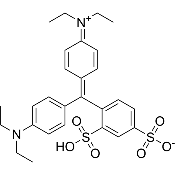
- HY-139792
-
|
SHR117887
|
Endogenous Metabolite
|
Metabolic Disease
|
|
Besigliptin tosylate (SHR117887) is a DPP-4 inhibitor with activity to improve metabolic control and β-cell function. Besigliptin tosylate can effectively reduce serum DPP-4 activity and improve oral glucose tolerance. Besigliptin tosylate significantly reduces fasting blood glucose levels and improves lipid profiles in a diabetic mouse model. The effect of besigliptin tosylate is comparable to that of the known compound vildagliptin (HY-14291) at the same concentration. Besigliptin tosylate increases insulin staining of pancreatic islet cells in chronic administration, indicating improved β-cell function .
|
-

- HY-D0938
-
|
CFSE; 5(6)-Carboxyfluorescein diacetate succinimidyl ester; 5(6)-CFDA N-succinmidyl ester
|
Fluorescent Dye
|
Others
|
|
CFDA-SE is a fluorescent dye that can penetrate the cell membrane. It can react with the free amine group in the cytoskeleton protein inside the cell, and finally form a protein complex with fluorescence. After entering the cell, CFDA-SE locates in the cell membrane, cytoplasm and nucleus, and the fluorescence staining is strongest in the nucleus .
CFDA-SE dye can be uniformly inherited by the cells with cell division and proliferation, and its attenuation is proportional to the number of cell divisions. This phenomenon can be detected and analyzed by flow cytometry under the excitation light of 488 nm, and can be used to detect the proliferation of cells .
|
-
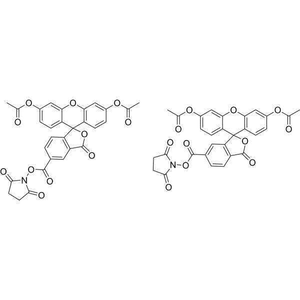
- HY-W129513
-
|
|
Biochemical Assay Reagents
|
Others
|
|
Sorbitan monopalmitate is a food-grade, non-ionic emulsifier that exhibits oral activity. Sorbitan monopalmitate functions as a crystallization modifier, promoting the aggregation of spherical mango butter crystals, introducing lattice defects, and regulating the crystallization rate of mango butter. Sorbitan monopalmitate serves as a component of precursor vesicle gels, enabling the controlled and sustained release of drugs and facilitating the preparation of stable transdermal delivery formulations .
|
-
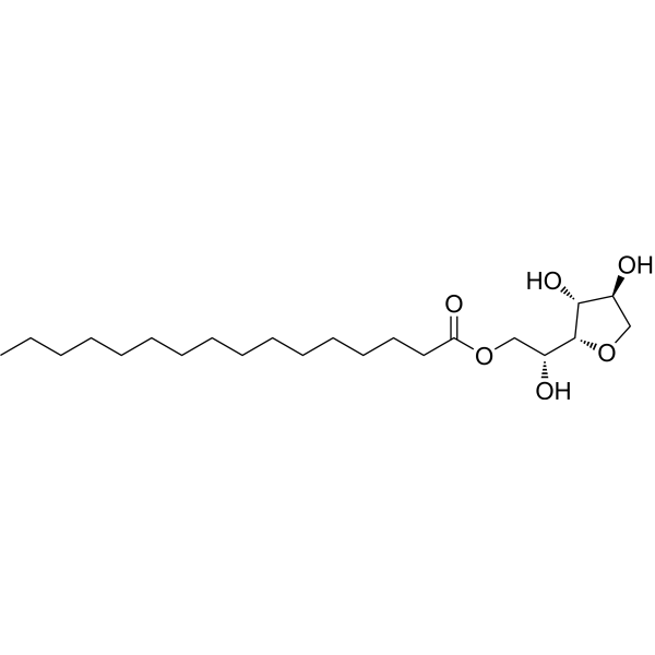
- HY-103240R
-
|
|
Reference Standards
Amyloid-β
|
Others
|
|
Methoxy-X04 (Standard) is the analytical standard of Methoxy-X04 (HY-103240). This product is intended for research and analytical applications. Methoxy-X04 is a fluorescent dye that crosses the blood-brain barrier and selectively binds to beta-pleated sheets found in dense core amyloid Aβ plaques. Methoxy-X04 retains in vitro binding affinity for amyloid b (Ab) fibrils (Ki= 26.8 nM). Methoxy-X04 is fluorescent and stains plaques, tangles, and cerebrovascular amyloid in postmortem sections of AD brain with good specificity .
|
-

- HY-D1396
-
Br-DAPI
3 Publications Verification
|
Fluorescent Dye
DNA Stain
|
Others
|
|
Br-DAPI is a marker dye in DAPI series. DAPI is a fluorescent dye that binds strongly to DNA. It binds to the AT base pair of the double-stranded DNA minor groove, and one DAPI molecule can occupy three base pair positions. The fluorescence intensity of DAPI molecules bound to double-stranded DNA is increased by about 20 times, and it is commonly observed with fluorescence microscopy, and the amount of DNA can be determined based on the intensity of fluorescence. In addition, because DAPI can pass through intact cell membranes, it can be used to stain both live and fixed cells . Storage: Keep away from light.
|
-
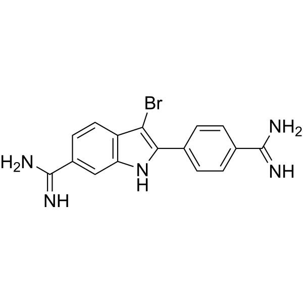
- HY-D1809
-
|
|
Fluorescent Dye
|
Others
|
Vari Fluor 405-Streptavidin is a dye marker of Vari Fluor-streptavidin consisting of labeling streptavidin with a Vari Fluor series of fluorescent probes. Streptavidin is a high-affinity tetramer protein, each tetramer consisting of four identical streptavidin subunits. Streptavidin binds to biotin specifically via a reversible non-covalent effect. Streptavidin can achieve rapid and efficient detection of biotin markers, and is often used in immunofluorescence (IF), enzyme-linked immunosorbent assay (ELISA), immunohistochemical staining (IFH), in situ hybridization (ISH) and other experiments. Ex/Em=405 nm/431 nm.
Product Concentration: 0.8 mg/mL
|
-

- HY-DY1026
-
|
|
Fluorescent Dye
|
Cancer
|
ER-Tracker dye is a derivative of BODIPY series dyes coupled with Glibenclamide (HY-15206) , highly selective binding to the endoplasmic reticulum, non-toxic to cells at low concentrations, this type of dye is an environmentally sensitive probe, and formaldehyde treatment can still retain part of the fluorescence, with high fluorescence life, good extinction coefficient and other characteristics. Glibenclamide is an atp-dependent K + channel blocker (Kir6, KATP) and CFTR Cl-channel blocker that binds in the endoplasmic reticulum. ER-Tracker is not suitable for staining cells after fixation. Ex/Em=587/615 nm .
Solvent and concentration: DMSO: 1 mM
|
-

- HY-DY1009
-
|
|
Fluorescent Dye
|
Others
|
CFDA-SE (solution) is a fluorescent dye that can penetrate the cell membrane. It can react with the free amine group in the cytoskeleton protein inside the cell, and finally form a protein complex with fluorescence. After entering the cell, CFDA-SE locates in the cell membrane, cytoplasm and nucleus, and the fluorescence staining is strongest in the nucleus . CFDA-SE dye can be uniformly inherited by the cells with cell division and proliferation, and its attenuation is proportional to the number of cell divisions. This phenomenon can be detected and analyzed by flow cytometry under the excitation light of 488 nm, and can be used to detect the proliferation of cells .
Solvent and concentration: DMSO: 5 mM
|
-

- HY-D1805
-
|
|
Fluorescent Dye
|
Others
|
Vari Fluor 647-Streptavidin is a dye marker of Vari Fluor-streptavidin consisting of labeling streptavidin with a Vari Fluor series of fluorescent probes. Streptavidin is a high-affinity tetramer protein, each tetramer consisting of four identical streptavidin subunits. Streptavidin binds to biotin specifically via a reversible non-covalent effect. Streptavidin can achieve rapid and efficient detection of biotin markers, and is often used in immunofluorescence (IF), enzyme-linked immunosorbent assay (ELISA), immunohistochemical staining (IFH), in situ hybridization (ISH) and other experiments. Ex/Em=650 nm/665 nm.
Product Concentration: 0.8 mg/mL
|
-

- HY-W110927
-
|
|
Biochemical Assay Reagents
Fluorescent Dye
|
Others
|
|
Alizarin red S indicator (C.I. 58005) is a reductively active (quinone-based) anthraquinone dye that forms complexes with metal ions (such as zirconium, calcium) or boric acid to label calcium deposition and perform electrochemical sensing functions. Alizarin Red S undergoes reversible redox reactions (for electrochemical detection) and irreversible chelation (for bone staining). Alizarin Red S is mainly used in bone metabolism research (labeling mineralized tissue), sugar detection (boric acid-sugar competition system) and metal ion sensing (such as zirconium ion detection), and can be used in osteoporosis and metabolic disease research .
|
-
- HY-D1808
-
|
|
Fluorescent Dye
|
Others
|
Vari Fluor 488-Streptavidin is a dye marker of Vari Fluor-streptavidin consisting of labeling streptavidin with a Vari Fluor series of fluorescent probes. Streptavidin is a high-affinity tetramer protein, each tetramer consisting of four identical streptavidin subunits. Streptavidin binds to biotin specifically via a reversible non-covalent effect. Streptavidin can achieve rapid and efficient detection of biotin markers, and is often used in immunofluorescence (IF), enzyme-linked immunosorbent assay (ELISA), immunohistochemical staining (IFH), in situ hybridization (ISH) and other experiments. Ex/Em=490 nm/515 nm.
Product Concentration: 0.8 mg/mL
|
-

- HY-121075R
-
|
|
Reference Standards
Reverse Transcriptase
Fluorescent Dye
HIV
RSV
|
Infection
Endocrinology
Cancer
|
|
Alizarin complexone (Standard) is the analytical standard of Alizarin complexone (HY-121075). This product is intended for research and analytical applications. Alizarin complexone is a calcium-binding fluorescent dye. Alizarin complexone stains mineralized areas of bone by binding to calcium crystals. Alizarin complexone inhibits the reverse transcriptase activity of RAV-2, HIV-1, and RSV with IC50 values of 3.8 μg/mL, 45 μg/mL, and 100 μg/mL, respectively. Alizarin complexone exhibits antiviral activity against HIV-1 and RSV. Alizarin complexone delays RSV-induced tumor induction in chickens .
|
-

- HY-D1804
-
|
|
Fluorescent Dye
|
Others
|
Vari Fluor 680-Streptavidin is a dye marker of Vari Fluor-streptavidin consisting of labeling streptavidin with a Vari Fluor series of fluorescent probes. Streptavidin is a high-affinity tetramer protein, each tetramer consisting of four identical streptavidin subunits. Streptavidin binds to biotin specifically via a reversible non-covalent effect. Streptavidin can achieve rapid and efficient detection of biotin markers, and is often used in immunofluorescence (IF), enzyme-linked immunosorbent assay (ELISA), immunohistochemical staining (IFH), in situ hybridization (ISH) and other experiments. Ex/Em=680 nm/701 nm.
Product Concentration: 0.8 mg/mL
|
-

- HY-118411
-
|
EdU
|
PROTAC Linkers
|
Cancer
|
|
5-Ethynyl-2'-deoxyuridine (EdU), a thymidine analogue, is incorporated into cellular DNA during DNA replication and the subsequent reaction of EdU with a fluorescent azide in a “Click” reaction. EdU staining is a fast, sensitive and reproducible method to study cell proliferation . 5-Ethynyl-2'-deoxyuridine is an alkyl chain-based PROTAC linker that can be used in the synthesis of PROTACs . 5-Ethynyl-2'-deoxyuridine is a click chemistry reagent, it contains an Alkyne group and can undergo copper-catalyzed azide-alkyne cycloaddition (CuAAc) with molecules containing Azide groups.
|
-
- HY-W009411S
-
|
|
Isotope-Labeled Compounds
Fluorescent Dye
|
Others
|
|
Dansyl hydrazine- 13C2 is the 13C-labeled Dansyl hydrazine (HY-W009411). Dansyl hydrazine is a carbohydrate-specific fluorescent dye (Ex/Em = 340 nm/525 nm). Dansyl hydrazine undergoes a condensation reaction with aldehyde groups generated by periodate oxidation on carbohydrate-containing structures to form fluorescent hydrazone compounds. Dansyl hydrazine selectively stains polysaccharides in formalin-fixed, paraffin-embedded human post-mortem brain tissues, revealing detailed structural features. Dansyl hydrazine is applicable to research related to Alzheimer's disease, Lafora disease, and polyglucosan body disease.
|
-

- HY-D1807
-
|
|
Fluorescent Dye
|
Others
|
Vari Fluor 555-Streptavidin is a dye marker of Vari Fluor-streptavidin consisting of labeling streptavidin with a Vari Fluor series of fluorescent probes. Streptavidin is a high-affinity tetramer protein, each tetramer consisting of four identical streptavidin subunits. Streptavidin binds to biotin specifically via a reversible non-covalent effect. Streptavidin can achieve rapid and efficient detection of biotin markers, and is often used in immunofluorescence (IF), enzyme-linked immunosorbent assay (ELISA), immunohistochemical staining (IFH), in situ hybridization (ISH) and other experiments. Ex/Em=555 nm/565 nm.
Product Concentration: 0.8 mg/mL
|
-

- HY-DY1015
-
|
|
Fluorescent Dye
|
Others
|
Dihydroethidium (solution) , also known as DHE, is a peroxide indicator. Dihydroethidium penetrates cell membranes to form a fluorescent protein complex with blue fluoresces. After entering the cells, Dihydroethidium is mainly localized in the cell membrane, cytoplasm and nucleus, and the staining effect is the strongest in the nucleus. Dihydroethidium produces inherent blue fluorescence with a maximum excitation wavelength of 370 nm and a maximum emission wavelength of 420 nm; after dehydrogenation, Dihydroethidium combines with RNA or DNA to produce red fluorescence with a maximum excitation wavelength of 300 nm and a maximum emission wavelength of 610 nm. 535 nm can also be used as the excitation wavelength for actual observation .
Solvent and concentration: DMSO: 5 mM
|
-

- HY-D1806
-
|
|
Fluorescent Dye
|
Others
|
Vari Fluor 594-Streptavidin is a dye marker of Vari Fluor-streptavidin consisting of labeling streptavidin with a Vari Fluor series of fluorescent probes. Streptavidin is a high-affinity tetramer protein, each tetramer consisting of four identical streptavidin subunits. Streptavidin binds to biotin specifically via a reversible non-covalent effect. Streptavidin can achieve rapid and efficient detection of biotin markers, and is often used in immunofluorescence (IF), enzyme-linked immunosorbent assay (ELISA), immunohistochemical staining (IFH), in situ hybridization (ISH) and other experiments. Ex/Em=590 nm/617 nm.
Product Concentration: 0.8 mg/mL
|
-

- HY-W800699
-
|
|
Fluorescent Dye
|
Others
|
|
BP Fluor 430 DBCO reacts with azides via a copper-free click chemistry reaction to form a stable triazole and does not require a Cu-catalyst or elevated temperatures. In applications where the presence of copper is a concern, BP Fluor 430 DBCO is an ideal alternative to copper requiring fluorescent alkynes.
BP Fluor 430 is a bright, photostable, green-fluorescent probe optimally excited near its absorption maximum at 432 nm. Its emission peak at 539 nm is pH independent over a wide pH range.
BP Fluor DBCO reagent is not suitable for staining intracellular components of fixed and permeabilized cells due to high backgrounds.
|
-

- HY-D1570
-
|
|
Fluorescent Dye
|
Others
|
|
BODIPYFL C12 is a BODIPY dye. BODIPY dye is a small molecule dye with strong ultraviolet absorption ability, its fluorescence peak is relatively sharp, and the quantum yield is high. They are relatively insensitive to the polarity and pH of the environment and are relatively stable under different physiological conditions. Due to its structural asymmetry, BODIPY derives a variety of structural products. BODIPY lipid droplet dyes can well pass through the cell membrane into the cell, and localize the neutral lipids in the cell to specifically stain the lipid droplets, which can be used for labeling of live cells and fixed cells . Maximum excitation/emission wavelength: 480/508 nm .
|
-
- HY-D0932
-
|
Solvent Red 24; C.I. 26105
|
Environmental Pollutants
Biochemical Assay Reagents
Cytochrome P450
Aryl Hydrocarbon Receptor
|
Metabolic Disease
|
|
Sudan IV is an agonist of the aryl hydrocarbon receptor (AhR) that activates downstream signaling pathways and induces CYP1A1 expression. Sudan IV promotes CYP1A1 gene transcription by activating AhR-ARNT heterodimers and binding to exogenous response elements (XREs) on DNA, thereby enhancing drug metabolizing enzyme activity. Sudan IV can be used to study the toxicity mechanisms of industrial dyes and the effects of interactions with serum proteins (such as bovine serum albumin (BSA)) on their distribution in vivo. Sudan IV is a fat-soluble diazo dye that can be used to stain lipids, triglycerides, and lipoproteins on frozen sections .
|
-
- HY-W090090
-
|
Pyrromethene 546
|
Fluorescent Dye
|
Others
|
|
BODIPY493/503 is a BODIPY dye. BODIPY dye is a small molecule dye with strong ultraviolet absorption ability, its fluorescence peak is relatively sharp, and the quantum yield is high. They are relatively insensitive to the polarity and pH of the environment and are relatively stable under different physiological conditions. Due to its structural asymmetry, BODIPY derives a variety of structural products. BODIPY lipid droplet dyes can well pass through the cell membrane into the cell, and localize the neutral lipids in the cell to specifically stain the lipid droplets, which can be used for labeling of live cells and fixed cells . Maximum excitation/emission wavelength: 493/503 nm .
|
-
- HY-D3179
-
|
|
Fluorescent Dye
BCRP
|
Neurological Disease
|
|
CDg13 is a fluorescent probe that specifically detects living neural stem/progenitor cells. CDg13 localizes to the endoplasmic reticulum via its dihexyl moiety, with no interaction with endoplasmic reticulum biomolecules. CDg13 undergoes selective efflux by active ABCG2 transporters, with increased intracellular accumulation following ABCG2 inhibition or knockdown. CDg13 isolates and enriches self-renewable neural stem/progenitor cells from embryonic mouse brain tissue, stains neural stem/progenitor cells brightly, and sorts ABCG2low cell populations from heterogeneous populations . (Ex/Em = 520/553 nm)
|
-

- HY-D1237
-
|
|
Fluorescent Dye
|
Metabolic Disease
|
|
BODIPY505/515 is a BODIPY dye. BODIPY dye is a small molecule dye with strong ultraviolet absorption ability, its fluorescence peak is relatively sharp, and the quantum yield is high. They are relatively insensitive to the polarity and pH of the environment and are relatively stable under different physiological conditions. Due to its structural asymmetry, BODIPY derives a variety of structural products. BODIPY lipid droplet dyes can well pass through the cell membrane into the cell, and localize the neutral lipids in the cell to specifically stain the lipid droplets, which can be used for labeling of live cells and fixed cells . Maximum excitation/emission wavelength: 505/515 nm .
|
-
- HY-138226
-
|
|
Fluorescent Dye
|
Others
|
|
BODIPY 558/568 C12 is a BODIPY dye. BODIPY dye is a small molecule dye with strong ultraviolet absorption ability, its fluorescence peak is relatively sharp, and the quantum yield is high. They are relatively insensitive to the polarity and pH of the environment and are relatively stable under different physiological conditions. Due to its structural asymmetry, BODIPY derives a variety of structural products. BODIPY lipid droplet dyes can well pass through the cell membrane into the cell, and localize the neutral lipids in the cell to specifically stain the lipid droplets, which can be used for labeling of live cells and fixed cells . Maximum excitation/emission wavelength: 558/568 nm .
|
-
- HY-W250147
-
|
Victoria blue B
|
Biochemical Assay Reagents
|
Others
|
|
Basic blue 26 (Victoria blue B) is a synthetic cationic dye belonging to the class of triarylmethane dyes. It has a bright blue color and is commonly used as a colorant for a variety of applications, including textiles, paper and leather. Basic Blue 26 is also used as a biological stain for DNA and protein detection in laboratories. Due to its ability to bind negatively charged materials, it can be used as an indicator of the presence of specific molecules in biological samples. However, Basic blue 26 has been reported to have potentially harmful effects on human health and the environment and its use is regulated in some countries. Proper handling and disposal procedures are necessary to minimize its impact on the environment.
|
-
- HY-D1614
-
|
|
Fluorescent Dye
|
Others
|
|
BODIPY493/503 methyl bromide is a BODIPY dye. BODIPY dye is a small molecule dye with strong ultraviolet absorption ability, its fluorescence peak is relatively sharp, and the quantum yield is high. They are relatively insensitive to the polarity and pH of the environment and are relatively stable under different physiological conditions. Due to its structural asymmetry, BODIPY derives a variety of structural products. BODIPY lipid droplet dyes can well pass through the cell membrane into the cell, and localize the neutral lipids in the cell to specifically stain the lipid droplets, which can be used for labeling of live cells and fixed cells . Maximum excitation/emission wavelength: 493/503 nm .
|
-
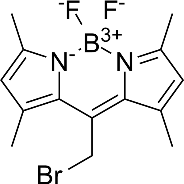
- HY-D3192
-
|
|
Fluorescent Dye
Bacterial
Amyloid-β
|
Infection
|
|
CDy11 is a fluorescent probe and amyloid-binding dye (λex=590 nm; λem=612 nm), with a Ka of 29 μM for Pseudomonas aeruginosa Fap. CDy11 specifically recognizes amyloid fibrils in bacterial biofilms and exhibits significantly enhanced fluorescence upon binding to the target. CDy11 shows no staining effect on amyloid-deficient mutant strains, planktonic cells or protein monomers. CDy11 supports in vivo imaging of Pseudomonas aeruginosa biofilms in mouse implant and corneal infection models. CDy11 is widely used in studies of Staphylococcus aureus biofilm infections, dental caries, and Pseudomonas aeruginosa-associated implant and corneal infections .
|
-

- HY-D3274
-
|
|
Fluorescent Dye
|
Others
|
|
PE-Cy5 is a tandem fluorescent dye commonly used in flow cytometry, immunofluorescence, and cell biology research. It is formed by the covalent linkage of two fluorescent molecules, namely phycoerythrin and CY5. PE-Cy5 binds to human FcγRI (CD64), and this binding can be blocked by human pooled serum, anti-CD64 monoclonal antibodies targeting the ligand-binding region, or aggregated IgG. PE-Cy5 supports three-color flow cytometry analysis, and whole blood staining can partially reduce its non-specific binding (Ex/Em = 450-500 nm/665 nm) .
|
-

- HY-DY1004
-
|
|
Fluorescent Dye
|
Others
|
BODIPY 493/503 (solution) is a BODIPY dye. BODIPY dye is a small molecule dye with strong ultraviolet absorption ability, its fluorescence peak is relatively sharp, and the quantum yield is high. They are relatively insensitive to the polarity and pH of the environment and are relatively stable under different physiological conditions. Due to its structural asymmetry, BODIPY derives a variety of structural products. BODIPY lipid droplet dyes can well pass through the cell membrane into the cell, and localize the neutral lipids in the cell to specifically stain the lipid droplets, which can be used for labeling of live cells and fixed cells . Maximum excitation/emission wavelength: 493/503 nm.
Solvent and concentration: DMSO: 5 mM.
|
-

- HY-DY1090
-
|
|
Fluorescent Dye
|
Others
|
BODIPY 493/503 methyl bromide (solution) is a BODIPY dye. BODIPY dye is a small molecule dye with strong ultraviolet absorption ability, its fluorescence peak is relatively sharp, and the quantum yield is high. They are relatively insensitive to the polarity and pH of the environment and are relatively stable under different physiological conditions. Due to its structural asymmetry, BODIPY derives a variety of structural products. BODIPY lipid droplet dyes can well pass through the cell membrane into the cell, and localize the neutral lipids in the cell to specifically stain the lipid droplets, which can be used for labeling of live cells and fixed cells . Maximum excitation/emission wavelength: 493/503 nm .
Solvent and concentration: DMSO: 5 mM
|
-

- HY-NP163A
-
|
WGA-AF488
|
Fluorescent Dye
|
Cancer
|
Wheat germ agglutinin-AF488 (WGA-AF488) is a cell membrane-specific staining agent prepared by conjugating wheat germ agglutinin with the Alexa Fluor 488 (HY-D1304) fluorescent dye, and it binds to cell surface glycoproteins with high affinity. Wheat germ agglutinin-AF488 is applied in fluorescence microscopy and confocal imaging techniques, and it can clearly label the membrane structures of various cells including breast cancer cells, enabling high-resolution visual observation. Wheat germ agglutinin-AF488 is used in studies of breast cancer and triple-negative breast cancer to observe cell morphology and membrane dynamic changes .
|
-

- HY-DY1061
-
|
|
Fluorescent Dye
|
Others
|
BODIPY 558/568 C12 (solution) is a BODIPY dye. BODIPY dye is a small molecule dye with strong ultraviolet absorption ability, its fluorescence peak is relatively sharp, and the quantum yield is high. They are relatively insensitive to the polarity and pH of the environment and are relatively stable under different physiological conditions. Due to its structural asymmetry, BODIPY derives a variety of structural products. BODIPY lipid droplet dyes can well pass through the cell membrane into the cell, and localize the neutral lipids in the cell to specifically stain the lipid droplets, which can be used for labeling of live cells and fixed cells . Maximum excitation/emission wavelength: 558/568 nm .
Solvent and concentration: DMSO: 2 mM
|
-

- HY-B2235B
-
|
L-α-Phosphatidylcholine (egg yolk, Type XVI-E), 99%, lyophilized powder; 1,2-Diacyl-sn-glycero-3-phosphocholine (egg yolk, Type XVI-E), 99%; egg yolk Lecithins, Type XVI-E, 99%
|
Environmental Pollutants
Biochemical Assay Reagents
Liposome
|
Others
|
|
L-α-Lecithin (egg yolk, Type XVI-E), 99% (L-α-Phosphatidylcholine (egg yolk, Type XVI-E), 99%) is an active biomaterial. L-α-Lecithin (egg yolk, Type XVI-E), 99% forms liposomes with compounds (PF or BA). L-α-Lecithin (egg yolk, Type XVI-E), 99% increases membrane fluidity and affects microemulsion stability and fluorescence intensity stained with Nile red (HY-D0718). L-α-Lecithin (egg yolk, Type XVI-E), 99% It can be used for cell membrane structure research, biological membrane potential research, and liposome research .
|
-

- HY-D0233
-
|
|
Fluorescent Dye
|
Others
|
|
Leucocrystal violet is a cationic triarylmethane dye. Leucocrystal violet generates a colored purple product in the presence of hydrogen peroxide and horseradish peroxidase, or when oxidized by periodate under the catalysis of horseradish peroxidase. Leucocrystal violet serves as a chromogenic reagent for the determination of submicrogram-level hydrogen peroxide, with no interference from tryptophan, persulfate or chromate. Leucocrystal violet reacts with blood to produce a purple/violet color in the presence of hydrogen peroxide; it forms a very weak fluorophore when acting with whole blood in an unbuffered solution, with a maximum absorption wavelength of 630 nm and a maximum fluorescence emission wavelength of 665 nm upon excitation at 630 nm. Leucocrystal violet is widely used for staining blood residues on porous and non-porous materials .
|
-
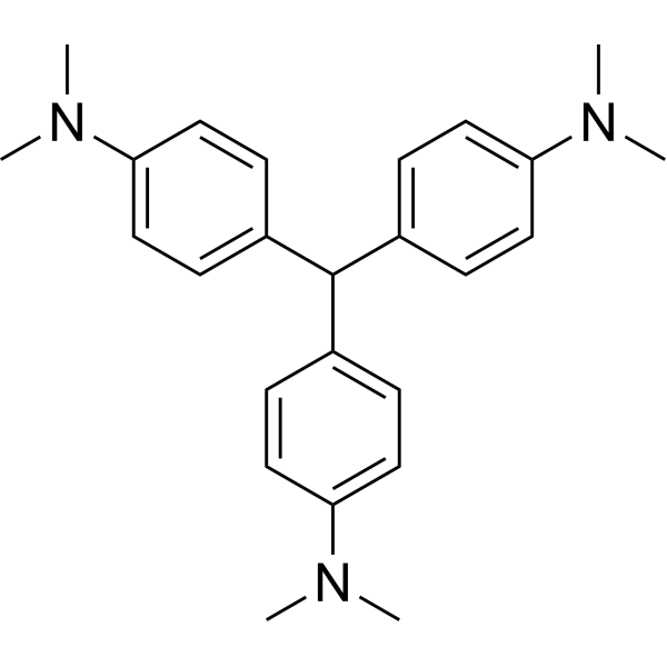
- HY-D0150
-
|
|
Fluorescent Dye
|
Others
|
|
Thiazole Orange is an asymmetric anthocyanin dye that can be coupled with oligonucleotides (ONs) to prepare fluorescent hybridization probes. Thiazole Orange has been widely used in biomolecular detection and staining of DNA/ RNA in gels and can be used for reticulocyte analysis. Thiazole orange generates a significant fluorescence enhancement and high quantum yield when it binds with nucleic acids, especially RNA. Thiazole orange can permeate living cell membranes. Thiazole orange can use UV light for detection, but can also be detected with blue light. The excitation and emission of Thiazole orange are λex = 510 nm (488 nm and 470 nm also show strong excitation) and λem = 527 nm, respectively .
|
-
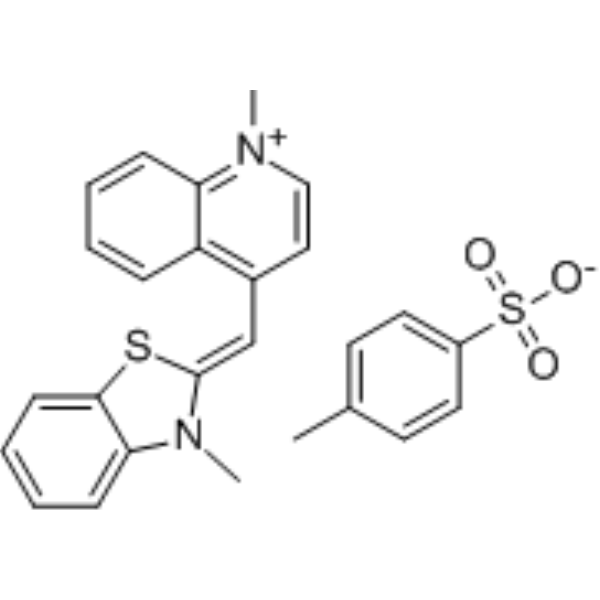
- HY-15560A
-
-

- HY-D0932R
-
|
Solvent Red 24 (Standard); C.I. 26105 (Standard)
|
Biochemical Assay Reagents
Aryl Hydrocarbon Receptor
Cytochrome P450
Reference Standards
|
Metabolic Disease
|
|
Sudan IV (Standard) (Solvent Red 24 (Standard)) is the analytical standard of Sudan IV (HY-D0932). This product is intended for research and analytical applications. Sudan IV is an agonist of the aryl hydrocarbon receptor (AhR) that activates downstream signaling pathways and induces CYP1A1 expression. Sudan IV promotes CYP1A1 gene transcription by activating AhR-ARNT heterodimers and binding to exogenous response elements (XREs) on DNA, thereby enhancing drug metabolizing enzyme activity. Sudan IV can be used to study the toxicity mechanisms of industrial dyes and the effects of interactions with serum proteins (such as bovine serum albumin (BSA)) on their distribution in vivo. Sudan IV is a fat-soluble diazo dye that can be used to stain lipids, triglycerides, and lipoproteins on frozen sections.
|
-

- HY-D1617
-
|
|
Fluorescent Dye
|
Others
|
|
BODIPY 500/510 C1, C12 is a BODIPY dye. BODIPY dye is a small molecule dye with strong ultraviolet absorption ability, its fluorescence peak is relatively sharp, and the quantum yield is high. They are relatively insensitive to the polarity and pH of the environment and are relatively stable under different physiological conditions. Due to its structural asymmetry, BODIPY derives a variety of structural products. BODIPY lipid droplet dyes can well pass through the cell membrane into the cell, and localize the neutral lipids in the cell to specifically stain the lipid droplets, which can be used for labeling of live cells and fixed cells . Maximum excitation/emission wavelength: 500/510 nm . Protect from light, stored at -20°C.
|
-
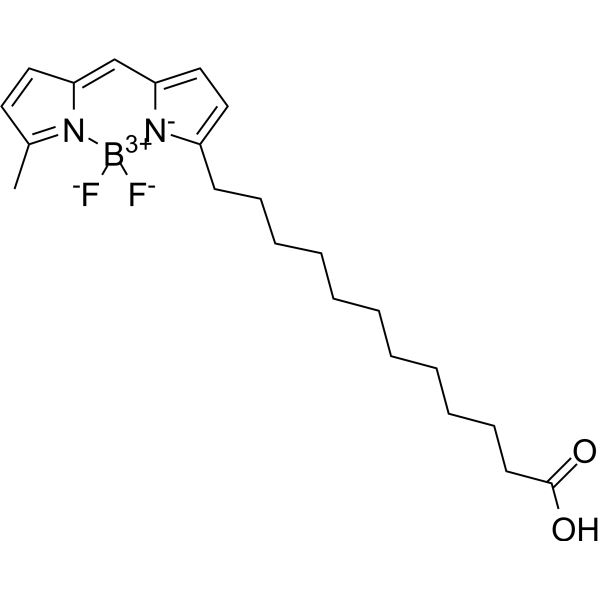
- HY-D0233S
-
|
|
Isotope-Labeled Compounds
Fluorescent Dye
|
Others
|
|
Leucocrystal violet-d6 is the deuterium labeled Leucocrystal violet (HY-D0233). Leucocrystal violet is a cationic triarylmethane dye. Leucocrystal violet generates a colored purple product in the presence of hydrogen peroxide and horseradish peroxidase, or when oxidized by periodate under the catalysis of horseradish peroxidase. Leucocrystal violet serves as a chromogenic reagent for the determination of submicrogram-level hydrogen peroxide, with no interference from tryptophan, persulfate or chromate. Leucocrystal violet reacts with blood to produce a purple/violet color in the presence of hydrogen peroxide; it forms a very weak fluorophore when acting with whole blood in an unbuffered solution, with a maximum absorption wavelength of 630 nm and a maximum fluorescence emission wavelength of 665 nm upon excitation at 630 nm. Leucocrystal violet is widely used for staining blood residues on porous and non-porous materials.
|
-
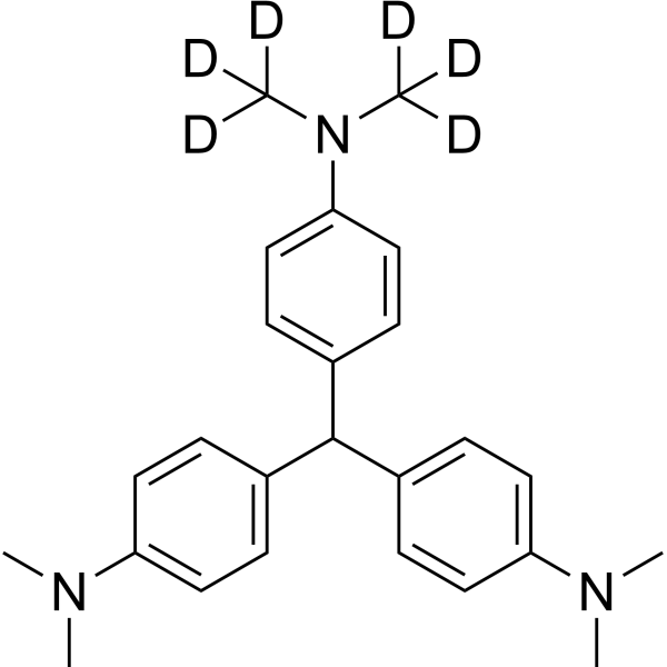
- HY-D3197
-
|
|
Fluorescent Dye
|
Inflammation/Immunology
|
|
CDg16 is a selective fluorescent dye targeting SLC18B1 (λabs/λem=458/544 nm) that is actively transported into lysosomal vesicles of activated macrophages independent of the endocytic pathway. CDg16 enables highly specific vesicle localization in live cells. CDg16 exhibits no cytotoxicity and accurately distinguishes activated M1 and M2 subsets from different origins. CDg16 shows low background staining in non-activated cells and normal organs, making it suitable for time-lapse imaging. In preclinical animal models of inflammatory sites, atherosclerotic plaques and liver inflammation, CDg16 allows visualization of activated macrophages. CDg16 can be used to study inflammation-related diseases and atherosclerosis .
|
-

- HY-118411S
-
|
EdU-13C
|
Isotope-Labeled Compounds
PROTAC Linkers
|
Cancer
|
|
5-Ethynyl-2'-deoxyuridine- 13C1 (EdU- 13C1) is the 13C-labeled 5-Ethynyl-2'-deoxyuridine (HY-118411). 5-Ethynyl-2'-deoxyuridine (EdU), a thymidine analogue, is incorporated into cellular DNA during DNA replication and the subsequent reaction of EdU with a fluorescent azide in a “Click” reaction. EdU staining is a fast, sensitive and reproducible method to study cell proliferation . 5-Ethynyl-2'-deoxyuridine is an alkyl chain-based PROTAC linker that can be used in the synthesis of PROTACs . 5-Ethynyl-2'-deoxyuridine is a click chemistry reagent, it contains an Alkyne group and can undergo copper-catalyzed azide-alkyne cycloaddition (CuAAc) with molecules containing Azide groups.
|
-

- HY-118411S1
-
|
EdU-13C2
|
Isotope-Labeled Compounds
PROTAC Linkers
|
Cancer
|
|
5-Ethynyl-2'-deoxyuridine- 13C2 (EdU- 13C2) is the 13C-labeled 5-Ethynyl-2'-deoxyuridine (HY-118411). 5-Ethynyl-2'-deoxyuridine (EdU), a thymidine analogue, is incorporated into cellular DNA during DNA replication and the subsequent reaction of EdU with a fluorescent azide in a “Click” reaction. EdU staining is a fast, sensitive and reproducible method to study cell proliferation . 5-Ethynyl-2'-deoxyuridine is an alkyl chain-based PROTAC linker that can be used in the synthesis of PROTACs . 5-Ethynyl-2'-deoxyuridine is a click chemistry reagent, it contains an Alkyne group and can undergo copper-catalyzed azide-alkyne cycloaddition (CuAAc) with molecules containing Azide groups.
|
-

- HY-D2772
-
|
|
Biochemical Assay Reagents
|
Others
|
|
5-TAMRA cadaverine can used to modify carboxylic acid group in the presence of activators (e.g. EDC, or DCC) or activated esters (e.g. NHS esters) through a stable amide bond. It also can be reversibly coupled to aldehydes and ketones to form a Schiff base – which can be reduced to a generate stable amine derivative by sodium borohydride (NaBH4) or sodium cyanoborohydride (NaCNH3).
Although the mixed isomers of 5(6)-TAMRA cadaverine is a preferred, routinely used orange-fluorescent dye for staining proteins, it is rearly used for labeling peptides and nucleotides. Purification of 5(6)-TAMRA labeled peptide and nucleotides might be troublesome due to significant signal broadening in HPLC purification. Peptides and nucleotides labeled with a single isomer TAMRA usually give better resolution in HPLC purification that is often required in the conjugation processes.
|
-

- HY-DY1097
-
|
|
Fluorescent Dye
|
Others
|
Thiazole Orange (solution) is an asymmetric anthocyanin dye that can be coupled with oligonucleotides (ONs) to prepare fluorescent hybridization probes. Thiazole Orange has been widely used in biomolecular detection and staining of DNA/ RNA in gels and can be used for reticulocyte analysis. Thiazole orange generates a significant fluorescence enhancement and high quantum yield when it binds with nucleic acids, especially RNA. Thiazole orange can permeate living cell membranes. Thiazole orange can use UV light for detection, but can also be detected with blue light. The excitation and emission of Thiazole orange are λex = 510 nm (488 nm and 470 nm also show strong excitation) and λem = 527 nm, respectively .
Solvent and concentration: DMSO: 10 mM
The 1 mL volume is defined as the base specification. All larger sizes correspond to incremental volumes of this base.
|
-

- HY-D0233R
-
|
|
Reference Standards
Fluorescent Dye
|
Others
|
|
Leucocrystal violet (Standard) is the analytical standard of Leucocrystal violet (HY-D0233). This product is intended for research and analytical applications. Leucocrystal violet is a cationic triarylmethane dye. Leucocrystal violet generates a colored purple product in the presence of hydrogen peroxide and horseradish peroxidase, or when oxidized by periodate under the catalysis of horseradish peroxidase. Leucocrystal violet serves as a chromogenic reagent for the determination of submicrogram-level hydrogen peroxide, with no interference from tryptophan, persulfate or chromate. Leucocrystal violet reacts with blood to produce a purple/violet color in the presence of hydrogen peroxide; it forms a very weak fluorophore when acting with whole blood in an unbuffered solution, with a maximum absorption wavelength of 630 nm and a maximum fluorescence emission wavelength of 665 nm upon excitation at 630 nm. Leucocrystal violet is widely used for staining blood residues on porous and non-porous materials.
|
-

- HY-D0932S
-
|
Solvent Red 24-d6; C.I. 26105-d6
|
Isotope-Labeled Compounds
Biochemical Assay Reagents
Aryl Hydrocarbon Receptor
Cytochrome P450
|
Metabolic Disease
|
|
Sudan IV-d6 (Solvent Red 24-d6) is the deuterium labeled Sudan IV(HY-D0932). Sudan IV is an agonist of the aryl hydrocarbon receptor (AhR) that activates downstream signaling pathways and induces CYP1A1 expression. Sudan IV promotes CYP1A1 gene transcription by activating AhR-ARNT heterodimers and binding to exogenous response elements (XREs) on DNA, thereby enhancing drug metabolizing enzyme activity. Sudan IV can be used to study the toxicity mechanisms of industrial dyes and the effects of interactions with serum proteins (such as bovine serum albumin (BSA)) on their distribution in vivo. Sudan IV is a fat-soluble diazo dye that can be used to stain lipids, triglycerides, and lipoproteins on frozen sections .
|
-

- HY-D1738
-
|
4',6-Diamidino-2-phenylindole dilactate
|
Fluorescent Dye
|
Others
|
|
DAPI (4',6-Diamidino-2-phenylindole) dilactate is a DAPI dye. DAPI is a fluorescent dye that binds strongly to DNA. It binds to the AT base pair of the double-stranded DNA minor groove, and one DAPI molecule can occupy three base pair positions. The fluorescence intensity of DAPI molecules bound to double-stranded DNA is increased by about 20 times, and it is commonly observed with fluorescence microscopy, and the amount of DNA can be determined based on the intensity of fluorescence. DAPI cannot penetrate intact cell membranes and is commonly used for staining both live and fixed cells . DAPI (Compound 3) is an acid-sensing ion channel 3 (ASIC3) inhibitor. DAPI binds to ASIC3 and blocks the channel function. DAPI can be used in the study of chronic pain treatment (Ex/Em = 356/451 nm) .
|
-
- HY-D2868
-
|
4',6-Diamidino-2-phenylindole
|
DNA Stain
Sodium Channel
|
Neurological Disease
|
|
DAPI (4',6-Diamidino-2-phenylindole) is a DAPI dye. DAPI is a fluorescent dye that binds strongly to DNA. It binds to the AT base pair of the double-stranded DNA minor groove, and one DAPI molecule can occupy three base pair positions. The fluorescence intensity of DAPI molecules bound to double-stranded DNA is increased by about 20 times, and it is commonly observed with fluorescence microscopy, and the amount of DNA can be determined based on the intensity of fluorescence. In addition, because DAPI can pass through intact cell membranes, it can be used to stain both live and fixed cells . DAPI (Compound 3) is an acid-sensing ion channel 3 (ASIC3) inhibitor. DAPI binds to ASIC3 and blocks the channel function. DAPI can be used in the study of chronic pain treatment (Ex/Em = 356/451 nm).
|
-

- HY-D0814
-
|
4',6-Diamidino-2-phenylindole dihydrochloride
|
DNA Stain
Sodium Channel
|
Neurological Disease
|
|
DAPI (4',6-Diamidino-2-phenylindole) dihydrochloride is a DAPI dye. DAPI is a fluorescent dye that binds strongly to DNA. It binds to the AT base pair of the double-stranded DNA minor groove, and one DAPI molecule can occupy three base pair positions. The fluorescence intensity of DAPI molecules bound to double-stranded DNA is increased by about 20 times, and it is commonly observed with fluorescence microscopy, and the amount of DNA can be determined based on the intensity of fluorescence. In addition, because DAPI can pass through intact cell membranes, it can be used to stain both live and fixed cells . DAPI (Compound 3) is an acid-sensing ion channel 3 (ASIC3) inhibitor. DAPI binds to ASIC3 and blocks the channel function. DAPI can be used in the study of chronic pain treatment (Ex/Em = 356/451 nm).
|
-
- HY-D1737
-
|
|
Bacterial
|
Infection
|
|
RADA is a fluorescent D-amino acid (FDAA) with high photostability and thermostability, which emits yellow-to-orange fluorescence. RADA shows low outer membrane permeability in wild-type Gram-negative Escherichia coli, but it targets penicillin-binding proteins and L,D-transpeptidases, mimics the interaction between acyl acceptors and enzyme intermediates, and integrates into peptidoglycan during biosynthesis. As a peptidoglycan labeling reagent, RADA metabolically integrates into the nascent peptidoglycan of live bacterial cells, labels the peptidoglycan at the poles and lateral walls of mycobacteria, and enables visualization of peptidoglycan synthesis and remodeling processes. RADA serves as a non-specific stain for fixed cells, is non-toxic to bacterial cells, and its red-shifted excitation/emission spectra reduce phototoxicity. RADA also supports virtual pulse-chase labeling experiments and stochastic optical reconstruction microscopy for sub-diffraction-limited imaging of bacterial cell walls .
|
-

- HY-182341
-
|
|
Drug Derivative
|
Infection
|
|
APD-209 is a sialic acid conjugate with a PEG4 polar linker and a PDA non-polar tail. APD-209 aggregates adenovirus type 37 particles, blocks the binding of viral particles to human corneal epithelial cells, and inhibits cellular entry of adenovirus type 37. APD-209 can be used for the research of epidemic keratoconjunctivitis .
|
-

- HY-DY1081
-
|
4',6-Diamidino-2-phenylindole dihydrochloride (solution)
|
Sodium Channel
DNA Stain
|
Neurological Disease
|
DAPI (4',6-Diamidino-2-phenylindole) dihydrochloride (solution) is a DAPI dye. DAPI is a fluorescent dye that binds strongly to DNA. It binds to the AT base pair of the double-stranded DNA minor groove, and one DAPI molecule can occupy three base pair positions. The fluorescence intensity of DAPI molecules bound to double-stranded DNA is increased by about 20 times, and it is commonly observed with fluorescence microscopy, and the amount of DNA can be determined based on the intensity of fluorescence. In addition, because DAPI can pass through intact cell membranes, it can be used to stain both live and fixed cells . DAPI (Compound 3) is an acid-sensing ion channel 3 (ASIC3) inhibitor. DAPI binds to ASIC3 and blocks the channel function. DAPI can be used in the study of chronic pain treatment (Ex/Em = 356/451 nm).
Solvent and concentration: ddH2O: 5 mg/mL
|
-

- HY-149003
-
|
|
PARP
Apoptosis
|
Cancer
|
|
PARP1-IN-10 (compound 12c) is a no-cytotoxicity and potent PARP1 inhibitor with an IC50 value of 50.62 nM in vitro. PARP1-IN-10 causes cell cycle arrest at G2/M phase and apoptosis, and enhances the cytotoxicity of temozolomide (TMZ) .
|
-
| Cat. No. |
Product Name |
Type |
-
- HY-D0815
-
|
|
Fluorescent Dyes
|
|
Propidium Iodide (PI) is a nuclear staining agent that stains DNA. Propidium Iodide is an analogue of ethidine bromide that emits red fluorescence upon embedding in double-stranded DNA. Propidium Iodide cannot pass through living cell membranes, but it can pass through damaged cell membranes to stain the nucleus. Propidium Iodide has a fluorescence wavelength of 493/617 nm and a wavelength of 536/635 nm after Mosaic with DNA. Propidium Iodide is commonly used in the detection of apoptosis (apoptosis) or necrosis (necrosis), and is often used in flow cytometry analysis.
|
-
- HY-D0718
-
|
Nile Blue A oxazone; Phenoxazone 9
|
Fluorescent Dyes
|
|
Nile red (Nile blue oxazone) is a lipophilic stain. Nile red has environment-sensitive fluorescence. Nile red is intensely fluorescent in a lipid-rich environment while it has minimal fluorescence in aqueous media. Nile red is an excellent vital stain for the detection of intracellular lipid droplets by fluorescence microscopy and flow cytof uorometry. Nile red stains intracellular lipid droplets red. The fluorescence wavelength is 559/635 nm .
|
-
- HY-B0324A
-
|
Basic Violet 3; Gentian Violet; Methyl Violet 10B
|
Fluorescent Dyes
|
|
Crystal Violet, also known as Gentian violet, methyl violet 10B, is a triphenyl-methane, an alkaline dye that binds to DNA in the nucleus of a cell, staining it a deep purple. It is often used for Gram staining to classify bacteria, or for cell or histological staining[1].
|
-
- HY-Y0016
-
|
Basic Violet 10; Brilliant Pink B; Rhodamine O; Tetraethylrhodamine
|
Fluorescent Dyes
|
|
Rhodamine B is a staining fluorescent dye, commonly used for dyeing textiles, paper, soap, leather, and agents.
|
-
- HY-D1168
-
|
|
Fluorescent Dyes
|
|
Oil Red O is a fat-soluble diazol dye, with a maximum absorption at 518 nm. Oil Red O stains neutral lipids and cholesteryl esters but not biological membranes. Oil Red O can be used for detecting and quantifying hepatic steatosis in mouse liver biopsies. Oil Red O staining efficiently helps to visualize the radical changes that occur in tissues as metabolic disease occurs and progresses .
|
-
- HY-D0021
-
|
EtBr; Homidium bromide
|
Fluorescent Dyes
|
|
EthD bromide is an intercalating agent commonly used as a fluorescent tag (nucleic acid stain) in molecular biology laboratories for techniques such as agarose gel electrophoresis.
|
-
- HY-D1020
-
|
7-AAD
|
Fluorescent Dyes
|
|
7-Aminoactinomycin D (7-AAD) a cell-impermeant fluorescent DNA stain, is a potent RNA polymerase inhibitor. 7-Aminoactinomycin D selectively binds to GC regions of the DNA. 7-Aminoactinomycin D also has antibacterial effects .
|
-
- HY-N0116
-
|
Natural Black 1; Haematoxylin
|
Fluorescent Dyes
|
|
Hematoxylin (Natural Black 1), a naturally occurring flavonoid compound derived from Caesalpinia sappan Linn.. Hematoxylin is a nuclear stain in histology and is also a potent Aβ42 fibrillogenesis inhibitor with an IC50 of 1.6 μM.
|
-
- HY-D0914
-
|
FD&C Green No. 3; Food green 3; C.I. 42053
|
Fluorescent Dyes
|
|
Fast Green FCF is a sea green triarylmethane food dye, with absorption maximum ranging from 622 to 626 nm. Fast Green FCF inhibits α-synuclein aggregation, as well as Aβ and P2X4 receptor, and TLR4/Myd88/NF-κB. Fast Green FCF is widely used as a staining agent like quantitative stain for histones at alkaline pH after acid extraction of DNA, and as a protein stain in electrophoresis. Fast Green FCF improves cognitive impairment, depression, relieves pain allergies, and promotes reproductive function .
|
-
- HY-12489
-
|
Acid Red 112
|
Fluorescent Dyes
|
|
Ponceau S (Acid Red 112) is a non-specific protein dye commonly used as a stain for Western blot. Ponceau S is used in an acidic aqueous solution that is compatible with antibody-antigen binding and dyes the proteins on the membrane red .
|
-
- HY-D0970
-
|
Direct Blue 14; Trypan Blue
|
Fluorescent Dyes
|
|
Diphenyl Blue (Trypan Blue) is a cell active dye, the most commonly used dye for the identification of dead cells, of en used to test cell membrane integrity and cell viability. Diphenyl Blue staining is one of the methods for tissue and cell culture. When cells are deactivated or have incomplete cell membranes, Diphenyl Blue can stain them Blue. Normal living cells with intact cell membranes reject Diphenyl blue and do not stain them blue. However, macrophages are capable of phagocytosis of Diphenyl Blue, so it can be used as a living stain for macrophages .
|
-
- HY-15922
-
Luminol
2 Publications Verification
Diogenes reagent
|
Fluorescent Dyes
|
|
Luminol is a chemical that exhibits chemiluminescence with pKa values of 6.74 and 15.1. Luminol exhibits chemiluminescence (CL) at 425 nm λmax. Luminol is commonly used in forensics as a diagnostic tool for the detection of blood stains .
|
-
- HY-D0987
-
|
|
Fluorescent Dyes
|
|
Stains-All, a cationic carbocyanine dye, is a convenient probe to study the structural features of the individual calcium-binding sites of calmodulin (CaM) and related calcium-binding proteins (CaBP) .
|
-
- HY-D0952
-
|
|
Fluorescent Dyes
|
|
Acridine Orange base is a cell-permeable fluorescent dye that stains organisms (bacteria, parasites, viruses, etc.) bright orange and, when used under appropriate conditions (pH=3.5, Ex=460 nm), distinguishes human cells in green for detection by fluorescence microscopy. Acridine Orange base fluoresces green when bound to dsDNA (Ex=488, Em=520-524) and red when bound to ssDNA (Ex=457, Em=630-644) or ssRNA (Ex=457, Em=630-644), also can be used in cell cycle assays .
|
-
- HY-D0367
-
|
Calcofluor White M2R
|
Fluorescent Dyes
|
|
Fluorescent brightener 28 is a fluorescent whitening agent commonly used in the padding process of the textile industry. Fluorescent brightener 28 is capable of staining polysaccharides such as cellulose, and when the plasma membrane ruptures, it also weakly stains the cytoplasm and strongly stains the cell nucleus. Additionally, Fluorescent brightener 28 can be utilized to detect intracellular chitin in living cells. Fluorescent Brightener 28 also is a visible light emitting diode (LED)-light sensitive photoinitiator for free radical photopolymerizations .
|
-
- HY-D0166
-
|
|
Fluorescent Dyes
|
|
Neutral Red, a nitrogenous pH-indicator with a pKi of 6.8, is an indicator for the internal acidification of thylakoids. Neutral Red stains lysosomes red .
|
-
- HY-D0944
-
|
|
Fluorescent Dyes
|
|
Giemsa stain is a composite dye composed of methylene azure, methylene blue, eosin and other components. Giemsa stain stains chromatin, nuclear membranes, specific cytoplasmic components and microorganisms. Giemsa stain is used in research on cytological and parasitological staining .
|
-
- HY-D0955
-
|
Thionine acetate
|
Fluorescent Dyes
|
|
Thionin acetate (Thionine acetate) is a compound present in the seeds, stems, roots, and leaves of many plant species with antibacterial activity. Thionin acetate is a metachromatic cationic histological dye widely used in biological staining .
|
-
- HY-D0004
-
|
Azure B chloride
|
Fluorescent Dyes
|
|
Azure B is a cationic dye and the major metabolite of Methylene blue. Azure B is used in making Azure eosin stains for blood smear staining. Azure B is a high-potency, selective and reversible inhibitor of monoamine oxidases (MAO)-A, with IC50s of 11 and 968 nM for recombinant human MAO-A and MAO-B, respectively. Azure B possesses significant antidepressant-like effects .
|
-
- HY-D0232
-
|
Brilliant Blue R
|
Fluorescent Dyes
|
|
Brilliant blue R250 (Brilliant Blue R), an anionic dye, is the most popular stain to detect proteins resolved in SDS-PAGE gels .
|
-
- HY-D0093
-
|
EthD-1
|
Fluorescent Dyes
|
|
Ethidium homodimer (EthD-1) is a high-affinity fluorescent nucleic acid dye commonly used to stain mammals, bacteria, yeast, and fungi. Ethidium homodimer binds to DNA or RNA, enhancing fluorescence more than 30 times. The Ethidium homodimer has a strong positive charge, so it cannot cross cell membranes and stain living cells; But the Ethidium homodimer can cross the disordered region of the dead cell membrane to reach the nucleus and embed the DNA double strand to produce red fluorescence. Therefore, Ethidium homodimer is a relatively sensitive nucleic acid stain that can accurately detect nucleic acids in solution or in decomposing cells. Ethidium homodimer binds DNA, Ex/Em=528/617 nm .
|
-
- HY-D0333
-
|
Sirius Red
|
Fluorescent Dyes
|
|
Direct Red 80 (Sirius Red) is a polyazo dye used principally in staining methods for collagen and amyloid. Direct Red 80 does not release benzidine upon degradation and is safer than many traditional direct dyes .
|
-
- HY-101900
-
|
Nile blue sulfate
|
Fluorescent Dyes
|
|
Nile Blue A (Nile blue sulfate) is used to differentiate melanins and lipofuscins. It is also useful for staining fats and preparation of an amperometric glucose sensor .
|
-
- HY-W094758A
-
|
|
Fluorescent Dyes
|
|
4-Di-1-ASP is a styryl dye used to stain glioma cells in living brain tissue for analysis of cell structure, viability, proliferation and endocytosis, cytokinesis and phagocytosis, as well as for observation of mitochondrial structures in living cells. 4-Di-1-ASP fluoresces green when imaged microscopically (λex /λem = 475/606 nm) .
|
-
- HY-N0335
-
Indigo
1 Publications Verification
|
Fluorescent Dyes
|
|
Indigo is a blue dye. Indigo stains cellulose fibers such as cotton. Indigo stains food or biological samples to visualize and quantify the uptake or distribution of substances by organisms .
|
-
- HY-101888
-
|
|
Fluorescent Dyes
|
|
Cresyl Violet acetate is a dye, which can be used to stain neurons.
|
-
- HY-D0003
-
|
|
Fluorescent Dyes
|
|
Methyl blue belongs to the group of triaminotriphenylmethane dyes. Methyl blue is widely used as antiseptic dye in polychrome staining method and has applications in histological and microbiological staining solutions. Methyl blue has been used as a model to study the effect of various catalysts on photodegradation of dyes .
|
-
- HY-B1539A
-
|
Magenta base monohydrochloride; Basic Fuchsin monohydrochloride; Rosaniline Base monohydrochloride
|
Fluorescent Dyes
|
|
Fuchsine base (Magenta base; Basic Fuchsin) monohydrochloride is a triaminotriphenylmethane dye. Fuchsine base monohydrochloride has anesthetic, bactericidal and fungicidal properties. Fuchsine base monohydrochloride can be used for the staining of collagen, muscle, mitochondria and tuberculosis. Fuchsine base monohydrochloride is commonly used as a counterstain in Gram staining .
|
-
- HY-D0166A
-
|
|
Fluorescent Dyes
|
|
Neutral Red (IND) is an organic dye commonly used in biology and cytology laboratories. It can be used to stain living cells, secreted proteins and other molecular structures, etc., and has a wide range of applications in cell imaging and staining. In addition, Neutral Red (IND) is widely used in industrial fields such as water treatment, food processing and paper manufacturing, for example as an indicator or colorant. Although the compound has no direct medical application, it has important application value in the fields of biology, chemistry and industry.
|
-
- HY-D0996
-
|
|
Fluorescent Dyes
|
|
Lds-751 is a nucleic acid stain that mainly detects DNA. Lds-751 is a nucleic acid stain that mainly detects DNA. Lds-751 has a high affinity for DNA and fluorescence is enhanced after binding, but the maximum emission wavelength is 670nm. Lds-751 and Thiazole orange can be used for the differentiation of red blood cells, platelets, reticulocytes, and nucleated cells and can be stimulated at 488nm. Studies have shown that LDS-751 binds almost exclusively to mitochondria when incubated with nucleated living cells. After nucleated Acridine Orange (HY-101879) staining and LDS-751 treatment of cells, confocal microscopy revealed almost no co-location of the cells. Staining with Rhodamine 123 (HY-D0816), a dye known to bind polarized mitochondria, was almost identical to the pattern observed with LDS-751 .
|
-
- HY-DY1008
-
|
|
Fluorescent Dyes
|
Nile Red (solution) is a lipophilic stain. Nile red has environment-sensitive fluorescence. Nile red is intensely fluorescent in a lipid-rich environment while it has minimal fluorescence in aqueous media. Nile red is an excellent vital stain for the detection of intracellular lipid droplets by fluorescence microscopy and flow cytof uorometry. Nile red stains intracellular lipid droplets red. The fluorescence wavelength is 559/635 nm .
Solvent and concentration: DMSO: 1 mM
|
-
- HY-DY1006
-
|
|
Fluorescent Dyes
|
Propidium Iodide (PI) (solution) is a nuclear staining agent that stains DNA. Propidium Iodide is an analogue of ethidine bromide that emits red fluorescence upon embedding in double-stranded DNA. Propidium Iodide cannot pass through living cell membranes, but it can pass through damaged cell membranes to stain the nucleus. Propidium Iodide has a fluorescence wavelength of 493/617 nm and a wavelength of 536/635 nm after Mosaic with DNA. Propidium Iodide is commonly used in the detection of apoptosis (apoptosis) or necrosis (necrosis) , and is often used in flow cytometry analysis.
Solvent and Concentration: Sterile water: 1 mg/mL
The 1 mL volume is defined as the base specification. All larger sizes correspond to incremental volumes of this base.
|
-
- HY-W356116
-
|
Nbd-ceramide
|
Fluorescent Dyes
|
|
C6 NBD Ceramide is a Golgi apparatus fluorescent probe with cell membrane permeability. C6 NBD Ceramide can be used for fast and convenient green fluorescent labeling of Golgi in living and fixed cells, and can be used to observe changes in Golgi morphology in living cells (Ex=466 nm, Em=536 nm). C6-NBD-ceramide is metabolized to fluorescent sphingomyelin and glucosylceramide, can be used for the study of sphingolipid transport and metabolic mechanism .
|
-
- HY-DY1050
-
|
|
Fluorescent Dyes
|
Ethidium bromide (solution) is an intercalating agent commonly used as a fluorescent tag (nucleic acid stain) in molecular biology laboratories for techniques such as agarose gel electrophoresis.
Solvent and Concentration: Sterile water: 10 mg/Ml
|
-
- HY-D0721
-
|
6-Carboxyfluorescein diacetate
|
Fluorescent Dyes
|
|
6-CFDA is a common aliphatic luciferin-line organism. CFDA conducts free diffusion into cells, and then it is hydrolyzed into carboxyl fluorescein (CF) by intracellular non-specific lipase. CF containing portion contains an additional negative charge so that it is better retained in cells, compared to fluorescein dyes .
|
-
- HY-15922A
-
|
|
Fluorescent Dyes
|
|
Luminol sodium salt is a chemical that exhibits chemiluminescence with pKa values of 6.74 and 15.1. Luminol sodium salt exhibits chemiluminescence (CL) at 425 nm λmax. Luminol sodium salt is commonly used in forensics as a diagnostic tool for the detection of blood stains .
|
-
- HY-D1122
-
|
|
Fluorescent Dyes
|
|
Janus green B is a supravital stain. Janus green B staining reaction is oxygen dependent, and is reversibly inhibited by cyanide. Janus green B has been used for staining peripheral nerves in live insects, lymphatic vessels of rabbits and mitochondria .
|
-
- HY-W040198
-
|
|
Fluorescent Dyes
|
|
Phenosafranine is a phenazine dye. Phenosafranine exhibits higher binding affinity for triple-stranded RNA than for double-stranded RNA, and binds to both types of RNA via intercalation. Phenosafranine interacts with hemoglobin. Phenosafranine can be used for plant cell staining, as well as the detection of hemoglobin, dopamine, serotonin, etc.
|
-
- HY-D0163
-
|
|
Fluorescent Dyes
|
|
Methyl Green is a non-intercalating fluorescent labeling agent that selectively binds to the major groove of DNA. Methyl Green electrostatically interacts with the major groove of DNA through positively charged groups, exhibiting key activities such as high affinity, resistance to photobleaching, and stable fluorescence emission. Methyl Green can be directly measured by microscopy and flow cytometry, with peaks at 633 and 677 nm. Methyl Green can be used for fluorescent labeling of the nuclei of embryonic tissues or cells, or DNA staining and cell activity detection in gel electrophoresis[1][2][3].
|
-
- HY-126367
-
|
Acid Green 5
|
Fluorescent Dyes
|
|
Light green SF yellowish (Acid Green 5) is a water-soluble triarylmethane dye. Light green SF yellowish serves as a histological stain that selectively labels mitochondria, collagen, and cartilage, while being an essential component of Papanicolaou staining. Light green SF yellowish is commonly used as a cytoplasmic counterstain for nuclear stains, and is applied in Masson's trichrome staining for collagen fibers, Pap staining, and cytological polychromatic staining in histopathology. Light green SF yellowish also induces growth inhibition and local fibrosarcomas in rats and exerts mild pulmonary tumorigenicity in mice .
|
-
- HY-DY2001
-
|
|
Fluorescent Dyes
|
Crystal Violet (solution) , also known as Gentian violet, methyl violet 10B, is a triphenyl-methane, an alkaline dye that binds to DNA in the nucleus of a cell, staining it a deep purple. It is often used for Gram staining to classify bacteria, or for cell or histological staining .
Solvent and Concentration: Sterile water: 5 mg/mL (0.5%)
|
-
- HY-W088068
-
|
|
Fluorescent Dyes
|
|
Wright's stain is a composite cell stain that mainly binds to intracellular nucleic acids, proteins and other components through thiazine dyes (such as methylene blue) and eosin. Wright's stain is pH-dependent (optimal pH 6.4-6.7) and achieves cell morphology resolution by differentially staining the cytoplasm and nucleus. Under alkaline conditions, thiazine dyes bind to nucleic acids to form purple, and acidic eosin binds to cytoplasmic proteins to form red, which can form contrasting cell morphological features. Wright's stain can clearly display the fine structures of blood cells and bone marrow cells (such as nuclear chromatin and granules) and quickly evaluate cell morphological abnormalities .
|
-
- HY-W247131
-
DASPEI
2 Publications Verification
|
Fluorescent Dyes
|
|
DASPEI is a cationic styrenyl mitochondrial dye with large Stokes shift. DASPEI has excitation and emission wavelength at 550/573 nm, which has good light chromogenic property. DASPEI can stain mitochondria in living cells with good labeling property. And DASPEI can also be used to stain presynaptic nerve endings independently of neuronal activity .
|
-
- HY-D0220A
-
|
Toluidine Blue O (purity 36%)
|
Fluorescent Dyes
|
|
Toluidine Blue (Toluidine Blue O) purity 36% is an alkaline quinonimine dye (vivo dye) with high affinity for acidic tissue components, staining nuclei blue and polysaccharides purple. Toluidine Blue purity 36% shows heterostaining properties for mast cells, mucins and chondrocytes. Toluidine Blue purity 36% can stain different components of plant tissues and cells in different colours. Toluidine Blue purity 36% is also used as a diagnostic aid to identify malignant lesions, such as cancer .
|
-
- HY-D0950A
-
|
|
Fluorescent Dyes
|
|
Methyl Green zinc chloride is a potent fluorescent dye. Methyl Green zinc chloride is a DNA stains of cells and electrophoretic gels. Methyl Green zinc chloride can be used as direct measuring of viability by both microscopy and flow cytometry, with peaks at 633 and 677 nm .
|
-
- HY-D0957
-
|
|
Fluorescent Dyes
|
|
Ethyl Violet is a triphenylmethane cationic dye with antibacterial activity. Ethyl Violet is applicable to research related to antibacterial therapy and histological staining .
|
-
- HY-D0127
-
|
|
Fluorescent Dyes
|
|
Merocyanin 540 is a fluorescent membrane probe that selectively stains the membranes of a wide variety of electrically excitable cells, but not those of nonexcitable cells (Ex/Em: 540/580 nm) .
|
-
- HY-D1543
-
|
|
Fluorescent Dyes
|
|
Pyronin B is an organic cationic dye used for the staining of bacteria, mycobacteria and ribonucleic acids. Pyronin B is also used as a small hydrophobic (SH) protein channel inhibitor .
|
-
- HY-117070
-
|
|
Fluorescent Dyes
|
|
TO-PRO-3 iodide is a cell-impermeant nucleic acid stain. TO-PRO-3 iodide is useful as a nuclear counterstain and dead cell indicator (Ex/Em = 642/661 nm).
|
-
- HY-137896
-
|
|
Fluorescent Dyes
|
|
4-Acetamido-4'-isothiocyanatostilbene-2,2'-disulfonic acid disodium is a fluorescent dye. 4-Acetamido-4'-isothiocyanatostilbene-2,2'-disulfonic acid disodium can be used to demonstrate retrograde axonal transport to label secondary antibodies and as a fluorescent whole cell stain .
|
- HY-D0954
-
|
|
Fluorescent Dyes
|
|
Jenner's Stain is a dye that is used in microscopy for staining blood smears. Jenner's Stain can be used for the chromosome stain by C-banding technique. Jenner's Stain can be used for the stain for routine blood examinations and malarial staining .
|
- HY-145612
-
|
|
Fluorescent Dyes
|
|
Sudan red 7B is a red non-fluorescent stain that can be used to stain fat bodies .
|
- HY-D0983
-
- HY-DY1077
-
|
|
Fluorescent Dyes
|
Rhodamine B (solution) is a staining fluorescent dye, commonly used for dyeing textiles, paper, soap, leather, and agents.
Solvent and concentration: DMSO: 10 mM
|
- HY-W250151
-
|
|
Fluorescent Dyes
|
|
Leishman's stain is an essential staining tool for for staining of the peripheral blood and bone marrow smears (displayed pale bluish-grey to deep blue under oil-immersion lens) .
|
- HY-D1174
-
|
|
Fluorescent Dyes
|
|
Rhodamine 700, a Lambdachrome laser dye, is one of the few rhodamine dyes with near infrared fluorescence. Rhodamine 700 can be used for mitochondrial staining .
|
- HY-D1075
-
|
HIDC
|
Fluorescent Dyes
|
|
1,1',3,3,3',3'-Hexamethylindodicarbocyanine iodide is a carbocyanine dye that stains mitochondria of live cells.
|
- HY-D0164
-
|
|
Fluorescent Dyes
|
|
Naphthol AS-BI is a substrate of β-glucuronidase and produces a bright red effect after staining biological tissues.
|
- HY-D2170
-
|
|
Fluorescent Dyes
|
|
AF488 streptavidin is a fluorescently labeled streptavidin. AF488 streptavidin is a streptavidin conjugated to Alexa Fluor 488, with 4 fluorophores per protein molecule, enabling stoichiometric fluorescent labeling of cell surface targets. AF488 streptavidin can form a complex with biotinylated E07 aptamer to stain cells expressing EGFR, and the staining is reversible after treatment with mA9 detoxifying oligonucleotides (Ex/Em = 470/520 nm) .
|
- HY-Y0016R
-
|
Basic Violet 10 (Standard); Brilliant Pink B (Standard); Rhodamine O (Standard); Tetraethylrhodamine (Standard)
|
Fluorescent Dyes
|
|
Rhodamine B (Standard) is the analytical standard of Rhodamine B. This product is intended for research and analytical applications. Rhodamine B is a staining fluorescent dye, commonly used for dyeing textiles, paper, soap, leather, and agents.
|
- HY-D1544
-
|
|
Fluorescent Dyes
|
|
Uniblue A sodium is a reactive protein stain that can be used in the covalent pre-gel staining of the protein (Ex=594 nM) .
|
- HY-118320
-
|
Mordant orange 1
|
Fluorescent Dyes
|
|
Alizarine Yellow R (Mordant orange 1), Salicylic acid derivative (HY-B0167), is an azo dye. Alizarine Yellow R is used as a pH indicator and a biological stain in chemical examinations and dyeing industries .
|
- HY-W250143
-
|
|
Fluorescent Dyes
|
|
Toluidine blue (ZnCl2) is a basic thiazine dye commonly used as a biological stain for microscopy. It has a deep bluish-purple color and is commonly used to stain nucleic acids such as DNA and RNA, as well as to stain mast cells, cartilage, and other connective tissues. Toluidine blue (ZnCl2) stains the acidic components of these tissues, such as sulfated or carboxylated mucopolysaccharides. It is frequently used in histology, cytology, and pathology applications to aid in the diagnosis of various diseases and conditions. The dye is usually applied to tissue sections prior to microscopic examination and can be differentiated using an acidic alcohol solution. Toluidine blue (ZnCl2) is a relatively simple and inexpensive stain with good reproducibility, making it a popular choice for many laboratories.
|
- HY-D1220
-
|
Indanthrene
|
Fluorescent Dyes
|
|
Pigment blue 60 is an excellent paint and ceramic stain.
|
- HY-D2582
-
|
|
Fluorescent Dyes
|
|
DMAO is a membrane-permeable DNA fluorescent dye stains live and dead bacteria. (Ex/Em = 490/540 nm) .
|
- HY-D0404
-
|
|
Fluorescent Dyes
|
|
Direct Red 254 is a bisazo dye for textile applications. Direct Red 254 can be used for amyloid tissue staining in basic research on neurodegenerative diseases (e.g., Alzheimer's disease) .
|
- HY-101889
-
|
Oxazine 9 perchlorate
|
Fluorescent Dyes
|
|
Cresyl Violet perchlorate is a red fluorescent stain, which can be used to stain neurons.
|
- HY-W250306
-
|
|
Fluorescent Dyes
|
|
Carbol fuchsin is a histological stain used in microbiology to distinguish acid-fast bacteria from non-acid-fast bacteria. It is a mixture of basic fuchsin, phenol, and water and is commonly used in the Ziehl-Neelsen staining technique for the detection of tuberculosis and other mycobacterial infections. Carbol fuchsin stains the cell walls of acid-fast bacteria bright red, while other cells are unstained or slightly stained. This makes it easier to see and identify these microbes under a microscope. Carbol fuchsin is also used in veterinary medicine and phytopathology for similar purposes.
|
- HY-D1491
-
|
|
Fluorescent Dyes
|
|
Fast Red Violet LB is a dye for staining tartrate resistant acid phosphatase (TRAP). Fast Red Violet LB can be used for alkaline phosphatase (ALP) activity staining .
|
- HY-D1585
-
|
|
Fluorescent Dyes
|
|
BODIPY TR methyl ester is a lipophilic GFP Counterstain. BODIPY TR methyl ester dye readily permeates cell membranes and localizes in endomembranous organelles but not localize strongly in plasma membranes. BODIPY TR methyl ester is an excellent red fluorescent vital dye (Ex=568 nm, Em=625 nm), can be used to reveal the location and shapes of cell nuclei, the shapes of cells within embryonic tissues, as well as the bound aries of organ-forming tissues within the whole embryo .
|
- HY-W053871
-
|
|
Fluorescent Dyes
|
|
4-Methylphthalimide is a stain with biological research applications.
|
- HY-D0004R
-
|
Azure B chloride (Standard)
|
Fluorescent Dyes
|
|
Azure B (Standard) is the analytical standard of Azure B. This product is intended for research and analytical applications. Azure B is a cationic dye and the major metabolite of Methylene blue. Azure B is used in making Azure eosin stains for blood smear staining. Azure B is a high-potency, selective and reversible inhibitor of monoamine oxidases (MAO)-A, with IC50s of 11 and 968 nM for recombinant human MAO-A and MAO-B, respectively. Azure B possesses significant antidepressant-like effects .
|
- HY-D1386
-
|
|
Fluorescent Dyes
|
|
JF526-Taxol (TFA) is a versatile scaffold for fluorogenic probes including ligands for self-labeling tags, stains for endogenous structures, and spontaneously blinking labels for super-resolution immunofluorescence .
|
- HY-D1246
-
|
|
Fluorescent Dyes
|
|
Ethidium monoazide bromide is a DNA intercalating fluorescent dye that enters bacteria with damaged membranes. Ethidium monoazide bromide can be covalently linked to DNA by photoactivation. Ethidium monoazide bromide stains only dead cells . Ethidium monoazide (bromide) is a click chemistry reagent, it contains an Azide group and can undergo copper-catalyzed azide-alkyne cycloaddition reaction (CuAAc) with molecules containing Alkyne groups. It can also undergo strain-promoted alkyne-azide cycloaddition (SPAAC) reactions with molecules containing DBCO or BCN groups.
|
- HY-D1102
-
|
|
Fluorescent Dyes
|
|
DNA intercalator 3 is a DNA intercalating agent that can be used for DNA staining.
|
- HY-D1173
-
|
|
Fluorescent Dyes
|
|
Acid blue 113 is an azo dye whose staining effect is effectively removed by nanoscale zero-valent iron (NZVI) particles.
|
- HY-D1615
-
|
|
Fluorescent Dyes
|
|
BODIPY 530/550 NHS ester can be used for the stain of protein. BODIPY 530/550 NHS ester can be used for fluorescence OIM (oblique illumination microscopic) image .
|
- HY-D1121
-
|
|
Fluorescent Dyes
|
|
Acid black 24 is a black agent whose staining effect is effectively removed by nanoscale zero-valent iron (NZVI) particles. The maximum unit removal capacity is 609.4 mg of dye per gram of NZVI.
|
- HY-D0914A
-
|
|
Fluorescent Dyes
|
|
Fast green FCF free acid is a dye that is acid-resistant. Fast Green FCF free acid inhibits α-synuclein aggregation, as well as Aβ, P2X4 receptor and TLR4/Myd88/NF-κB. Fast Green FCF free acid is widely used as a staining agent like quantitative stain for histones at alkaline pH after acid extraction of DNA, and as a protein stain in electrophoresis. Fast Green FCF free acid improves cognitive impairment, depression, relieves pain allergies, and promotes reproductive function .
|
- HY-W441766
-
|
|
Fluorescent Dyes
|
|
3-Nitrobenzaldoxime is a stain with biological research applications.
|
- HY-D1385
-
|
|
Fluorescent Dyes
|
|
JF526–Pepstatin A TFA is a fluorescent dye that can be used for lysosomal staining in live cells. The excitation maximum is 530 nm and the emission maximum is 549 nm .
|
- HY-D1493
-
|
|
Fluorescent Dyes
|
|
FIM-1 is a fluorescent PKC (protein kinase C) probe that can be used for mitochondrial staining. FIM-1 inhibits PKC and acts as ATP-competitive catalytic site inhibitor .
|
- HY-D1536
-
|
|
Fluorescent Dyes
|
|
Glycine cresol red is a complexometric indicator. Glycine cresol red forms coloured complexes with Al 3+, Ga 3+ and In 3+ ions in aqueous solutions. Glycine cresol red can been used for the spectrophotometric determination of inorganic ions. Glycine cresol red can be used as a stain in neurohistology .
|
- HY-D1771
-
|
|
Fluorescent Dyes
|
|
Thionin perchlorate is a general nuclear stain for chromatin and mucin.
|
- HY-D0371
-
|
|
Fluorescent Dyes
|
|
Acid blue 62 is an acid dye whose staining effect can be removed by sepiolite
|
- HY-D1101
-
|
|
Fluorescent Dyes
|
|
DNA intercalator 2 is a DNA intercalating agent that can be used for DNA staining.
|
- HY-D1100
-
|
|
Fluorescent Dyes
|
|
DNA intercalator 1 is a DNA intercalating agent that can be used for DNA staining.
|
- HY-D1203
-
|
|
Fluorescent Dyes
|
|
Acid Brown 4 is a brown dye whose staining effects can be removed by the electrocoagulation (EC) process.
|
- HY-D0389
-
|
Echt Brown M; AB14
|
Fluorescent Dyes
|
|
Acid Brown 14 is a brown dye whose staining effects can be removed by the electrocoagulation (EC) process.
|
- HY-D0659
-
|
|
Fluorescent Dyes
|
|
Acid brown 58 is a brown dye whose staining effects can be removed by the electrocoagulation (EC) process.
|
- HY-D0388
-
|
|
Fluorescent Dyes
|
|
Acid Brown 5 is a brown dye whose staining effects can be removed by the electrocoagulation (EC) process.
|
- HY-D1511
-
|
|
Fluorescent Dyes
|
|
Oxonol Blue is a staining dye. Oxonol Blue can be used as a monitor of membrane potential .
|
- HY-DY1099
-
|
7-AAD (solution)
|
Fluorescent Dyes
|
7-Aminoactinomycin D (solution) (7-AAD (solution)) a cell-impermeant fluorescent DNA stain, is a potent RNA polymerase inhibitor. 7-Aminoactinomycin D selectively binds to GC regions of the DNA. 7-Aminoactinomycin D also has antibacterial effects .
Solvent and concentration: DMSO: 1 mg/mL
|
- HY-W713888
-
|
|
Fluorescent Dyes
|
|
Aniline blue diammonium is a component of commonly used polychrome stains. Aniline blue diammonium is used to stain collagen fibers in tissue sections using Masson′s trichrome protocol for staining multiple components. Collagen is stained blue by this method. The dye is suitable for selective staining of callose in plant specimens and staining histones for assessing nuclear maturity. Aniline blue diammonium is used in Gomori′s one-step trichrome stain and Mallory′s connective tissue stain for tissue including kidney and intestine.
|
- HY-203233
-
|
|
Fluorescent Dyes
|
|
Rhodamine-DHPE is a fluorescently labeled phosphatidylethanolamine lipid that labels phospholipid bilayers. Rhodamine-DHPE serves as a fluorescence quenching substrate and membrane stain. The fluorescence lifetime of Rhodamine-DHPE decreases significantly in the presence of Cu 2+-PS complexes. Rhodamine-DHPE effectively stains the membranes of human red blood cells and mouse fibroblasts, and supports lifetime-resolved imaging via pump-probe fluorescence microscopy .
|
- HY-172754
-
|
|
Fluorescent Dyes
|
|
ICAAc is a solvatochromic fluorophore with reduced basicity. ICAAc demonstrates significant solvatochromic behavior across solvents of varying polarity, with a large dipole moment difference and low quantum yield in water, making it a tunable solvatochromic fluorophore. ICAAc enables pH sensing via UV-vis/fluorescence detection and in microenvironments including sodium lauryl sulfate micelle Stern layers. ICAAc acts as a supravital cell stain for epifluorescence imaging of live cancer cells .
|
- HY-D2733
-
|
|
Fluorescent Dyes
|
|
N14G is a fluorescent dye that can be used to stain bacteria such as Mycobacterium tuberculosis .
|
- HY-116234
-
|
|
Fluorescent Dyes
|
|
Solvent Green 3 is an anthraquinone-based fluorescent dye. Solvent Green 3 can be used for staining of cell, bacterial and tissue samples .
|
- HY-D3172
-
|
|
Fluorescent Dyes
|
|
BD-105 is a glucagon-binding fluorescent probe with a Ka value of 13.3 μM. BD-105 exhibits changes in fluorescence intensity upon interaction with glucagon, and colocalizes with glucagon in cells and tissues. BD-105 is a selective cell stain that labels glucagon-secreting cells without staining insulin-secreting cells or non-endocrine control cells. BD-105 serves as an imaging reagent for glucagon in live cells and tissues .
|
- HY-101888R
-
|
|
Fluorescent Dyes
|
|
Cresyl Violet acetate (Standard) is the analytical standard of Cresyl Violet acetate (HY-101888). This product is intended for research and analytical applications. Cresyl Violet acetate is a dye, which can be used to stain neurons.
|
- HY-Y0700
-
|
|
Fluorescent Dyes
|
|
Calconcarboxylic acid, an azo dye, acts as a silver-ion sensitizer to stain protein in SDS-PAGE gels. Calconcarboxylic acid increases silver binding on protein bands or spots by the formation of a silver-dye complex and also increases the reducing power o
|
- HY-15925
-
|
NBT
|
Fluorescent Dyes
|
|
Nitro blue tetrazolium chloride (NBT) is a substrate for dehydrogenases; is used with the alkaline phosphatase substrate 5-Bromo-4-Chloro-3-Indolyl Phosphate (BCIP) in western blotting and immunohistological staining procedures .
|
- HY-D1353
-
|
|
Fluorescent Dyes
|
|
LipidGreen 2 is a second generation small molecule probe for lipid imaging. LipidGreen 2 has a better fluorescence signal compared with the previous LipidGreen, and selectively stains neutral lipids in cells and fat deposits in live zebrafish .
|
- HY-D1144
-
|
|
Fluorescent Dyes
|
|
Acid blue 260 is an azo dye whose staining effect is effectively removed by multi-walled carbon nanotubes (MWCNTs). At 298 K, the adsorption capacity of MWCNT is 233.34 mg/g; and increases with the increase of dye concentration and temperature.
|
- HY-Y0695
-
|
Naphthol Blue Black
|
Fluorescent Dyes
|
|
Amido Black 10B (Naphthol Blue Black) is a highly toxic azo dye. Amido Black 10B is genotoxic and mutagenic. Amido Black 10B can cause respiratory problems. Amido Black 10B is used for amino acid dyeing .
|
- HY-D1630
-
|
|
Fluorescent Dyes
|
|
4-Di-10-ASP is a fluorescent lipophilic tracer (Excitation 485 nm; Emission 620 nm). 4-Di-10-ASP can be used to stain phospholipid membranes in a specific manner .
|
- HY-D3334
-
|
|
Fluorescent Dyes
|
|
PE-CF594 is a labeled monoclonal antibody conjugate that specifically binds to HLA-DR on the surface of monocytes and B cells, while acting as a signal attenuator. Through steric hindrance and a possible fluorescence resonance energy transfer mechanism, PE-CF594 specifically reduces the fluorescence intensity of PE-CD124 staining, but does not interfere with the staining of other PE-labeled antibodies such as CD40, CD4 or CD14. PE-CF594 can also be used to detect the emission signal of mt-Keima after excitation with a 561-nm laser, thereby effectively evaluating mitophagy activity .
|
- HY-D3410
-
|
|
Fluorescent Dyes
|
|
CDr17 is a GLUT1 substrate and selective fluorescent dye staining M1 microphages. CDr17 utilizes the Gating-Oriented Live-cell Distinction (GOLD) mechanism to enter M1 macrophages (Ex/Em = 646/662 nm) .
|
- HY-W440916
-
|
|
Fluorescent Dyes
|
|
DSPE-PEG3400-FITC is a fluorescein attached PEG lipid. It can be used to prepare liposomes as drug carrier in targeted drug delivery. The polymer is modified with fluorescein (green) dye which can be used for staining cells, tissues, biomarkers, or nanoparticles.
|
- HY-D1426
-
|
|
Fluorescent Dyes
|
|
Di-12-ANEPPQ is a fast-responding membrane potential dye. Di-12-ANEPPQ, the lipophilic dye, shows cell-specific loading and Golgi-like staining patterns with minimal background fluorescence in the slices of neocortex and hippocampus .
|
- HY-D0001
-
|
|
Fluorescent Dyes
|
|
Alcian Blue 8GX is a commonly used phthalocyanine dye that binds to glycoproteins and glycosaminoglycans. Alcian Blue 8GX has a wide range of applications in biological staining, including proteins in brain tumors and DNA in cells and tissues .
|
- HY-D1074
-
|
3,3'-Dipropyloxacarbocyanine iodide
|
Fluorescent Dyes
|
|
DiOC3(3) (3,3'-Dipropyloxacarbocyanine iodide) is a green fluorescent lipophilic dye with cell membrane permeability. DiOC3(3) can be used to stain cell membranes and other lipid-soluble biological structures .
|
- HY-DY1074
-
|
|
Fluorescent Dyes
|
ER-Tracker dye is a derivative of BODIPY series dyes coupled with Glibenclamide (HY-15206) , highly selective binding to the endoplasmic reticulum, non-toxic to cells at low concentrations, this type of dye is an environmentally sensitive probe, and formaldehyde treatment can still retain part of the fluorescence, with high fluorescence life, good extinction coefficient and other characteristics. Glibenclamide is an atp-dependent K + channel blocker (Kir6, KATP) and CFTR Cl-channel blocker that binds in the endoplasmic reticulum. ER-Tracker is not suitable for staining fixed cells. Staining followed by fixation is possible, but cells fixed with aldehydes will only retain partial fluorescence .
Solvent and concentration: DMSO: 1 mM
|
- HY-D1429
-
|
|
Fluorescent Dyes
|
|
ER-Tracker dye is a derivative of BODIPY series dyes coupled with Glibenclamide (HY-15206), highly selective binding to the endoplasmic reticulum, non-toxic to cells at low concentrations, this type of dye is an environmentally sensitive probe, and formaldehyde treatment can still retain part of the fluorescence, with high fluorescence life, good extinction coefficient and other characteristics. Glibenclamide is an atp-dependent K + channel blocker (Kir6, KATP) and CFTR Cl-channel blocker that binds in the endoplasmic reticulum. ER-Tracker is not suitable for staining fixed cells. Staining followed by fixation is possible, but cells fixed with aldehydes will only retain partial fluorescence (Ex/Em = 374/ 430-640 nm) .
|
- HY-D1775
-
|
|
Fluorescent Dyes
|
|
LysoTracker blue DND-22 is a blue fluorescent dye (Ex/Em: 373/422 nm). LysoTracker blue DND-22 stains acidic regions in living cells. LysoTracker blue DND-22 is used in the researches of neurodegenerative diseases and leukemia .
|
- HY-D2972
-
|
|
Fluorescent Dyes
|
|
Apotracker Red is a fluorogenic peptide (excitation/emission: 561/610 nm). Apotracker Red binds to PtdSer on the surface of cells. Apotracker Red rapidly and selectively stains Apoptotic cells but not viable cells. Apotracker Red can be used to detect cancer cell death in real time .
|
- HY-D1249
-
|
|
Fluorescent Dyes
|
|
Calcein (mixture of isomers), Calcein (HY-D0040) Derivative, is a fluorescent dye (Ex/Em = 495/515 nm). Calcein (mixture of isomers) can be used for the researches for live cell staining, calcium ion detection, bone fluorescence labeling and membrane permeability leakage detection.
|
- HY-D0933
-
|
|
Fluorescent Dyes
|
|
Auramine O is a yellow fluorescent dye. Auramine O is used to detect amyloid fibrils. Auramine O promotes lung malignancy. Auramine O is also used to determine algal cell viability and stain acid-fast bacteria .
|
- HY-DY1017
-
|
|
Fluorescent Dyes
|
Filipin complex (solution) is a potent polyene macrolide antifungal antibiotic. Filipin complex inserts into membranes and sequester cholesterol into complexes and inhibits PRRSV entry. The Filipin complex consists of about 75.8% Filipin III (HY-N6718) , 10.8% Filipin IV, 9.1% Filipin II, and 1.2% Filipin I (Ex/Em = 380/430 nm) .
Solvent and concentration: DMSO: 5 mg/mL
|
- HY-N6716
-
|
|
Fluorescent Dyes
|
|
Filipin complex is a potent polyene macrolide antifungal antibiotic. Filipin complex inserts into membranes and sequester cholesterol into complexes and inhibits PRRSV entry. The Filipin complex consists of about 75.8% Filipin III (HY-N6718), 10.8% Filipin IV, 9.1% Filipin II, and 1.2% Filipin I (Ex/Em = 380/430 nm) .
|
- HY-W133919
-
|
|
Fluorescent Dyes
|
|
Aniline Blue sodium is a water-soluble dye commonly used as a biological stain for the detection of nucleic acids and proteins in various laboratory procedures such as electrophoresis and microscopy. Aniline Blue sodium has unique chemical properties that allow it to bind to specific cellular components, producing a color change that facilitates their visualization and analysis.
|
- HY-135056
-
|
|
Fluorescent Dyes
|
|
Mito-Tracker Green is a green fluorescent dye that selectively accumulates in the mitochondrial matrix. MitoTracker Green FM covalently binds mitochondrial proteins by reacting with free mercaptan of cysteine residues, allowing staining of mitochondrial membrane potential independent of membrane potential. Excitation/emission wavelength 490/523 nm.
|
- HY-101900R
-
|
Nile blue sulfate (Standard)
|
Fluorescent Dyes
|
|
Nile Blue A sulfate (Standard) is the analytical standard of Nile Blue A sulfate (HY-101900). This product is intended for research and analytical applications. Nile Blue A (Nile blue sulfate) is used to differentiate melanins and lipofuscins. It is also useful for staining fats and preparation of an amperometric glucose sensor .
|
- HY-D0023
-
|
HPTS; Solvent Green 7
|
Fluorescent Dyes
|
|
Pyranine (HPTS; Solvent Green 7) is a cell-impermeant pH-sensitive fluorescent indicator. Pyranine acts as a class of fluorescent chemosensor for the Cu + ion. Pyranine is used as a coloring agent, biological stain, optical detecting reagent, and pH indicator (Ex/Em = 450/510 nm) .
|
- HY-D3276
-
|
|
Fluorescent Dyes
|
|
PE-Cy7 is a tandem fluorescent dye composed of R-phycoerythrin (PE) coupled with the near-infrared dye Cy7. It is primarily excited by a 488 nm laser, with a maximum emission wavelength of approximately 780 nm. PE-Cy7 is widely used in flow cytometry and immunofluorescence staining.
|
- HY-D0023A
-
|
HPTS hydrate; Solvent Green 7 hydrate
|
Fluorescent Dyes
|
|
Pyranine (HPTS; Solvent Green 7) hydrate is a cell-impermeant pH-sensitive fluorescent indicator. Pyranine hydrate acts as a class of fluorescent chemosensor for the Cu + ion. Pyranine hydrate is used as a coloring agent, biological stain, optical detecting reagent, and pH indicator (Ex/Em = 450/510 nm) .
|
- HY-D1359
-
|
|
Fluorescent Dyes
|
|
Mito Red is a vital dye and mitochondrial stain that can be used to detect and evaluate mitochondrial function and status. Mito Red accumulates in mitochondria, and its fluorescence intensity is positively correlated with mitochondrial membrane potential. When the mitochondrial membrane potential increases, the fluorescence signal of Mito Red increases .
|
- HY-D0116
-
|
HPTS free acid; Solvent Green 7 free acid
|
Fluorescent Dyes
|
|
Pyranine (HPTS; Solvent Green 7) free acid is a pH-sensitive fluorescent indicator. Pyranine free acid acts as a class of fluorescent chemosensor for the Cu + ion. Pyranine free acid is used as a coloring agent, biological stain, optical detecting reagent, and pH indicator (Ex/Em = 450/510 nm) .
|
- HY-D0286
-
|
2,8-Dimethylnaphtho[3,2,1-kl]xanthene
|
Fluorescent Dyes
|
|
Fluorol Yellow 088 is a fluorescent dye that can be used as a biological material or an organic compound for life science research. Fluorol Yellow 088 is suitable for lipid staining in plant tissues, particularly for the fluorescence imaging of suberized cell walls and hydrophobic structures, with an excitation wavelength of 365 nm and an emission wavelength of ≥420 nm .
|
- HY-D2222
-
|
|
Fluorescent Dyes
|
|
5-HMSiR-Hoechst is a DNA probe. 5-HMSiR-Hoechst comprises Hoechst 33258 and spontaneously blinking far-red hydroxymethyl silicon-rhodamine (HMSiR). 5-HMSiR-Hoechst can stain living cells. Ex/Em=640 nm/675 nm .
|
- HY-D3173
-
|
TPG-456
|
Fluorescent Dyes
|
|
TP-α is a glucagon-selective two-photon fluorescent probe with a Kd of 65 μM. TP-α directly interacts with glucagon to produce significant fluorescence enhancement. TP-α selectively stains viable glucagon-secreting α cells in pancreatic islets, enabling visualization of their localization, distribution and availability. TP-α is applicable to diabetes research .
|
- HY-DY1032
-
|
|
Fluorescent Dyes
|
Mito-Tracker Green (solution) is a green fluorescent dye that selectively accumulates in the mitochondrial matrix. MitoTracker Green FM covalently binds mitochondrial proteins by reacting with free mercaptan of cysteine residues, allowing staining of mitochondrial membrane potential independent of membrane potential. Excitation/emission wavelength 490/523 nm.
Solvent and concentration: DMSO: 1 mM
|
- HY-135009
-
|
DASPI
|
Fluorescent Dyes
|
|
2-Di-1-ASP (DASPI; Compound 18a) is a mono-stryryl dye, and widely used as mitochondrial stain and groove-binding fluorescent probes for double-stranded DNA. 2-Di-1-ASP is selective for G-quadruplex (G4) and double-stranded DNA .
|
- HY-D3419
-
|
|
Fluorescent Dyes
|
|
Neuro-DiO 4-chlorobenzenesulfonate is a hydrophobic C18 alkyl chain carbocyanine dye with green fluorescence, commonly used as a vascular marker, cell internalizer and deposition agent. Neuro-DiO 4-chlorobenzenesulfonate inserts its alkyl chain into the endothelial plasma membrane via liposome-mediated perfusion to achieve vascular labeling. Neuro-DiO chlorobenzenesulfonate can also stain the cell membrane and cytoplasm of cancer cells to assist in confocal microscopy observations. Neuro-DiO chlorobenzenesulfonate can be released from nanosponges and accumulate on the surface of mouse retina, then internalize into retinal ganglion cells, which is applicable to researches related to glaucoma and other diseases. It should be noted that during liposome-mediated vascular staining in mice, Neuro-DiO 4-chlorobenzenesulfonate may cause leakage of airway lavage fluid .
|
- HY-122266
-
|
|
Fluorescent Dyes
|
|
Orcein is an irreversible stain that specifically targets elastic fibers and can interact hydrophobically with the protein components in elastic fibers. Orcein makes elastic fibers in tissues appear purple or purple-red. Orcein can be used for morphological studies of Drosophila polytene chromosomes and for qualitative and quantitative analysis of elastic fibers, collagen fibers and other components in atherosclerotic plaques .
|
- HY-D2208
-
|
|
Fluorescent Dyes
|
|
SYTM Green is a cell impermeant DNA dye that fluoresces green by binding to dsDNA (Ex/Em=503/530 nm). SYTM Green can also be used to stain bacteria, including Gram-positive and Gram-negative bacteria. Note: SYTM Green cannot penetrate living cells, and this product is equivalent to (Ex/Em=488/530 nm) .
|
- HY-D2346
-
|
|
Fluorescent Dyes
|
|
HBmito Crimson is a deep red fluorescent probe (λex: 658 nm, λem: 678 nm) for the inner mitochondrial membrane. HBmito Crimson is a cell membrane-permeable probe with high selectivity for the mitochondrial inner membrane, suitable for specific fluorescence staining of the inner mitochondrial membrane in living cells. HBmito Crimson has high photostability and brightness, suitable for long-term dynamic fluorescence imaging.
|
- HY-D0128
-
|
7-Methoxy-4-methylcoumarin
|
Fluorescent Dyes
|
|
4-Methylherniarin (7-Methoxy-4-methylcoumarin) is a coumarin derivative and fluorescent label, has an antimicrobial activitiy against both gram positive and gram negative bacterial stains. 4-Methylherniarin displays good activity against B. subtilis and S.sonnei with IC50 values of 11.76 μg/ml and 13.47 μg/ml .
|
- HY-15625
-
|
|
Fluorescent Dyes
|
|
Hoechst 33258 analog 3 is a marker dye in Hoechst series. Hoechst is A live nuclear marker dye. Hoechst binds to the grooves in the DNA double strand, which tends to be A/ T-rich DNA strand. Although it binds to all nucleic acids, the A/ T-rich double strand DNA significantly enhances fluorescence intensity Therefore,Hoechst dye can be used for living cell labeling. The fluorescence intensity of Hoechst dye increases with the increase of pH of solution .
|
- HY-15559
-
|
bisBenzimide H 33342; HOE 33342
|
Fluorescent Dyes
|
|
Hoechst 33342 is a marker dye in Hoechst series. Hoechst is A live nuclear marker dye. Hoechst binds to the grooves in the DNA double strand, which tends to be A/ T-rich DNA strand. Although it binds to all nucleic acids, the A/ T-rich double strand DNA significantly enhances fluorescence intensity Therefore,Hoechst dye can be used for living cell labeling. The fluorescence intensity of Hoechst dye increases with the increase of pH of solution .
|
- HY-15422
-
|
|
Fluorescent Dyes
|
|
Dye 937, substituted unsymmetrical cyanine dyes with selected permeability, useful in the detection of DNA in electrophoretic gels.
|
- HY-15562
-
|
|
Fluorescent Dyes
|
|
HOE 32021 is a marker dye in Hoechst series. Hoechst is A live nuclear marker dye. Hoechst binds to the grooves in the DNA double strand, which tends to be A/ T-rich DNA strand. Although it binds to all nucleic acids, the A/ T-rich double strand DNA significantly enhances fluorescence intensity Therefore,Hoechst dye can be used for living cell labeling. The fluorescence intensity of Hoechst dye increases with the increase of pH of solution .
|
- HY-15561
-
|
meta-Hoechst
|
Fluorescent Dyes
|
|
HOE-S 785026 is a marker dye in Hoechst series. Hoechst is A live nuclear marker dye. Hoechst binds to the grooves in the DNA double strand, which tends to be A/ T-rich DNA strand. Although it binds to all nucleic acids, the A/ T-rich double strand DNA significantly enhances fluorescence intensity Therefore,Hoechst dye can be used for living cell labeling. The fluorescence intensity of Hoechst dye increases with the increase of pH of solution .
|
- HY-15559A
-
|
bisBenzimide H 33342 trihydrochloride; HOE 33342 trihydrochloride
|
Fluorescent Dyes
|
|
Hoechst 33342 trihydrochloride is a marker dye in Hoechst series. Hoechst is A live nuclear marker dye. Hoechst binds to the grooves in the DNA double strand, which tends to be A/ T-rich DNA strand. Although it binds to all nucleic acids, the A/ T-rich double strand DNA significantly enhances fluorescence intensity Therefore,Hoechst dye can be used for living cell labeling. The fluorescence intensity of Hoechst dye increases with the increase of pH of solution .
|
- HY-15619
-
|
Nuclear yellow
|
Fluorescent Dyes
|
|
Hoechst S 769121 is a marker dye in Hoechst series. Hoechst is A live nuclear marker dye. Hoechst binds to the grooves in the DNA double strand, which tends to be A/ T-rich DNA strand. Although it binds to all nucleic acids, the A/ T-rich double strand DNA significantly enhances fluorescence intensity Therefore,Hoechst dye can be used for living cell labeling. The fluorescence intensity of Hoechst dye increases with the increase of pH of solution .
|
- HY-15560
-
|
HOE 34580
|
Fluorescent Dyes
|
|
Hoechst 34580 is a marker dye in Hoechst series. Hoechst is A live nuclear marker dye. Hoechst binds to the grooves in the DNA double strand, which tends to be A/ T-rich DNA strand. Although it binds to all nucleic acids, the A/ T-rich double strand DNA significantly enhances fluorescence intensity Therefore,Hoechst dye can be used for living cell labeling. The fluorescence intensity of Hoechst dye increases with the increase of pH of solution .
|
- HY-D0917
-
|
|
Fluorescent Dyes
|
|
NIR-Red Dead Cell-1 Dye is a DNA-binding fluorescent dye for non-living cells (Ex/Em=515 nm/531 nm). NIR-Red Dead Cell-1 Dye can intercalate into base pairs of double-stranded DNA and produce stronger fluorescence. NIR-Red Dead Cell-1 Dye is suitable for necrotic cells or late apoptotic cells with damaged cell membranes, showing green fluorescence under fluorescence microscopy or flow cytometry. NIR-Red Dead Cell-1 Dye can be used to distinguish live cells from dead cells and distinguish cell membrane integrity. NIR-Red Dead Cell-1 Dye can be attached to the surface of Feraheme (FH) nanoparticles (NPs) to obtain fluorescent dye-functionalized NPs for drug delivery studies .
|
- HY-15630A
-
|
|
Fluorescent Dyes
|
|
Hoechst 33342 analog 2 trihydrochloride is a marker dye in Hoechst series. Hoechst is A live nuclear marker dye. Hoechst binds to the grooves in the DNA double strand, which tends to be A/ T-rich DNA strand. Although it binds to all nucleic acids, the A/ T-rich double strand DNA significantly enhances fluorescence intensity Therefore,Hoechst dye can be used for living cell labeling. The fluorescence intensity of Hoechst dye increases with the increase of pH of solution .
|
- HY-108166A
-
|
|
Fluorescent Dyes
|
|
Hydroxystilbamidine bis(methanesulfonate) is a dye that can bind to DNA and RNA; it's a fluorescent cationic dye, often used as a retrograde neuronal tracer and has also been found to be a potent inhibitor of cellular ribonucleases.
|
- HY-15630
-
|
|
Fluorescent Dyes
|
|
Hoechst 33342 analog 2 is a marker dye in Hoechst series. Hoechst is A live nuclear marker dye. Hoechst binds to the grooves in the DNA double strand, which tends to be A/ T-rich DNA strand. Although it binds to all nucleic acids, the A/ T-rich double strand DNA significantly enhances fluorescence intensity Therefore,Hoechst dye can be used for living cell labeling. The fluorescence intensity of Hoechst dye increases with the increase of pH of solution .
|
- HY-15563
-
|
|
Fluorescent Dyes
|
|
HOE 33187 is a marker dye in Hoechst series. Hoechst is A live nuclear marker dye. Hoechst binds to the grooves in the DNA double strand, which tends to be A/ T-rich DNA strand. Although it binds to all nucleic acids, the A/ T-rich double strand DNA significantly enhances fluorescence intensity Therefore,Hoechst dye can be used for living cell labeling. The fluorescence intensity of Hoechst dye increases with the increase of pH of solution .
|
- HY-15631
-
|
|
Fluorescent Dyes
|
|
Hoechst 33258 analog 6 is a marker dye in Hoechst series. Hoechst is A live nuclear marker dye. Hoechst binds to the grooves in the DNA double strand, which tends to be A/ T-rich DNA strand. Although it binds to all nucleic acids, the A/ T-rich double strand DNA significantly enhances fluorescence intensity Therefore,Hoechst dye can be used for living cell labeling. The fluorescence intensity of Hoechst dye increases with the increase of pH of solution (Ex/Em = 356/451 nm) .
|
- HY-15558A
-
|
bisBenzimide H 33258 trihydrochloride; H 33258 trihydrochloride
|
Fluorescent Dyes
|
|
Hoechst 33258 trihydrochloride is a marker dye in Hoechst series. Hoechst is A live nuclear marker dye. Hoechst binds to the grooves in the DNA double strand, which tends to be A/ T-rich DNA strand. Although it binds to all nucleic acids, the A/ T-rich double strand DNA significantly enhances fluorescence intensity Therefore,Hoechst dye can be used for living cell labeling. The fluorescence intensity of Hoechst dye increases with the increase of pH of solution .
|
- HY-15561B
-
|
meta-Hoechst trihydrochloride
|
Fluorescent Dyes
|
|
HOE-S 785026 trihydrochloride is a marker dye in Hoechst series. Hoechst is A live nuclear marker dye. Hoechst binds to the grooves in the DNA double strand, which tends to be A/ T-rich DNA strand. Although it binds to all nucleic acids, the A/ T-rich double strand DNA significantly enhances fluorescence intensity Therefore,Hoechst dye can be used for living cell labeling. The fluorescence intensity of Hoechst dye increases with the increase of pH of solution .
|
- HY-15560B
-
|
HOE 34580 tetrahydrochloride
|
Fluorescent Dyes
|
|
Hoechst 34580 tetrahydrochloride is a marker dye in Hoechst series. Hoechst is A live nuclear marker dye. Hoechst binds to the grooves in the DNA double strand, which tends to be A/ T-rich DNA strand. Although it binds to all nucleic acids, the A/ T-rich double strand DNA significantly enhances fluorescence intensity Therefore,Hoechst dye can be used for living cell labeling. The fluorescence intensity of Hoechst dye increases with the increase of pH of solution .
|
- HY-15626
-
|
|
Fluorescent Dyes
|
|
ortho-iodoHoechst 33258 is a marker dye in Hoechst series. Hoechst is A live nuclear marker dye. Hoechst binds to the grooves in the DNA double strand, which tends to be A/ T-rich DNA strand. Although it binds to all nucleic acids, the A/ T-rich double strand DNA significantly enhances fluorescence intensity Therefore,Hoechst dye can be used for living cell labeling. The fluorescence intensity of Hoechst dye increases with the increase of pH of solution .
|
- HY-15627
-
|
|
Fluorescent Dyes
|
|
Hoechst 33342 analog is a marker dye in Hoechst series. Hoechst is A live nuclear marker dye. Hoechst binds to the grooves in the DNA double strand, which tends to be A/ T-rich DNA strand. Although it binds to all nucleic acids, the A/ T-rich double strand DNA significantly enhances fluorescence intensity Therefore,Hoechst dye can be used for living cell labeling. The fluorescence intensity of Hoechst dye increases with the increase of pH of solution .
|
- HY-D1539
-
|
CY 5.5 azide; Lumiprobe CY 5.5 azide
|
Fluorescent Dyes
|
|
Cyanine 5.5 azide (CY 5.5 azide) is a potent fluorescent dye. Cyanine 5.5 azide can label DNA. Cyanine 5.5 azide can be used for NIR live organism imaging. (λex=684 nm, λem=710 nm) . Cyanine 5.5 azide is a click chemistry reagent, it contains an Azide group and can undergo copper-catalyzed azide-alkyne cycloaddition reaction (CuAAc) with molecules containing Alkyne groups. It can also undergo strain-promoted alkyne-azide cycloaddition (SPAAC) reactions with molecules containing DBCO or BCN groups.
|
- HY-D1444
-
|
|
Fluorescent Dyes
|
|
Propidium monoazide is a cell-impermeant photoreactive DNA-binding dye that preferentially binds to dsDNA. Propidium monoazide (PMA) prevents DNA from dead bacteria from being amplified during the PCR. PMA-PCR enhanced both the specificity and the sensitivity of PCR . Propidium monoazide is a click chemistry reagent, it contains an Azide group and can undergo copper-catalyzed azide-alkyne cycloaddition reaction (CuAAc) with molecules containing Alkyne groups. It can also undergo strain-promoted alkyne-azide cycloaddition (SPAAC) reactions with molecules containing DBCO or BCN groups.
|
- HY-D0942
-
|
Euchrysine 3RX
|
Fluorescent Dyes
|
|
Acridine Orange (Euchrysine 3RX) zinc chloride salt is a cell-penetrable nucleic acid-selective fluorescent dye. Acridine Orange zinc chloride salt produces orange fluorescence when it binds to ssDNA or RNA, and green fluorescence when it binds to dsDNA (Ex: 488 nM; Em: green fluorescence at 530 nm, orange fluorescence at 640 nm) .
|
- HY-15629
-
|
|
Fluorescent Dyes
|
|
HOE 32020 is a marker dye in Hoechst series. Hoechst is A live nuclear marker dye. Hoechst binds to the grooves in the DNA double strand, which tends to be A/ T-rich DNA strand. Although it binds to all nucleic acids, the A/ T-rich double strand DNA significantly enhances fluorescence intensity Therefore,Hoechst dye can be used for living cell labeling. The fluorescence intensity of Hoechst dye increases with the increase of pH of solution .
|
- HY-15623
-
|
|
Fluorescent Dyes
|
|
Hoechst 33258 analog is a marker dye in Hoechst series. Hoechst is A live nuclear marker dye. Hoechst binds to the grooves in the DNA double strand, which tends to be A/ T-rich DNA strand. Although it binds to all nucleic acids, the A/ T-rich double strand DNA significantly enhances fluorescence intensity Therefore,Hoechst dye can be used for living cell labeling. The fluorescence intensity of Hoechst dye increases with the increase of pH of solution .
|
- HY-15622
-
|
|
Fluorescent Dyes
|
|
meta-iodoHoechst 33258 is a marker dye in Hoechst series. Hoechst is A live nuclear marker dye. Hoechst binds to the grooves in the DNA double strand, which tends to be A/ T-rich DNA strand. Although it binds to all nucleic acids, the A/ T-rich double strand DNA significantly enhances fluorescence intensity Therefore,Hoechst dye can be used for living cell labeling. The fluorescence intensity of Hoechst dye increases with the increase of pH of solution .
|
- HY-15621A
-
|
|
Fluorescent Dyes
|
|
DMA trihydrochloride is a fluorescent compound (λex=340 nm, λem=478 nm).
|
- HY-15628
-
|
|
Fluorescent Dyes
|
|
Hoechst 33258 analog 5 is a marker dye in Hoechst series. Hoechst is A live nuclear marker dye. Hoechst binds to the grooves in the DNA double strand, which tends to be A/ T-rich DNA strand. Although it binds to all nucleic acids, the A/ T-rich double strand DNA significantly enhances fluorescence intensity Therefore,Hoechst dye can be used for living cell labeling. The fluorescence intensity of Hoechst dye increases with the increase of pH of solution .
|
- HY-15624
-
|
|
Fluorescent Dyes
|
|
Hoechst 33258 analog 2 is a marker dye in Hoechst series. Hoechst is A live nuclear marker dye. Hoechst binds to the grooves in the DNA double strand, which tends to be A/ T-rich DNA strand. Although it binds to all nucleic acids, the A/ T-rich double strand DNA significantly enhances fluorescence intensity Therefore,Hoechst dye can be used for living cell labeling. The fluorescence intensity of Hoechst dye increases with the increase of pH of solution .
|
- HY-15632
-
|
|
Fluorescent Dyes
|
|
para-iodoHoechst 33258 is a marker dye in Hoechst series. Hoechst is A live nuclear marker dye. Hoechst binds to the grooves in the DNA double strand, which tends to be A/ T-rich DNA strand. Although it binds to all nucleic acids, the A/ T-rich double strand DNA significantly enhances fluorescence intensity Therefore,Hoechst dye can be used for living cell labeling. The fluorescence intensity of Hoechst dye increases with the increase of pH of solution .
|
- HY-15621
-
- HY-15558
-
|
bisBenzimide H 33258; H 33258
|
Fluorescent Dyes
|
|
Hoechst 33258 is a blue to blue-green fluorescent live cell dye that can label DNA. Hoechst 33258 can specifically bind to the minor groove of DNA (and tends to bind to A/T-rich DNA), resulting in a significant increase in fluorescence intensity. Hoechst 33258 can cross the cell membrane and cause changes in DNA structure, such as G2/M phase arrest. Hoechst 33258 can bind to live or fixed cells, and the fluorescence intensity increases with increasing solution pH. As a DNA-specific probe, Hoechst 33258 can be used to detect DNA content, analyze cell cycle, etc. The excitation wavelength of Hoechst 33258 is 350-365 nm, and the emission wavelength is 460-490 nm .
|
- HY-D1691
-
|
|
Fluorescent Dyes
|
|
BODIPY-581/591 NHS ester is a bright, red fluorescent dye (excitation: 581 nm; emission: 591 nm). BODIPY-581/591 NHS ester can be used for the labeling of amine containing biomolecules .
|
- HY-D1421
-
|
|
Fluorescent Dyes
|
|
PKH67 is a fluorescent cell binding dye with green fluorescence. PKH67 can stain the cell membrane and the Ex/Em is 490/502 nm. PKH67 is often used in combination with the non-specific red fluorescent dye PKH26 (Ex/Em=551/567 nm) to label cells, detect cell proliferation in vitro, and trace cells in vitro and in vivo .
|
- HY-D0945
-
|
|
Fluorescent Dyes
|
|
Xylene Cyanol FF is an acid triphenylmethane dye. Xylene Cyanol FF can be used for histochemical staining of hemoglobin peroxidase or as a tracking dye for DNA sequencing in electrophoresis. Xylene Cyanol FF will be catalyzed by Fe and Al to accelerate oxidation under the addition of double oxidant hydrogen peroxide and potassium periodate. Xylene Cyanol FF thus enables the spectrophotometric determination of Fe and Al in the solution to be tested .
|
- HY-D3255
-
|
|
Fluorescent Dyes
|
|
CTAP-1 is a monovalent copper ion probe. CTAP-1 binds to monovalent copper Cu (I) to inhibit photoinduced electron transfer, thereby enhancing fluorescence emission. CTAP-1 produces copper-dependent perinuclear staining that colocalizes with mitochondria and the Golgi apparatus. CTAP-1 can be used for imaging Cu (I) pools, excitation/emission Wavelength: 365/480 nm .
|
- HY-D1346
-
|
|
Fluorescent Dyes
|
|
610CP is a new type of actin labeling dye. It dissolves in organic solvents. In DMSO the 610CP excitation/emission wavelength is between 609 and 634 nm. 610CP is a fluorescent dye that penetrates living cells. Upon cell entry, 610CP binds to Bromo-des-methyl-Jasplakinolide Therefore, 610CP dye can be used to stain actin fluorescence images with low background and high resolution.
|
- HY-D3375
-
|
|
Fluorescent Dyes
|
|
RAPID DiI is a cyanine dye with orange-red fluorescenc (EX≈550 nm; Em≈565 nm). RAPID DiI is a lipophilic dye that labels cell membranes by inserting its two long hydrocarbon (C18 carbon) chains into the lipid bilayer. RAPID DiI is weakly fluorescent until incorporated into membranes. RAPID DiI diffuses laterally to stain the entire cell, allowing it to be used as an anterograde and retrograde tracer of neurons.
|
- HY-D3372
-
|
Dilinoleyl DiO
|
Fluorescent Dyes
|
|
RAPID DiO (Dilinoleyl DiO) is a cyanine dye with green fluorescence (EX≈490 nm; Em≈505 nm). RAPID DiO is a lipophilic dye that labels cell membranes by inserting its two long hydrocarbon (C18 carbon) chains into the lipid bilayer. RAPID DiO is weakly fluorescent until incorporated into membranes. RAPID DiO diffuses laterally to stain the entire cell, allowing it to be used as an anterograde and retrograde tracer of neurons.
|
- HY-D0992
-
|
Peridinin-chlorophyll-protein complex
|
Fluorescent Dyes
|
|
PerCP is a peridinin-chlorophyll protein complex derived from the dinoflagellate *Glenodinium*, suitable for immunofluorescence staining of formalin-fixed, paraffin-embedded human tumor sections. As a red fluorescent dye, PerCP enables clear differentiation between antibody-bound tumor regions and yellow-green autofluorescence of tissues. PerCP allows simultaneous observation of tissue morphology without counterstaining. PerCP can be used in cancer research (Ex/Em = 482/677 nm) .
|
- HY-D2769
-
|
Difluorocarboxyfluorescein azide, 6-isomer
|
Fluorescent Dyes
|
|
Oregon green 488 azide (Difluorocarboxyfluorescein azide, 6-isomer) is a bright green fluorescent azide-activated probe that reacts with terminal alkynes via copper-catalyzed azide-alkyne cycloaddition (CuAAC). Oregon green 488 azide can label goat anti-mouse IgG and exhibits excellent luminescence efficiency. Oregon green 488 azide, as a streptavidin conjugate, is used for flow cytometry staining of macrophages .
|
- HY-12489A
-
|
Acid Red 112, BS
|
Fluorescent Dyes
|
|
Both Ponceau S and Ponceau BS are synthetic dyes commonly used in biological research. They are commonly used as protein stains to visualize proteins in western blots and other protein detection analyses. Ponceau S is a red dye, while Ponceau BS is a blue shade of the same dye. Both dyes bind to positively charged amino acid residues in proteins for easy visualization. However, Ponceau S is more commonly used due to its higher sensitivity.
|
- HY-103240
-
|
|
Fluorescent Dyes
|
|
Methoxy-X04 is a fluorescent dye that crosses the blood-brain barrier and selectively binds to beta-pleated sheets found in dense core amyloid Aβ plaques. Methoxy-X04 retains in vitro binding affinity for amyloid b (Ab) fibrils (Ki= 26.8 nM). Methoxy-X04 is fluorescent and stains plaques, tangles, and cerebrovascular amyloid in postmortem sections of AD brain with good specificity .
|
- HY-D0014
-
|
|
Fluorescent Dyes
|
|
Brilliant Blue G-250 is a dye commonly used for the visualization of proteins separated by SDS-PAGE, offering a simple staining procedure and high quantitation. In the Bradford protein assay, protein concentrations are determined by the absorbance at 595 nm due to the binding of Brilliant Blue G-250 to proteins. Brilliant Blue G-250 is a safe highly selective P2×7R antagonist with promising consequent inactivation of NLRP3 inflammasome .
|
- HY-D2639
-
|
|
Fluorescent Dyes
|
|
TPE-PY is a fluorescent dye targeting mitochondria, which has cationic and lipophilic characteristics, enabling specific targeting of mitochondria. TPE-PY functions through the aggregation-induced emission (AIE) mechanism. TPE-PY mechanism of action is based on the restriction of intramolecular rotation (RIR), which enhances fluorescence in the aggregated state. TPE-PY is mainly used in the field of biological imaging, especially for the specific staining of mitochondria in living cells, facilitating the observation of the location, morphology, and quantity of mitochondria .
|
- HY-138658
-
|
JF526, SE; JF526, NHS
|
Fluorescent Dyes
|
|
Janelia Fluor 526, SE (JF526,SE) is a fluorogenic yellow fluorescent dye that contains NHS ester group. JF526 is a versatile scaffold for fluorogenic ligands, including labels for genetically encoded self-labeling protein tags and stains for endogenous structures . Janelia Fluor products are licensed under U.S. Pat. Nos. 9,933,417, 10,018,624 and 10,161,932 and other patents from Howard Hughes Medical Institute.
|
- HY-W127703
-
|
|
Fluorescent Dyes
|
|
Octadecyl Rhodamine B chloride is an amphiphilic fluorescent probe with both membrane-binding ability and fluorescent properties, which easily forms aggregates in aqueous phase leading to self-quenching. Octadecyl Rhodamine B chloride can be used for cell membrane staining and further applied in membrane biology research. The maximum absorption/emission wavelengths of Octadecyl Rhodamine B chloride monomer in Triton X-100 are 565 nm/585 nm; the absorption peaks of aggregates in water are blue-shifted to 524 nm and 574 nm, with significantly quenched fluorescence .
|
- HY-DY1045
-
|
|
Fluorescent Dyes
|
Methoxy-X04 (solution) is a fluorescent dye that crosses the blood-brain barrier and selectively binds to beta-pleated sheets found in dense core amyloid Aβ plaques. Methoxy-X04 retains in vitro binding affinity for amyloid b (Ab) fibrils (Ki= 26.8 nM). Methoxy-X04 is fluorescent and stains plaques, tangles, and cerebrovascular amyloid in postmortem sections of AD brain with good specificity .
Solvent and concentration: DMSO: 5 mg/mL
|
- HY-D3430
-
|
|
Fluorescent Dyes
|
|
PTPA-QM is a low-cytotoxicity Aβ protein probe that can be used for live-cell imaging and tissue section staining (Lys. Ex/Em = 385/615 nm). PTPA-QM can intercalate into the β-sheet layered structure of β-amyloid fibrils, form intermolecular interactions with amino acid residues, restrict intramolecular rotation and trigger fluorescence activation for imaging purposes. PTPA-QM is applicable to Alzheimer's disease-related research. Maximum excitation/emission wavelength: 448/605 nm .
|
- HY-171179
-
|
|
Fluorescent Dyes
|
|
BD-Oligo is an oligomer-specific fluorescent chemical probe. BD-Oligo preferentially identifies Aβ oligomer assemblies over monomers or fibrils by using diversity-directed fluorescent library (DOFL) screening and computational techniques. BD-Oligo exhibits dynamic oligomer monitoring capabilities during Aβ peptide fibril formation as Aβ is induced to form oligomers and ultimately fibrils over time. BD-Oligo also exhibits blood-brain barrier permeability with the ability to stain Aβ oligomers in vivo .
|
- HY-D1297
-
|
|
Fluorescent Dyes
|
|
ER-Tracker dye is a derivative of BODIPY series dyes coupled with Glibenclamide (HY-15206), highly selective binding to the endoplasmic reticulum, non-toxic to cells at low concentrations, this type of dye is an environmentally sensitive probe, and formaldehyde treatment can still retain part of the fluorescence, with high fluorescence life, good extinction coefficient and other characteristics. Glibenclamide is an atp-dependent K + channel blocker (Kir6, KATP) and CFTR Cl-channel blocker that binds in the endoplasmic reticulum. ER-Tracker is not suitable for staining cells after fixation .
|
- HY-151801
-
|
|
Fluorescent Dyes
|
|
DIBA-Cy5 is a fluorescent DIBA antagonist made up be DIBA-alkyne binding Cyanine5 fluorophores (Cy5) and polyethylene glycol (PEG) biomolecules. DIBA-Cy5 can serve as a fluorescent ligand, suitable for probe attachment through click chemistry. DIBA-Cy5 exerts a high binding affinity to type-2 mAChR (M2R) with the Kd value of 1.80 nM, can directly stain M2R receptors in the sinoatrial node of a mouse heart .
|
- HY-D3405
-
|
|
Fluorescent Dyes
|
|
BO-TAP-3 is an orange fluorescent carbocyanine monomer dye, and it is also a cell impermeable nucleic acid staining agent. When there is no nucleic acid present, BO-TAP-3 shows a non-fluorescent state. However, when it binds to double-stranded DNA, it exhibits multiple fluorescence enhancement effects. BO-TAP-3 can be used for cell nucleus and cytoplasm imaging, bacterial and sperm labeling, and can also be applied in nucleic acid detection and amplification research. It can also be used as a temperature sensor (Ex/Em = 564/600 nM).
|
- HY-D1068
-
|
DBCO-Sulfo-Cy5
|
Fluorescent Dyes
|
|
Cy5-DBCO (DBCO-Sulfo-Cy5) is a near-infrared (NIR)red fluorescent dye with λabsand λem of 646nm and 670 nm, respectively. Cy5-DBCO (DBCO-Sulfo-Cy5) is not suitable for staining intracellular components of permeabilezed cell, it may exhibits a high background. Cy5-DBCO is a click chemistry reagent, it contains a DBCO group that can undergo strain-promoted alkyne-azide cycloaddition (SPAAC) with molecules containing Azide groups.
|
- HY-D1106
-
|
|
Fluorescent Dyes
|
|
BODIPY dye is a small molecule dye with strong ultraviolet absorption ability, its fluorescence peak is relatively sharp, and the quantum yield is high. They are relatively insensitive to the polarity and pH of the environment and are relatively stable under different physiological conditions . Due to its structural asymmetry, BODIPY derives a variety of structural products. BODIPY lipid droplet dyes can well pass through the cell membrane into the cell, and localize the neutral lipids in the cell to specifically stain the lipid droplets, which can be used for labeling of live cells and fixed cells .
|
- HY-D1431
-
|
|
Fluorescent Dyes
|
|
ER-Tracker dye is a derivative of BODIPY series dyes coupled with Glibenclamide (HY-15206), highly selective binding to the endoplasmic reticulum, non-toxic to cells at low concentrations, this type of dye is an environmentally sensitive probe, and formaldehyde treatment can still retain part of the fluorescence, with high fluorescence life, good extinction coefficient and other characteristics. Glibenclamide is an atp-dependent K + channel blocker (Kir6, KATP) and CFTR Cl-channel blocker that binds in the endoplasmic reticulum. ER-Tracker is not suitable for staining cells after fixation. Ex/Em=587/615 nm .
|
- HY-D0079
-
|
Hydroethidine; PD-MY 003
|
Fluorescent Dyes
|
|
Dihydroethidium, also known as DHE, is a peroxide indicator. Dihydroethidium penetrates cell membranes to form a fluorescent protein complex with blue fluoresces. After entering the cells, Dihydroethidium is mainly localized in the cell membrane, cytoplasm and nucleus, and the staining effect is the strongest in the nucleus. Dihydroethidium produces inherent blue fluorescence with a maximum excitation wavelength of 370 nm and a maximum emission wavelength of 420 nm; after dehydrogenation, Dihydroethidium combines with RNA or DNA to produce red fluorescence with a maximum excitation wavelength of 300 nm and a maximum emission wavelength of 610 nm. 535 nm can also be used as the excitation wavelength for actual observation .
|
- HY-D0006
-
|
|
Fluorescent Dyes
|
|
Bathophenanthroline is a nitrogen-containing organic ligand that can form stable complexes with transition metal ions such as Ru 2+. In proteomics, Bathophenanthroline is often used for in-gel protein staining. The negatively charged sulfonic acid groups of Bathophenanthroline interact electrostatically with the positively charged amino acid residues of proteins, thereby achieving specific binding to proteins and enhancing the fluorescent signal of detection. Bathophenanthroline can be used for protein separation and detection in SDS-PAGE gels in the field of proteomics, as well as other fluorescence studies[1]. Ex/Em=532 nm/610 nm[1].
|
- HY-DY1043
-
|
|
Fluorescent Dyes
|
ER-Tracker dye is a derivative of BODIPY series dyes coupled with Glibenclamide (HY-15206) , highly selective binding to the endoplasmic reticulum, non-toxic to cells at low concentrations, this type of dye is an environmentally sensitive probe, and formaldehyde treatment can still retain part of the fluorescence, with high fluorescence life, good extinction coefficient and other characteristics. Glibenclamide is an atp-dependent K + channel blocker (Kir6, KATP) and CFTR Cl-channel blocker that binds in the endoplasmic reticulum. ER-Tracker is not suitable for staining cells after fixation .
Solvent and concentration: DMSO: 1 mM
|
- HY-D0293
-
|
Xylene blue free acid; C.I. Acid blue 1 free acid
|
Fluorescent Dyes
|
|
Acid blue 1 free acid (Patent Blue V) is a novel biological dye that can be used as an intraocular dye for retinectomy. Retinectomy refers to the removal of the translucent inner limiting membrane (ILM). The application of appropriate dyes in vitreoretinal surgery can achieve the purpose of complete removal. Acid blue 1 free acid can be used to stain retinal premembranous structures. Spectral analysis shows that Acid blue 1 free acid has strong absorption below 450 nm and above 600 nm, showing a blue-green color. Acid blue 1 free acid is also used as a marker in lymphangiography for resection of neoplastic lymph nodes .
|
- HY-D0938
-
|
CFSE; 5(6)-Carboxyfluorescein diacetate succinimidyl ester; 5(6)-CFDA N-succinmidyl ester
|
Fluorescent Dyes
|
|
CFDA-SE is a fluorescent dye that can penetrate the cell membrane. It can react with the free amine group in the cytoskeleton protein inside the cell, and finally form a protein complex with fluorescence. After entering the cell, CFDA-SE locates in the cell membrane, cytoplasm and nucleus, and the fluorescence staining is strongest in the nucleus .
CFDA-SE dye can be uniformly inherited by the cells with cell division and proliferation, and its attenuation is proportional to the number of cell divisions. This phenomenon can be detected and analyzed by flow cytometry under the excitation light of 488 nm, and can be used to detect the proliferation of cells .
|
- HY-103240R
-
|
|
Fluorescent Dyes
|
|
Methoxy-X04 (Standard) is the analytical standard of Methoxy-X04 (HY-103240). This product is intended for research and analytical applications. Methoxy-X04 is a fluorescent dye that crosses the blood-brain barrier and selectively binds to beta-pleated sheets found in dense core amyloid Aβ plaques. Methoxy-X04 retains in vitro binding affinity for amyloid b (Ab) fibrils (Ki= 26.8 nM). Methoxy-X04 is fluorescent and stains plaques, tangles, and cerebrovascular amyloid in postmortem sections of AD brain with good specificity .
|
- HY-D1809
-
|
|
Fluorescent Dyes
|
Vari Fluor 405-Streptavidin is a dye marker of Vari Fluor-streptavidin consisting of labeling streptavidin with a Vari Fluor series of fluorescent probes. Streptavidin is a high-affinity tetramer protein, each tetramer consisting of four identical streptavidin subunits. Streptavidin binds to biotin specifically via a reversible non-covalent effect. Streptavidin can achieve rapid and efficient detection of biotin markers, and is often used in immunofluorescence (IF), enzyme-linked immunosorbent assay (ELISA), immunohistochemical staining (IFH), in situ hybridization (ISH) and other experiments. Ex/Em=405 nm/431 nm.
Product Concentration: 0.8 mg/mL
|
- HY-DY1026
-
|
|
Fluorescent Dyes
|
ER-Tracker dye is a derivative of BODIPY series dyes coupled with Glibenclamide (HY-15206) , highly selective binding to the endoplasmic reticulum, non-toxic to cells at low concentrations, this type of dye is an environmentally sensitive probe, and formaldehyde treatment can still retain part of the fluorescence, with high fluorescence life, good extinction coefficient and other characteristics. Glibenclamide is an atp-dependent K + channel blocker (Kir6, KATP) and CFTR Cl-channel blocker that binds in the endoplasmic reticulum. ER-Tracker is not suitable for staining cells after fixation. Ex/Em=587/615 nm .
Solvent and concentration: DMSO: 1 mM
|
- HY-DY1009
-
|
|
Fluorescent Dyes
|
CFDA-SE (solution) is a fluorescent dye that can penetrate the cell membrane. It can react with the free amine group in the cytoskeleton protein inside the cell, and finally form a protein complex with fluorescence. After entering the cell, CFDA-SE locates in the cell membrane, cytoplasm and nucleus, and the fluorescence staining is strongest in the nucleus . CFDA-SE dye can be uniformly inherited by the cells with cell division and proliferation, and its attenuation is proportional to the number of cell divisions. This phenomenon can be detected and analyzed by flow cytometry under the excitation light of 488 nm, and can be used to detect the proliferation of cells .
Solvent and concentration: DMSO: 5 mM
|
- HY-D1805
-
|
|
Fluorescent Dyes
|
Vari Fluor 647-Streptavidin is a dye marker of Vari Fluor-streptavidin consisting of labeling streptavidin with a Vari Fluor series of fluorescent probes. Streptavidin is a high-affinity tetramer protein, each tetramer consisting of four identical streptavidin subunits. Streptavidin binds to biotin specifically via a reversible non-covalent effect. Streptavidin can achieve rapid and efficient detection of biotin markers, and is often used in immunofluorescence (IF), enzyme-linked immunosorbent assay (ELISA), immunohistochemical staining (IFH), in situ hybridization (ISH) and other experiments. Ex/Em=650 nm/665 nm.
Product Concentration: 0.8 mg/mL
|
- HY-D1808
-
|
|
Fluorescent Dyes
|
Vari Fluor 488-Streptavidin is a dye marker of Vari Fluor-streptavidin consisting of labeling streptavidin with a Vari Fluor series of fluorescent probes. Streptavidin is a high-affinity tetramer protein, each tetramer consisting of four identical streptavidin subunits. Streptavidin binds to biotin specifically via a reversible non-covalent effect. Streptavidin can achieve rapid and efficient detection of biotin markers, and is often used in immunofluorescence (IF), enzyme-linked immunosorbent assay (ELISA), immunohistochemical staining (IFH), in situ hybridization (ISH) and other experiments. Ex/Em=490 nm/515 nm.
Product Concentration: 0.8 mg/mL
|
- HY-D1804
-
|
|
Fluorescent Dyes
|
Vari Fluor 680-Streptavidin is a dye marker of Vari Fluor-streptavidin consisting of labeling streptavidin with a Vari Fluor series of fluorescent probes. Streptavidin is a high-affinity tetramer protein, each tetramer consisting of four identical streptavidin subunits. Streptavidin binds to biotin specifically via a reversible non-covalent effect. Streptavidin can achieve rapid and efficient detection of biotin markers, and is often used in immunofluorescence (IF), enzyme-linked immunosorbent assay (ELISA), immunohistochemical staining (IFH), in situ hybridization (ISH) and other experiments. Ex/Em=680 nm/701 nm.
Product Concentration: 0.8 mg/mL
|
- HY-D1807
-
|
|
Fluorescent Dyes
|
Vari Fluor 555-Streptavidin is a dye marker of Vari Fluor-streptavidin consisting of labeling streptavidin with a Vari Fluor series of fluorescent probes. Streptavidin is a high-affinity tetramer protein, each tetramer consisting of four identical streptavidin subunits. Streptavidin binds to biotin specifically via a reversible non-covalent effect. Streptavidin can achieve rapid and efficient detection of biotin markers, and is often used in immunofluorescence (IF), enzyme-linked immunosorbent assay (ELISA), immunohistochemical staining (IFH), in situ hybridization (ISH) and other experiments. Ex/Em=555 nm/565 nm.
Product Concentration: 0.8 mg/mL
|
- HY-DY1015
-
|
|
Fluorescent Dyes
|
Dihydroethidium (solution) , also known as DHE, is a peroxide indicator. Dihydroethidium penetrates cell membranes to form a fluorescent protein complex with blue fluoresces. After entering the cells, Dihydroethidium is mainly localized in the cell membrane, cytoplasm and nucleus, and the staining effect is the strongest in the nucleus. Dihydroethidium produces inherent blue fluorescence with a maximum excitation wavelength of 370 nm and a maximum emission wavelength of 420 nm; after dehydrogenation, Dihydroethidium combines with RNA or DNA to produce red fluorescence with a maximum excitation wavelength of 300 nm and a maximum emission wavelength of 610 nm. 535 nm can also be used as the excitation wavelength for actual observation .
Solvent and concentration: DMSO: 5 mM
|
- HY-D1806
-
|
|
Fluorescent Dyes
|
Vari Fluor 594-Streptavidin is a dye marker of Vari Fluor-streptavidin consisting of labeling streptavidin with a Vari Fluor series of fluorescent probes. Streptavidin is a high-affinity tetramer protein, each tetramer consisting of four identical streptavidin subunits. Streptavidin binds to biotin specifically via a reversible non-covalent effect. Streptavidin can achieve rapid and efficient detection of biotin markers, and is often used in immunofluorescence (IF), enzyme-linked immunosorbent assay (ELISA), immunohistochemical staining (IFH), in situ hybridization (ISH) and other experiments. Ex/Em=590 nm/617 nm.
Product Concentration: 0.8 mg/mL
|
- HY-W800699
-
|
|
Fluorescent Dyes
|
|
BP Fluor 430 DBCO reacts with azides via a copper-free click chemistry reaction to form a stable triazole and does not require a Cu-catalyst or elevated temperatures. In applications where the presence of copper is a concern, BP Fluor 430 DBCO is an ideal alternative to copper requiring fluorescent alkynes.
BP Fluor 430 is a bright, photostable, green-fluorescent probe optimally excited near its absorption maximum at 432 nm. Its emission peak at 539 nm is pH independent over a wide pH range.
BP Fluor DBCO reagent is not suitable for staining intracellular components of fixed and permeabilized cells due to high backgrounds.
|
- HY-D1570
-
|
|
Fluorescent Dyes
|
|
BODIPYFL C12 is a BODIPY dye. BODIPY dye is a small molecule dye with strong ultraviolet absorption ability, its fluorescence peak is relatively sharp, and the quantum yield is high. They are relatively insensitive to the polarity and pH of the environment and are relatively stable under different physiological conditions. Due to its structural asymmetry, BODIPY derives a variety of structural products. BODIPY lipid droplet dyes can well pass through the cell membrane into the cell, and localize the neutral lipids in the cell to specifically stain the lipid droplets, which can be used for labeling of live cells and fixed cells . Maximum excitation/emission wavelength: 480/508 nm .
|
- HY-D0932
-
|
Solvent Red 24; C.I. 26105
|
Fluorescent Dyes
|
|
Sudan IV is an agonist of the aryl hydrocarbon receptor (AhR) that activates downstream signaling pathways and induces CYP1A1 expression. Sudan IV promotes CYP1A1 gene transcription by activating AhR-ARNT heterodimers and binding to exogenous response elements (XREs) on DNA, thereby enhancing drug metabolizing enzyme activity. Sudan IV can be used to study the toxicity mechanisms of industrial dyes and the effects of interactions with serum proteins (such as bovine serum albumin (BSA)) on their distribution in vivo. Sudan IV is a fat-soluble diazo dye that can be used to stain lipids, triglycerides, and lipoproteins on frozen sections .
|
- HY-W090090
-
|
Pyrromethene 546
|
Fluorescent Dyes
|
|
BODIPY493/503 is a BODIPY dye. BODIPY dye is a small molecule dye with strong ultraviolet absorption ability, its fluorescence peak is relatively sharp, and the quantum yield is high. They are relatively insensitive to the polarity and pH of the environment and are relatively stable under different physiological conditions. Due to its structural asymmetry, BODIPY derives a variety of structural products. BODIPY lipid droplet dyes can well pass through the cell membrane into the cell, and localize the neutral lipids in the cell to specifically stain the lipid droplets, which can be used for labeling of live cells and fixed cells . Maximum excitation/emission wavelength: 493/503 nm .
|
- HY-D3179
-
|
|
Fluorescent Dyes
|
|
CDg13 is a fluorescent probe that specifically detects living neural stem/progenitor cells. CDg13 localizes to the endoplasmic reticulum via its dihexyl moiety, with no interaction with endoplasmic reticulum biomolecules. CDg13 undergoes selective efflux by active ABCG2 transporters, with increased intracellular accumulation following ABCG2 inhibition or knockdown. CDg13 isolates and enriches self-renewable neural stem/progenitor cells from embryonic mouse brain tissue, stains neural stem/progenitor cells brightly, and sorts ABCG2low cell populations from heterogeneous populations . (Ex/Em = 520/553 nm)
|
- HY-D1237
-
|
|
Fluorescent Dyes
|
|
BODIPY505/515 is a BODIPY dye. BODIPY dye is a small molecule dye with strong ultraviolet absorption ability, its fluorescence peak is relatively sharp, and the quantum yield is high. They are relatively insensitive to the polarity and pH of the environment and are relatively stable under different physiological conditions. Due to its structural asymmetry, BODIPY derives a variety of structural products. BODIPY lipid droplet dyes can well pass through the cell membrane into the cell, and localize the neutral lipids in the cell to specifically stain the lipid droplets, which can be used for labeling of live cells and fixed cells . Maximum excitation/emission wavelength: 505/515 nm .
|
- HY-138226
-
|
|
Fluorescent Dyes
|
|
BODIPY 558/568 C12 is a BODIPY dye. BODIPY dye is a small molecule dye with strong ultraviolet absorption ability, its fluorescence peak is relatively sharp, and the quantum yield is high. They are relatively insensitive to the polarity and pH of the environment and are relatively stable under different physiological conditions. Due to its structural asymmetry, BODIPY derives a variety of structural products. BODIPY lipid droplet dyes can well pass through the cell membrane into the cell, and localize the neutral lipids in the cell to specifically stain the lipid droplets, which can be used for labeling of live cells and fixed cells . Maximum excitation/emission wavelength: 558/568 nm .
|
- HY-W250147
-
|
Victoria blue B
|
Fluorescent Dyes
|
|
Basic blue 26 (Victoria blue B) is a synthetic cationic dye belonging to the class of triarylmethane dyes. It has a bright blue color and is commonly used as a colorant for a variety of applications, including textiles, paper and leather. Basic Blue 26 is also used as a biological stain for DNA and protein detection in laboratories. Due to its ability to bind negatively charged materials, it can be used as an indicator of the presence of specific molecules in biological samples. However, Basic blue 26 has been reported to have potentially harmful effects on human health and the environment and its use is regulated in some countries. Proper handling and disposal procedures are necessary to minimize its impact on the environment.
|
- HY-D3192
-
|
|
Fluorescent Dyes
|
|
CDy11 is a fluorescent probe and amyloid-binding dye (λex=590 nm; λem=612 nm), with a Ka of 29 μM for Pseudomonas aeruginosa Fap. CDy11 specifically recognizes amyloid fibrils in bacterial biofilms and exhibits significantly enhanced fluorescence upon binding to the target. CDy11 shows no staining effect on amyloid-deficient mutant strains, planktonic cells or protein monomers. CDy11 supports in vivo imaging of Pseudomonas aeruginosa biofilms in mouse implant and corneal infection models. CDy11 is widely used in studies of Staphylococcus aureus biofilm infections, dental caries, and Pseudomonas aeruginosa-associated implant and corneal infections .
|
- HY-D3274
-
|
|
Fluorescent Dyes
|
|
PE-Cy5 is a tandem fluorescent dye commonly used in flow cytometry, immunofluorescence, and cell biology research. It is formed by the covalent linkage of two fluorescent molecules, namely phycoerythrin and CY5. PE-Cy5 binds to human FcγRI (CD64), and this binding can be blocked by human pooled serum, anti-CD64 monoclonal antibodies targeting the ligand-binding region, or aggregated IgG. PE-Cy5 supports three-color flow cytometry analysis, and whole blood staining can partially reduce its non-specific binding (Ex/Em = 450-500 nm/665 nm) .
|
- HY-DY1004
-
|
|
Fluorescent Dyes
|
BODIPY 493/503 (solution) is a BODIPY dye. BODIPY dye is a small molecule dye with strong ultraviolet absorption ability, its fluorescence peak is relatively sharp, and the quantum yield is high. They are relatively insensitive to the polarity and pH of the environment and are relatively stable under different physiological conditions. Due to its structural asymmetry, BODIPY derives a variety of structural products. BODIPY lipid droplet dyes can well pass through the cell membrane into the cell, and localize the neutral lipids in the cell to specifically stain the lipid droplets, which can be used for labeling of live cells and fixed cells . Maximum excitation/emission wavelength: 493/503 nm.
Solvent and concentration: DMSO: 5 mM.
|
- HY-DY1090
-
|
|
Fluorescent Dyes
|
BODIPY 493/503 methyl bromide (solution) is a BODIPY dye. BODIPY dye is a small molecule dye with strong ultraviolet absorption ability, its fluorescence peak is relatively sharp, and the quantum yield is high. They are relatively insensitive to the polarity and pH of the environment and are relatively stable under different physiological conditions. Due to its structural asymmetry, BODIPY derives a variety of structural products. BODIPY lipid droplet dyes can well pass through the cell membrane into the cell, and localize the neutral lipids in the cell to specifically stain the lipid droplets, which can be used for labeling of live cells and fixed cells . Maximum excitation/emission wavelength: 493/503 nm .
Solvent and concentration: DMSO: 5 mM
|
- HY-DY1061
-
|
|
Fluorescent Dyes
|
BODIPY 558/568 C12 (solution) is a BODIPY dye. BODIPY dye is a small molecule dye with strong ultraviolet absorption ability, its fluorescence peak is relatively sharp, and the quantum yield is high. They are relatively insensitive to the polarity and pH of the environment and are relatively stable under different physiological conditions. Due to its structural asymmetry, BODIPY derives a variety of structural products. BODIPY lipid droplet dyes can well pass through the cell membrane into the cell, and localize the neutral lipids in the cell to specifically stain the lipid droplets, which can be used for labeling of live cells and fixed cells . Maximum excitation/emission wavelength: 558/568 nm .
Solvent and concentration: DMSO: 2 mM
|
- HY-D0233
-
|
|
Fluorescent Dyes
|
|
Leucocrystal violet is a cationic triarylmethane dye. Leucocrystal violet generates a colored purple product in the presence of hydrogen peroxide and horseradish peroxidase, or when oxidized by periodate under the catalysis of horseradish peroxidase. Leucocrystal violet serves as a chromogenic reagent for the determination of submicrogram-level hydrogen peroxide, with no interference from tryptophan, persulfate or chromate. Leucocrystal violet reacts with blood to produce a purple/violet color in the presence of hydrogen peroxide; it forms a very weak fluorophore when acting with whole blood in an unbuffered solution, with a maximum absorption wavelength of 630 nm and a maximum fluorescence emission wavelength of 665 nm upon excitation at 630 nm. Leucocrystal violet is widely used for staining blood residues on porous and non-porous materials .
|
- HY-D0150
-
|
|
Fluorescent Dyes
|
|
Thiazole Orange is an asymmetric anthocyanin dye that can be coupled with oligonucleotides (ONs) to prepare fluorescent hybridization probes. Thiazole Orange has been widely used in biomolecular detection and staining of DNA/ RNA in gels and can be used for reticulocyte analysis. Thiazole orange generates a significant fluorescence enhancement and high quantum yield when it binds with nucleic acids, especially RNA. Thiazole orange can permeate living cell membranes. Thiazole orange can use UV light for detection, but can also be detected with blue light. The excitation and emission of Thiazole orange are λex = 510 nm (488 nm and 470 nm also show strong excitation) and λem = 527 nm, respectively .
|
- HY-15560A
-
|
|
Fluorescent Dyes
|
|
Hoechst 34580 (trihydrochloride) is a biochemical assay reagent.
|
- HY-D0932R
-
|
Solvent Red 24 (Standard); C.I. 26105 (Standard)
|
Fluorescent Dyes
|
|
Sudan IV (Standard) (Solvent Red 24 (Standard)) is the analytical standard of Sudan IV (HY-D0932). This product is intended for research and analytical applications. Sudan IV is an agonist of the aryl hydrocarbon receptor (AhR) that activates downstream signaling pathways and induces CYP1A1 expression. Sudan IV promotes CYP1A1 gene transcription by activating AhR-ARNT heterodimers and binding to exogenous response elements (XREs) on DNA, thereby enhancing drug metabolizing enzyme activity. Sudan IV can be used to study the toxicity mechanisms of industrial dyes and the effects of interactions with serum proteins (such as bovine serum albumin (BSA)) on their distribution in vivo. Sudan IV is a fat-soluble diazo dye that can be used to stain lipids, triglycerides, and lipoproteins on frozen sections.
|
- HY-D1617
-
|
|
Fluorescent Dyes
|
|
BODIPY 500/510 C1, C12 is a BODIPY dye. BODIPY dye is a small molecule dye with strong ultraviolet absorption ability, its fluorescence peak is relatively sharp, and the quantum yield is high. They are relatively insensitive to the polarity and pH of the environment and are relatively stable under different physiological conditions. Due to its structural asymmetry, BODIPY derives a variety of structural products. BODIPY lipid droplet dyes can well pass through the cell membrane into the cell, and localize the neutral lipids in the cell to specifically stain the lipid droplets, which can be used for labeling of live cells and fixed cells . Maximum excitation/emission wavelength: 500/510 nm . Protect from light, stored at -20°C.
|
- HY-D3197
-
|
|
Fluorescent Dyes
|
|
CDg16 is a selective fluorescent dye targeting SLC18B1 (λabs/λem=458/544 nm) that is actively transported into lysosomal vesicles of activated macrophages independent of the endocytic pathway. CDg16 enables highly specific vesicle localization in live cells. CDg16 exhibits no cytotoxicity and accurately distinguishes activated M1 and M2 subsets from different origins. CDg16 shows low background staining in non-activated cells and normal organs, making it suitable for time-lapse imaging. In preclinical animal models of inflammatory sites, atherosclerotic plaques and liver inflammation, CDg16 allows visualization of activated macrophages. CDg16 can be used to study inflammation-related diseases and atherosclerosis .
|
- HY-D2772
-
|
|
Fluorescent Dyes
|
|
5-TAMRA cadaverine can used to modify carboxylic acid group in the presence of activators (e.g. EDC, or DCC) or activated esters (e.g. NHS esters) through a stable amide bond. It also can be reversibly coupled to aldehydes and ketones to form a Schiff base – which can be reduced to a generate stable amine derivative by sodium borohydride (NaBH4) or sodium cyanoborohydride (NaCNH3).
Although the mixed isomers of 5(6)-TAMRA cadaverine is a preferred, routinely used orange-fluorescent dye for staining proteins, it is rearly used for labeling peptides and nucleotides. Purification of 5(6)-TAMRA labeled peptide and nucleotides might be troublesome due to significant signal broadening in HPLC purification. Peptides and nucleotides labeled with a single isomer TAMRA usually give better resolution in HPLC purification that is often required in the conjugation processes.
|
- HY-DY1097
-
|
|
Fluorescent Dyes
|
Thiazole Orange (solution) is an asymmetric anthocyanin dye that can be coupled with oligonucleotides (ONs) to prepare fluorescent hybridization probes. Thiazole Orange has been widely used in biomolecular detection and staining of DNA/ RNA in gels and can be used for reticulocyte analysis. Thiazole orange generates a significant fluorescence enhancement and high quantum yield when it binds with nucleic acids, especially RNA. Thiazole orange can permeate living cell membranes. Thiazole orange can use UV light for detection, but can also be detected with blue light. The excitation and emission of Thiazole orange are λex = 510 nm (488 nm and 470 nm also show strong excitation) and λem = 527 nm, respectively .
Solvent and concentration: DMSO: 10 mM
The 1 mL volume is defined as the base specification. All larger sizes correspond to incremental volumes of this base.
|
- HY-D0233R
-
|
|
Fluorescent Dyes
|
|
Leucocrystal violet (Standard) is the analytical standard of Leucocrystal violet (HY-D0233). This product is intended for research and analytical applications. Leucocrystal violet is a cationic triarylmethane dye. Leucocrystal violet generates a colored purple product in the presence of hydrogen peroxide and horseradish peroxidase, or when oxidized by periodate under the catalysis of horseradish peroxidase. Leucocrystal violet serves as a chromogenic reagent for the determination of submicrogram-level hydrogen peroxide, with no interference from tryptophan, persulfate or chromate. Leucocrystal violet reacts with blood to produce a purple/violet color in the presence of hydrogen peroxide; it forms a very weak fluorophore when acting with whole blood in an unbuffered solution, with a maximum absorption wavelength of 630 nm and a maximum fluorescence emission wavelength of 665 nm upon excitation at 630 nm. Leucocrystal violet is widely used for staining blood residues on porous and non-porous materials.
|
- HY-D1738
-
|
4',6-Diamidino-2-phenylindole dilactate
|
Fluorescent Dyes
|
|
DAPI (4',6-Diamidino-2-phenylindole) dilactate is a DAPI dye. DAPI is a fluorescent dye that binds strongly to DNA. It binds to the AT base pair of the double-stranded DNA minor groove, and one DAPI molecule can occupy three base pair positions. The fluorescence intensity of DAPI molecules bound to double-stranded DNA is increased by about 20 times, and it is commonly observed with fluorescence microscopy, and the amount of DNA can be determined based on the intensity of fluorescence. DAPI cannot penetrate intact cell membranes and is commonly used for staining both live and fixed cells . DAPI (Compound 3) is an acid-sensing ion channel 3 (ASIC3) inhibitor. DAPI binds to ASIC3 and blocks the channel function. DAPI can be used in the study of chronic pain treatment (Ex/Em = 356/451 nm) .
|
- HY-D2868
-
|
4',6-Diamidino-2-phenylindole
|
Fluorescent Dyes
|
|
DAPI (4',6-Diamidino-2-phenylindole) is a DAPI dye. DAPI is a fluorescent dye that binds strongly to DNA. It binds to the AT base pair of the double-stranded DNA minor groove, and one DAPI molecule can occupy three base pair positions. The fluorescence intensity of DAPI molecules bound to double-stranded DNA is increased by about 20 times, and it is commonly observed with fluorescence microscopy, and the amount of DNA can be determined based on the intensity of fluorescence. In addition, because DAPI can pass through intact cell membranes, it can be used to stain both live and fixed cells . DAPI (Compound 3) is an acid-sensing ion channel 3 (ASIC3) inhibitor. DAPI binds to ASIC3 and blocks the channel function. DAPI can be used in the study of chronic pain treatment (Ex/Em = 356/451 nm).
|
- HY-D0814
-
|
4',6-Diamidino-2-phenylindole dihydrochloride
|
Fluorescent Dyes
|
|
DAPI (4',6-Diamidino-2-phenylindole) dihydrochloride is a DAPI dye. DAPI is a fluorescent dye that binds strongly to DNA. It binds to the AT base pair of the double-stranded DNA minor groove, and one DAPI molecule can occupy three base pair positions. The fluorescence intensity of DAPI molecules bound to double-stranded DNA is increased by about 20 times, and it is commonly observed with fluorescence microscopy, and the amount of DNA can be determined based on the intensity of fluorescence. In addition, because DAPI can pass through intact cell membranes, it can be used to stain both live and fixed cells . DAPI (Compound 3) is an acid-sensing ion channel 3 (ASIC3) inhibitor. DAPI binds to ASIC3 and blocks the channel function. DAPI can be used in the study of chronic pain treatment (Ex/Em = 356/451 nm).
|
- HY-D1737
-
|
|
Fluorescent Dyes
|
|
RADA is a fluorescent D-amino acid (FDAA) with high photostability and thermostability, which emits yellow-to-orange fluorescence. RADA shows low outer membrane permeability in wild-type Gram-negative Escherichia coli, but it targets penicillin-binding proteins and L,D-transpeptidases, mimics the interaction between acyl acceptors and enzyme intermediates, and integrates into peptidoglycan during biosynthesis. As a peptidoglycan labeling reagent, RADA metabolically integrates into the nascent peptidoglycan of live bacterial cells, labels the peptidoglycan at the poles and lateral walls of mycobacteria, and enables visualization of peptidoglycan synthesis and remodeling processes. RADA serves as a non-specific stain for fixed cells, is non-toxic to bacterial cells, and its red-shifted excitation/emission spectra reduce phototoxicity. RADA also supports virtual pulse-chase labeling experiments and stochastic optical reconstruction microscopy for sub-diffraction-limited imaging of bacterial cell walls .
|
- HY-DY1081
-
|
4',6-Diamidino-2-phenylindole dihydrochloride (solution)
|
Fluorescent Dyes
|
DAPI (4',6-Diamidino-2-phenylindole) dihydrochloride (solution) is a DAPI dye. DAPI is a fluorescent dye that binds strongly to DNA. It binds to the AT base pair of the double-stranded DNA minor groove, and one DAPI molecule can occupy three base pair positions. The fluorescence intensity of DAPI molecules bound to double-stranded DNA is increased by about 20 times, and it is commonly observed with fluorescence microscopy, and the amount of DNA can be determined based on the intensity of fluorescence. In addition, because DAPI can pass through intact cell membranes, it can be used to stain both live and fixed cells . DAPI (Compound 3) is an acid-sensing ion channel 3 (ASIC3) inhibitor. DAPI binds to ASIC3 and blocks the channel function. DAPI can be used in the study of chronic pain treatment (Ex/Em = 356/451 nm).
Solvent and concentration: ddH2O: 5 mg/mL
|
| Cat. No. |
Product Name |
Type |
-
- HY-D0714
-
|
2,3,5-Triphenyltetrazolium chloride; TPTZ; TTC
|
Biochemical Assay Reagents
|
|
Tetrazolium Red (2,3,5-Triphenyltetrazolium chloride; TTC) is a not brain-penetrant, colorless, water-soluble dye that is reduced by mitochondrial enzymes to a deep red, water-insoluble compound (formazan) mainly in the mitochondria of living cells. Tetrazolium Red is used to observe the activity of dehydrogenase, and it turns colorless to red when exposed to hydrogen. Tetrazolium Red distinguishes between surviving and infarcted brain tissue after stroke. Tetrazolium Red has been used to stain heart tissue to measure the extent of acute lesions and also used to stain brain tissue to detect the size of the infarcted area. The absorption wavelength of Tetrazolium Red is 570 nm .
|
-
- HY-B2235B
-
|
L-α-Phosphatidylcholine (egg yolk, Type XVI-E), 99%, lyophilized powder; 1,2-Diacyl-sn-glycero-3-phosphocholine (egg yolk, Type XVI-E), 99%; egg yolk Lecithins, Type XVI-E, 99%
|
Biochemical Assay Reagents
|
|
L-α-Lecithin (egg yolk, Type XVI-E), 99% (L-α-Phosphatidylcholine (egg yolk, Type XVI-E), 99%) is an active biomaterial. L-α-Lecithin (egg yolk, Type XVI-E), 99% forms liposomes with compounds (PF or BA). L-α-Lecithin (egg yolk, Type XVI-E), 99% increases membrane fluidity and affects microemulsion stability and fluorescence intensity stained with Nile red (HY-D0718). L-α-Lecithin (egg yolk, Type XVI-E), 99% It can be used for cell membrane structure research, biological membrane potential research, and liposome research .
|
-
- HY-D0232
-
|
Brilliant Blue R
|
Biochemical Assay Reagents
|
|
Brilliant blue R250 (Brilliant Blue R), an anionic dye, is the most popular stain to detect proteins resolved in SDS-PAGE gels .
|
-
- HY-D0286
-
|
2,8-Dimethylnaphtho[3,2,1-kl]xanthene
|
Biochemical Assay Reagents
|
|
Fluorol Yellow 088 is a fluorescent dye that can be used as a biological material or an organic compound for life science research. Fluorol Yellow 088 is suitable for lipid staining in plant tissues, particularly for the fluorescence imaging of suberized cell walls and hydrophobic structures, with an excitation wavelength of 365 nm and an emission wavelength of ≥420 nm .
|
-
- HY-W110798
-
|
|
Biochemical Assay Reagents
|
|
Bromophenol blue indicator (3.0-4.6) is a synthetic dye commonly used as an acid-base indicator with a transition range of pH 3.0-4.6. Bromophenol blue indicator (3.0-4.6) is water soluble and changes color from yellow to blue as the pH of the solution changes from acidic to basic. Its unique chemical properties make it an important ingredient in a variety of scientific applications, especially in biochemistry and molecular biology. In addition, it can be used as a stain in microbiology and histology. However, Bromophenol blue indicator (3.0-4.6) has potential irritating and staining properties.
|
-
- HY-W110927
-
|
|
Biochemical Assay Reagents
|
|
Alizarin red S indicator (C.I. 58005) is a reductively active (quinone-based) anthraquinone dye that forms complexes with metal ions (such as zirconium, calcium) or boric acid to label calcium deposition and perform electrochemical sensing functions. Alizarin Red S undergoes reversible redox reactions (for electrochemical detection) and irreversible chelation (for bone staining). Alizarin Red S is mainly used in bone metabolism research (labeling mineralized tissue), sugar detection (boric acid-sugar competition system) and metal ion sensing (such as zirconium ion detection), and can be used in osteoporosis and metabolic disease research .
|
-
- HY-D0942
-
|
Euchrysine 3RX
|
Biochemical Assay Reagents
|
|
Acridine Orange (Euchrysine 3RX) zinc chloride salt is a cell-penetrable nucleic acid-selective fluorescent dye. Acridine Orange zinc chloride salt produces orange fluorescence when it binds to ssDNA or RNA, and green fluorescence when it binds to dsDNA (Ex: 488 nM; Em: green fluorescence at 530 nm, orange fluorescence at 640 nm) .
|
-
- HY-D0945
-
|
|
Biochemical Assay Reagents
|
|
Xylene Cyanol FF is an acid triphenylmethane dye. Xylene Cyanol FF can be used for histochemical staining of hemoglobin peroxidase or as a tracking dye for DNA sequencing in electrophoresis. Xylene Cyanol FF will be catalyzed by Fe and Al to accelerate oxidation under the addition of double oxidant hydrogen peroxide and potassium periodate. Xylene Cyanol FF thus enables the spectrophotometric determination of Fe and Al in the solution to be tested .
|
-
- HY-NP163C
-
|
WGA-AF555
|
Biochemical Assay Reagents
|
|
Wheat germ agglutinin-AF555 (WGA-AF555) is a membrane-staining lectin conjugate that combines wheat germ agglutinin with the Alexa Fluor 555 fluorescent dye. Wheat germ agglutinin-AF555 is used for precise staining and contour delineation of cell membranes. Wheat germ agglutinin-AF555 also effectively distinguishes between surface vimentin and intracellular vimentin in cells .
|
-
- HY-W440917
-
|
|
Biochemical Assay Reagents
|
|
DSPE-PEG5000-FITC is a fluorescein attached PEG lipid. It can be used to prepare liposomes as drug carrier in targeted drug delivery. The polymer is modified with fluorescein (green) dye which can be used for staining cells, tissues, biomarkers, or nanoparticles.
|
-
- HY-NP0147
-
|
WGA (Fluorescein)
|
Biochemical Assay Reagents
|
|
Wheat Germ Agglutinin (WGA) Fluorescein is a classic fluorescent label that specifically binds to sugar residues such as N-acetylglucosamine, N-acetylneuraminic acid and sialic acid. Wheat Germ Agglutinin Fluorescein performs regionally differential fluorescent staining of the ocular surface epithelial glycocalyx to assess its integrity, and causes no damage to the eye at safe concentrations. Wheat Germ Agglutinin Fluorescein is also used for staining structures including red blood cells, cultured cells, bacteria and pine wood nematodes, and facilitates the isolation of wheat-associated plant-growth-promoting rhizobacterial strains. Wheat Germ Agglutinin Fluorescein can be applied to the detection of ocular glycocalyx integrity and the research of related diseases such as pine wilt disease .
|
-
- HY-W110793
-
|
Diphenylthiocarbazone
|
Biochemical Assay Reagents
|
|
Dithizone can be toxic to pancreatic islet cells and cause experimental diabetes. Dithizone can be used for in vitro in vivo staining of transplanted islets and cytoplasmic granules of bone marrow cells. Dithizone can be used as a chelating agent and colorimetric indicator for the detection and extraction of various heavy metals, including lead and mercury, in analytical and biochemical applications. Dithizone forms stable colored complexes with metal ions and can be quantitatively analyzed by methods such as spectrophotometry .
|
-
- HY-D0014
-
|
|
Biochemical Assay Reagents
|
|
Brilliant Blue G-250 is a dye commonly used for the visualization of proteins separated by SDS-PAGE, offering a simple staining procedure and high quantitation. In the Bradford protein assay, protein concentrations are determined by the absorbance at 595 nm due to the binding of Brilliant Blue G-250 to proteins. Brilliant Blue G-250 is a safe highly selective P2×7R antagonist with promising consequent inactivation of NLRP3 inflammasome .
|
-
- HY-W540972
-
|
Primuline
|
Biochemical Assay Reagents
|
|
Primulin is a versatile fluorescent dye and bioactive compound widely used in analytical, biological, botanical and virological studies. Primulin acts as a versatile stain that labels plant cell walls and differentiates live and dead spermatozoa via distinct fluorescence patterns. Primulin exhibits strong albumin‑binding capacity. Primulin acts as a retrograde axonal tracer in neurobiological investigations. Primulin and its derivatives inhibit HCV NS3, block dengue virus NS3-mediated ATP hydrolysis, and disrupt HCV replicase assembly .
|
-
- HY-NP163A
-
|
WGA-AF488
|
Biochemical Assay Reagents
|
Wheat germ agglutinin-AF488 (WGA-AF488) is a cell membrane-specific staining agent prepared by conjugating wheat germ agglutinin with the Alexa Fluor 488 (HY-D1304) fluorescent dye, and it binds to cell surface glycoproteins with high affinity. Wheat germ agglutinin-AF488 is applied in fluorescence microscopy and confocal imaging techniques, and it can clearly label the membrane structures of various cells including breast cancer cells, enabling high-resolution visual observation. Wheat germ agglutinin-AF488 is used in studies of breast cancer and triple-negative breast cancer to observe cell morphology and membrane dynamic changes .
|
-
- HY-W440916
-
|
|
Biochemical Assay Reagents
|
|
DSPE-PEG3400-FITC is a fluorescein attached PEG lipid. It can be used to prepare liposomes as drug carrier in targeted drug delivery. The polymer is modified with fluorescein (green) dye which can be used for staining cells, tissues, biomarkers, or nanoparticles.
|
-
- HY-W440915
-
|
|
Biochemical Assay Reagents
|
|
DSPE-PEG2000-FITC is a PEG lipid conjugated with fluorescein. FITC is a green dye with a peak absorption at 494 nm and a maximum emission at 520 nm, which is used for staining biological samples or nanoparticles. DSPE-PEG2000-FITC spontaneously forms lipid bilayers or micelles in water and can be used to prepare liposomes for delivering substances such as mRNA vaccines .
|
-
- HY-B2241A
-
|
Aluminum potassium sulfate dodecahydrate, for cell culture
|
Biochemical Assay Reagents
|
|
Potassiumalum, for cell culture (Aluminum potassium sulfate dodecahydrate, for cell culture) is an egg white adjuvant. Potassiumalum, for cell culture acts as a mordant to help dyes bind to tissue components, enhancing the staining effect and stability during staining. Potassiumalum, for cell culture can induce allergic reactions in mice. Potassium alum, for cell culture can be used in research for bacterial and tissue staining .
|
-
- HY-W009411
-
|
|
Biochemical Assay Reagents
|
|
Dansyl hydrazine is a carbohydrate-specific fluorescent dye (Ex/Em = 340 nm/525 nm). Dansyl hydrazine undergoes a condensation reaction with aldehyde groups generated by periodate oxidation on carbohydrate-containing structures to form fluorescent hydrazone compounds. Dansyl hydrazine selectively stains polysaccharides in formalin-fixed, paraffin-embedded human post-mortem brain tissues, revealing detailed structural features. Dansyl hydrazine is applicable to research related to Alzheimer's disease, Lafora disease, and polyglucosan body disease .
|
-
- HY-W011831
-
|
3-(Benzyldimethylammonio)propane-1-sulfonate; PDA
|
Biochemical Assay Reagents
|
|
NDSB-256 is a non-stain remover sulfabetaine. NDSB-256 prevents protein aggregation and promotes denaturation of chemically and thermally denatured proteins .
|
-
- HY-107629
-
|
|
Biochemical Assay Reagents
|
|
Flutax 1 is a fluorescent Paclitaxel (HY-B0015) derivative, which can be used for microtubule staining (Ex/Em = 495 nm/520 nm) .
|
-
- HY-NP0158
-
|
UEA I (Rhodamine)
|
Biochemical Assay Reagents
|
|
Ulex Europaeus Agglutinin I Rhodamine (UEA I Rhodamine) is a fluorescently labeled lectin. Ulex Europaeus Agglutinin I Rhodamine exhibits specificity for cell surface glycoconjugates containing α-L-fucosyl groups. Ulex Europaeus Agglutinin I Rhodamine can be used in combination with other lectins for double staining .
|
-
- HY-W440939
-
|
|
Biochemical Assay Reagents
|
|
Stearic acid-PEG3400-FITC is a PEG lipid which forms micelles in water and can be used for drug delivery applications. The FITC fluorescent can be easily traced by miscroscopy. FITC is a green dye with peak absorption at 494 nm and maximum emission at 520 nm and can be used for staining biological samples or nanoparticles. FITC can be easily traced by fluorescence microscopy.
|
-
- HY-NP078A
-
|
PSA (FITC)
|
Biochemical Assay Reagents
|
|
Pisum sativum Agglutinin (PSA) FITC is a plant lectin conjugated with FITC (HY-66019). Pisum sativum Agglutinin FITC serves as an acrosome stain for detecting the acrosomal status of sperm. Pisum sativum Agglutinin exhibits lymphocyte mitogenic and immunomodulatory activities .
|
-
- HY-D0946
-
-
- HY-W342930
-
|
Acid Red 17
|
Biochemical Assay Reagents
|
|
Bordeaux Red (Acid Red 17) is a redox indicator that can be used for cytoplasm staining, such as spleen, testis, and liver slice staining. Bordeaux Red is a kind of biological materials or organic compounds that are widely used in life science research .
|
-
- HY-W341583
-
|
Pontacyl green B
|
Biochemical Assay Reagents
|
|
Acid green 3 (Pontacyl green B) is an organic compound that can be used as a biological stain. Acid green 3 is a kind of biological materials or organic compounds that are widely used in life science research .
|
-
- HY-W020211
-
-
- HY-W440934
-
|
|
Biochemical Assay Reagents
|
|
Stearic acid-PEG2000-Rhodamine is an amphiphilic PEG polymer which can form micelles in water. The rhodamine can be used for staining sample and easily traced by fluorescence microscopy. Rhodamine has maximum absorption at 570 nm and emission around 595 nm.
|
-
- HY-W440938
-
|
|
Biochemical Assay Reagents
|
|
Stearic acid-PEG2000-FITC is an amphiphatic polyPEG which can self assemble to form micelles in water. The polymer can be used to encapsulate therapeutic agent. FITC is a green dye with peak absorption at 494 nm and maximum emission at 520 nm and can be used for staining biological samples or nanoparticles. FITC can be easily traced by fluorescence microscopy.
|
-
- HY-W440940
-
|
|
Biochemical Assay Reagents
|
|
Stearic acid-PEG5000-FITC is a PEG lipid which forms micelles in water and can be used for drug delivery applications. The FITC fluorescent can be easily traced by miscroscopy. FITC is a green dye with peak absorption at 494 nm and maximum emission at 520 nm and can be used for staining biological samples or nanoparticles. FITC can be easily traced by fluorescence microscopy.
|
-
- HY-W320523
-
|
|
Biochemical Assay Reagents
|
|
ALizarin complexone dihydrate is the dehydrate of Alizarin complexone (HY-121075). Alizarin complexone is a calcium-binding fluorescent dye. Alizarin complexone stains mineralized areas of bone by binding to calcium crystals. Alizarin complexone inhibits the reverse transcriptase activity of RAV-2, HIV-1, and RSV with IC50 values of 3.8 μg/mL, 45 μg/mL, and 100 μg/mL, respectively. Alizarin complexone exhibits antiviral activity against HIV-1 and RSV. Alizarin complexone delays RSV-induced tumor induction in chickens .
|
-
- HY-15560A
-
| Cat. No. |
Product Name |
Target |
Research Area |
-
- HY-P2632A
-
|
RADA16 hydrochloride
|
Peptides
|
Neurological Disease
|
|
RAD16-I hydrochloride, a soft nanofibrous self-assembling peptide, is a suitable microenvironment for human mesenchymal stem cells’ (hMSC) proliferation and differentiation into chondrocytes . RAD16-I is a well-studied ionic complementary peptide was used as a model to check potential amyloid-like staining properties of SAPNFs .
|
-
- HY-P3430
-
|
|
Biochemical Assay Reagents
|
Cancer
|
|
JM3A is a highly specific peptoid reagent that targets newly appears cell surface vimentin (CSV) on tumor-transformed early lung cancer cells. JM3A can detect and stain CSV by coupling with fluorophores .
|
-
- HY-P11714
-
|
|
EBV
MHC
|
Infection
Cancer
|
|
EBNA3 246-254 (RYSIFFDYM) is an EBV peptide. EBNA3 246-254 is submitted by HLA-A24. EBNA3 246-254 is amplified in vitro, followed by HLA multimer staining to measure EBV-specific CTLs. EBNA3 246-254 can be used for the research of lung cancer .
|
-
- HY-K0315A
-
|
|
|
MCE Hematoxylin-Eosin Staining Kit (Including Differentiation and Bluing Solutions) contains staining solutions, differentiation solution, and bluing solution. Through optimized formulations, it helps to adjust the staining effect, ensuring proper staining intensity and further improving experimental accuracy.
|
-
- HY-K0315
-
|
|
|
MCE Hematoxylin-Eosin Staining Kit can be used for cell staining, the combination of hematoxylin and eosin results in a staining pattern where the cytoplasm appears red or pink and the nucleus takes on a blue or blue-violet.
|
-
- HY-K1007
-
4 Publications Verification
|
|
MCE Red Nucleic Acid Gel Stain (10,000×) is a nucleic acid stain that can be used as a safer alternative to the traditional ethidium bromide (EB) stain for detecting nucleic acids in agarose gels or polyacrylamide gels.
|
-
- HY-K0612
-
|
|
|
MCE Weigert Elastin Staining Kit combines Weigert Resorcinol Fuchsin Staining Solution with Van Gieson (VG) Staining Solution. Weigert Resorcinol Fuchsin Solution is mainly used for staining elastic fibers, Van Gieson (VG) Solution is used for collagen fiber staining. The principle of VG staining is based on differences in the size of anionic dye molecules and the permeability of tissues. Picric acid (PA) has the smallest molecular weight and preferentially enters dense structures. Ponceau Red or acid fuchsin has a relatively larger molecular weight and primarily binds to collagen fibers, while light green has the largest molecular weight and stains other tissue components. After VG staining, collagen fibers appear red, while muscle fibers and cytoplasm appear yellow, enabling a clear distinction between tissue components.
|
-
- HY-K1004
-
|
|
|
MCE SYBR Green I Nucleic Acid Gel Stain is one of the most sensitive stains available for detecting double-stranded DNA (dsDNA) in agarose and polyacrylamide gels.
|
-
- HY-K0605
-
|
|
|
MCE AB—PAS Staining Kit for Glycogen and Mucopolysaccharides combines Alcian Blue staining and PAS staining, allowing the simultaneous detection and differentiation of different types of mucins within the same tissue section. In this method, acidic mucins are typically stained blue, neutral mucins appear red or magenta, and mucins containing both acidic and neutral components may appear purple or bluish-purple. This technique is widely used in histological and pathological studies for the identification and distribution analysis of mucin types within tissues.
|
-
- HY-K0608
-
|
|
|
MCE Masson Staining Kit can simultaneously stain various tissue components, such as cell nuclei, collagen fibers, and muscle fibers. It features low toxicity, environmental friendliness, simple operation, and stable performance. The staining results show clear coloration and high contrast. The stained sections can be stored for long periods with minimal fading, facilitating long-term preservation and image analysis. This kit is widely used in studies of connective tissue, muscle tissue, and collagen fibers, and is suitable for histological observation and related pathological analyses.
|
-
- HY-K1109
-
1 Publications Verification
|
|
MCE Ponceau S staining solution (ready-to-use, 1×) is a ready-to-use membrane staining solution that can be used for protein detection on PVDF membranes, nitrocellulose membranes, and cellulose acetate membranes.
|
-
- HY-K0609
-
|
|
|
MCE Modified Masson Staining Kit employs Celestine Blue hematoxylin for light nuclear staining, offering shorter differentiation time compared to conventional methods. It features low toxicity, environmental friendliness, simple operation, and stable performance. The staining results show clear coloration and high contrast. The stained sections can be stored for long periods with minimal fading, facilitating long-term preservation and image analysis. This kit is widely used in studies of connective tissue, muscle tissue, and collagen fibers, and is suitable for histological observation and related pathological analyses.
|
-
- HY-K0610
-
|
|
|
MCE Sirius Red Staining Kit consists of hematoxylin staining solution and Sirius Red staining solution and is mainly used to observe abnormal collagen deposition or fibrosis in various pathological tissues. Under a conventional light microscope, collagen fibers in tissues such as the heart and blood vessels appear red. Under polarized light microscopy, this method is useful for the classification and evaluation of different types of fibrotic lesions.
|
-
- HY-K0611
-
|
|
|
MCE Modified Van Gieson Staining Kit employs Celestine Blue and Mayer’s hematoxylin for nuclear staining, providing clearer and more stable nuclear visualization while facilitating longer preservation of stained sections. Ponceau S is used for collagen fiber staining, offering stable coloration and strong resistance to fading. This method enables effective differentiation between collagen fibers and muscle fibers, and can assist in distinguishing collagen fiber–derived tumors from myogenic tumors to a certain extent. It is also suitable for observing tissue or organ injury, repair processes, and the degree of fibrosis.
|
-
- HY-K0613
-
|
|
|
MCE Oil Red O Staining Kit for Cultured Cells consists of Oil Red O staining solution and hematoxylin. It is used to demonstrate fat degeneration and abnormal lipid deposition in cultured cells, particularly when multiple neutral fat droplets are present within the cells. This kit helps in identifying lipid changes and their nature in cultured cells. It is important to note that samples should not be fixed with ethanol-containing fixatives, as ethanol may interfere with lipid staining. The positive staining result for fat typically appears orange-yellow to red, with the exact color depending on the lipid concentration.
|
-
- HY-K0614
-
|
|
|
MCE Oil Red O Staining Kit for Cell Smears effectively stains lipid droplets of various sizes, including smaller lipid droplets, and preferentially adsorbs dye from the solvent. It is suitable for staining oil red O in cell smears, bone marrow smears, fluid smears, blood smears, and other samples. When using the kit, specimens should not be fixed with fixatives containing ethanol. If fixation is required, 10% formalin can be used. The positive staining result for fat typically appears orange-yellow to red, with the exact color varying depending on the lipid concentration.
|
-
- HY-K0603
-
|
|
|
MCE Alcian Blue Staining Solution (pH 1.0) is commonly used for selective detection of sulfated mucins in histological research, including cartilage staining, mucin type differentiation, and aiding in the identification of mucinous epithelial tumors.
|
-
- HY-K0607
-
|
|
|
MCE Modified Safranin O–Fast Green Cartilage Staining Kit through an optimized staining system, cartilage matrix and bone tissue in histological sections can be clearly distinguished. This method provides high staining contrast, good sensitivity, simple operation, and good reproducibility, and is widely used for cartilage morphology observation, studies of cartilage injury and degeneration, osteochondral tissue development research, and related histological analyses.
|
-
- HY-K0602
-
|
|
|
MCE Alcian Blue Staining Solution (pH 2.5) is commonly used for the detection of acidic mucopolysaccharides and mucins in histological studies. It is suitable for cartilage staining, visualization of mucin distribution, evaluation of intestinal metaplasia in gastrointestinal tissues, and the auxiliary identification of mucin-producing epithelial tumors.
|
-
- HY-K1097
-
|
|
|
MCE Gram Stain Kit can be used to differentiate between Gram-negative and Gram-positive bacteria.
|
-
- HY-K1089
-
|
|
|
MCE Cell Senescence β-Galactosidase Staining Kit can be used to evaluate the senescence of cells or tissues.
|
-
- HY-K1072
-
2 Publications Verification
|
|
MCE Cell Apoptosis Analysis Kit (Hoechst staining) provides a rapid and convenient method to detect cell apoptosis.
|
-
- HY-K0606
-
|
|
|
MCE Methenamine Silver Stain Kit For Basement Membrane (PASM) is developed based on the classical staining principle. Through an optimized staining system and standardized reagent combination, it enables clear visualization of basement membranes and related reticular structures in tissue sections. This method is particularly widely used in renal pathology research, where it is commonly applied to examine morphological alterations of the glomerular capillary basement membrane, such as thickening, rupture, folding, double-contour (tram-track) appearance, or abnormal proliferation caused by inflammatory injury. In addition, this method can also be applied to the histological investigation and morphological observation of glomerular diseases, diabetic nephropathy, and other basement membrane–associated pathological changes.
|
-
- HY-K1071
-
|
|
|
MCE Cell Cycle and Apoptosis Analysis Kit (PI staining) provides a convenient method to detect cell cycle and cell apoptosis.
|
-
- HY-K1103
-
1 Publications Verification
|
|
MCE Coomassie Blue Fast Staining Solution (No Heating, 10×) uses Coomassie Brilliant Blue G250 as the dye. It can be used for rapid, sensitive staining of protein gels in SDS-PAGE or Native-PAGE without contamination, or for detecting residual proteins on PAGE gels after Western Blot transfer. The 25 mL is defined as the base specification. All larger sizes correspond to incremental volumes of this base.
|
-
- HY-K1107
-
|
|
|
MCE DAB Peroxidase Substrate Kit (Purple-Blue Color) can be used for staining and color development detection in experiments such as immunohistochemistry of cells or tissues, in situ hybridization, Western blotting, and for visualizing endogenous HRP in cells or tissues.
|
-
- HY-K0604
-
|
|
|
MCE Periodic Acid-Schiff (PAS) Staining Kit is developed based on the classical Periodic Acid–Schiff (PAS) reaction principle and is suitable for the detection of glycogen and polysaccharide-containing substances in paraffin-embedded or frozen tissue sections.
|
-
- HY-K1078
-
|
|
|
MCE One Step TUNEL Apoptosis Detection Kit (FITC) provides a rapid and convenient method to detect cell apoptosis. After staining cells with this kit, live cells have no fluorescence, apoptosis cells show green fluorescence.
|
-
- HY-K1076
-
|
|
|
Annexin V-mCherry Apoptosis Detection Kit provides a rapid and convenient method to detect cell apoptosis and necrosis. After staining, live cells show little or no fluorescence, apoptosis cells and necrosis cells show red fluorescence.
|
-
- HY-K1079
-
|
|
|
MCE One Step TUNEL Apoptosis Detection Kit (Cyanine 3) provides a rapid and convenient method to detect cell apoptosis. After staining cells with this kit, live cells have no fluorescence, apoptosis cells show red fluorescence.
|
-
- HY-K1075
-
1 Publications Verification
|
|
MCE Annexin V-PE Apoptosis Detection Kit provides a rapid and convenient method to detect cell apoptosis and necrosis. After staining, live cells show little or no fluorescence, apoptosis cells and necrosis cells show red fluorescence.
|
-
- HY-K2004
-
|
|
|
MCE Trypan Blue Staining Solution (0.4%) is suitable for viability assessment of most adherent and suspension cell lines. It is commonly used for cell culture quality control, evaluation of cellular responses following drug treatment, and viability measurements after cell thawing and recovery.
|
-
- HY-K1077
-
2 Publications Verification
|
|
Annexin V-mCherry/SYTOX Green Apoptosis Detection Kit provides a rapid and convenient method to detect cell apoptosis and necrosis. After staining, live cells show little or no fluorescence, apoptosis cells show red fluorescence, necrosis cells show red and green fluorescence.
|
-
- HY-K1095
-
2 Publications Verification
|
|
MCE Live & Dead Bacterial Viability and Counting Kit is based on dual-fluorescence staining technology, which can effectively differentiate live bacteria from dead bacteria and enable quantitative detection using flow cytometry. It can also be used for quantitative analysis of various bacterial mixtures.
|
-
- HY-K1094
-
|
|
|
MCE Viability/Cytotoxicity Assay Kit for Live & Dead Cells (Calcein/PI) enables dual fluorescent staining of both live and dead cells, making it suitable for assessing cell viability and cytotoxicity. The 500 T is defined as the base specification. All larger sizes correspond to incremental volumes of this base.
|
-
- HY-K0615
-
|
|
|
MCE ATPase Staining Kit for Muscle Fibers (Calcium Activation Method) is based on the hydrolysis of ATP by ATPase, which generates ADP and inorganic phosphate. Phosphate combines with calcium ions to form colorless calcium phosphate precipitates. Calcium phosphate is then treated with cobalt chloride to form cobalt phosphate, which, after treatment with sulfide solution, results in the formation of brown-black cobalt sulfide precipitates at the enzyme activity sites.
|
-
- HY-K1073
-
Maximum Cited Publications
66 Publications Verification
|
|
MCE Annexin V-FITC/PI Apoptosis Detection Kit provides a rapid and convenient method to detect cell apoptosis and necrosis. After staining, live cells show little or no fluorescence (Annexin V-/PI-), early apoptosis cells show green fluorescence(Annexin V+/PI-), late apoptosis cells and necrosis cells show red and green fluorescence (Annexin V+/PI+).
|
-
- HY-K1080
-
1 Publications Verification
|
|
MCE Annexin V-iFluor 488/PI Apoptosis Detection Kit provides a rapid and convenient method to detect cell apoptosis and necrosis. After staining, live cells show little or no fluorescence (Annexin V-/PI-),, early apoptosis cells show green fluorescence (Annexin V+/PI-), late apoptosis cells and necrosis cells show red and green fluorescence (Annexin V+/PI+).
|
| Cat. No. |
Product Name |
Target |
Research Area |
Image |
-
- HY-P99977
-
|
|
Inhibitory Antibodies
|
Inflammation/Immunology
|
|
Mouse IgG1 kappa, Isotype Control is a negative control reagent/isotype control for mouse-derived IgG1κ antibodies. Mouse IgG1 kappa, Isotype Control, by maintaining the same immunoglobulin structure (including isotype, light chain, etc.) as the specific mouse IgG1κ subtype primary antibody, eliminates false positive signals caused by non-specific binding in immunological experiments, thus playing a core role in verifying the specificity of the experiment. Mouse IgG1 kappa, Isotype Control can be applied to immunolocalization experiments in the field of cell biology, such as immunofluorescence staining and immunohistochemistry, assisting in research on protein subcellular localization and cell structure analysis, ensuring the reliability of experimental results .
|
-

(5)
-
- HY-P99978
-
|
|
Inhibitory Antibodies
|
Inflammation/Immunology
|
|
Mouse IgG2a kappa, Isotype Control is a negative control reagent/isotype control for mouse-derived IgG2aκ antibodies. Mouse IgG2a kappa, Isotype Control maintains the same immunoglobulin structure (including isotype, light chain, etc.) as the specific mouse IgG2aκ subtype primary antibody, thus eliminating false positive signals caused by non-specific adsorption in immunological experiments and playing a core role in verifying experimental specificity. Mouse IgG2a kappa, Isotype Control can be applied to immunolocalization experiments such as immunofluorescence staining and immunohistochemistry in the field of cell biology, assisting in protein subcellular localization, cell structure analysis, and other research to ensure the reliability of experimental results .
|
-
(5)
-
- HY-P990810
-
|
|
Arenavirus
|
Infection
|
|
Anti-LCMV Nucleoprotein Antibody (VL-4) is rat-derived IgG2a κ type antibody inhibitor, targeting to LCMV nucleoprotein. Anti-LCMV Nucleoprotein Antibody (VL-4) reacts with lymphocytic choriomeningitis virus (LCMV) nucleoprotein. Anti-LCMV Nucleoprotein Antibody (VL-4) stain LCMV-infected cell internally with no surface staining. Anti-LCMV Nucleoprotein Antibody (VL-4) can be used for the detections of immunohistochemistry and flow cytometry in LCMV infection .
|
-
(5)
| Cat. No. |
Product Name |
Category |
Target |
Chemical Structure |
| Cat. No. |
Product Name |
Chemical Structure |
-
- HY-B0324AS
-
|
|
|
Crystal Violet-d6 is the deuterium labeled Crystal Violet (HY-B0324A). Crystal Violet, also known as Gentian violet, methyl violet 10B, is a triphenyl-methane, an alkaline dye that binds to DNA in the nucleus of a cell, staining it a deep purple. It is often used for Gram staining to classify bacteria, or for cell or histological staining .
|
-
-
- HY-145612S
-
|
|
|
Sudan Red 7B-d5 is a deuterated labeled Sudan red 7B . Sudan red 7B is a red non-fluorescent stain that can be used to stain fat bodies .
|
-
-
- HY-D0021S
-
|
|
|
EthD-d5 bromide is the deuterium labeled Ethidium bromide. EthD bromide is an intercalating agent commonly used as a fluorescent tag (nucleic acid stain) in molecular biology laboratories for techniques such as agarose gel electrophoresis.
|
-
-
- HY-D0233S
-
|
|
|
Leucocrystal violet-d6 is the deuterium labeled Leucocrystal violet (HY-D0233). Leucocrystal violet is a cationic triarylmethane dye. Leucocrystal violet generates a colored purple product in the presence of hydrogen peroxide and horseradish peroxidase, or when oxidized by periodate under the catalysis of horseradish peroxidase. Leucocrystal violet serves as a chromogenic reagent for the determination of submicrogram-level hydrogen peroxide, with no interference from tryptophan, persulfate or chromate. Leucocrystal violet reacts with blood to produce a purple/violet color in the presence of hydrogen peroxide; it forms a very weak fluorophore when acting with whole blood in an unbuffered solution, with a maximum absorption wavelength of 630 nm and a maximum fluorescence emission wavelength of 665 nm upon excitation at 630 nm. Leucocrystal violet is widely used for staining blood residues on porous and non-porous materials.
|
-
-
- HY-118411S
-
|
|
|
5-Ethynyl-2'-deoxyuridine- 13C1 (EdU- 13C1) is the 13C-labeled 5-Ethynyl-2'-deoxyuridine (HY-118411). 5-Ethynyl-2'-deoxyuridine (EdU), a thymidine analogue, is incorporated into cellular DNA during DNA replication and the subsequent reaction of EdU with a fluorescent azide in a “Click” reaction. EdU staining is a fast, sensitive and reproducible method to study cell proliferation . 5-Ethynyl-2'-deoxyuridine is an alkyl chain-based PROTAC linker that can be used in the synthesis of PROTACs . 5-Ethynyl-2'-deoxyuridine is a click chemistry reagent, it contains an Alkyne group and can undergo copper-catalyzed azide-alkyne cycloaddition (CuAAc) with molecules containing Azide groups.
|
-
-
- HY-D0932S
-
|
|
|
Sudan IV-d6 (Solvent Red 24-d6) is the deuterium labeled Sudan IV(HY-D0932). Sudan IV is an agonist of the aryl hydrocarbon receptor (AhR) that activates downstream signaling pathways and induces CYP1A1 expression. Sudan IV promotes CYP1A1 gene transcription by activating AhR-ARNT heterodimers and binding to exogenous response elements (XREs) on DNA, thereby enhancing drug metabolizing enzyme activity. Sudan IV can be used to study the toxicity mechanisms of industrial dyes and the effects of interactions with serum proteins (such as bovine serum albumin (BSA)) on their distribution in vivo. Sudan IV is a fat-soluble diazo dye that can be used to stain lipids, triglycerides, and lipoproteins on frozen sections .
|
-
-
- HY-W009411S
-
|
|
|
Dansyl hydrazine- 13C2 is the 13C-labeled Dansyl hydrazine (HY-W009411). Dansyl hydrazine is a carbohydrate-specific fluorescent dye (Ex/Em = 340 nm/525 nm). Dansyl hydrazine undergoes a condensation reaction with aldehyde groups generated by periodate oxidation on carbohydrate-containing structures to form fluorescent hydrazone compounds. Dansyl hydrazine selectively stains polysaccharides in formalin-fixed, paraffin-embedded human post-mortem brain tissues, revealing detailed structural features. Dansyl hydrazine is applicable to research related to Alzheimer's disease, Lafora disease, and polyglucosan body disease.
|
-
-
- HY-118411S1
-
|
|
|
5-Ethynyl-2'-deoxyuridine- 13C2 (EdU- 13C2) is the 13C-labeled 5-Ethynyl-2'-deoxyuridine (HY-118411). 5-Ethynyl-2'-deoxyuridine (EdU), a thymidine analogue, is incorporated into cellular DNA during DNA replication and the subsequent reaction of EdU with a fluorescent azide in a “Click” reaction. EdU staining is a fast, sensitive and reproducible method to study cell proliferation . 5-Ethynyl-2'-deoxyuridine is an alkyl chain-based PROTAC linker that can be used in the synthesis of PROTACs . 5-Ethynyl-2'-deoxyuridine is a click chemistry reagent, it contains an Alkyne group and can undergo copper-catalyzed azide-alkyne cycloaddition (CuAAc) with molecules containing Azide groups.
|
-
| Cat. No. |
Product Name |
|
Classification |
-
- HY-118411
-
|
EdU
|
|
PROTAC Synthesis
Alkynes
|
|
5-Ethynyl-2'-deoxyuridine (EdU), a thymidine analogue, is incorporated into cellular DNA during DNA replication and the subsequent reaction of EdU with a fluorescent azide in a “Click” reaction. EdU staining is a fast, sensitive and reproducible method to study cell proliferation . 5-Ethynyl-2'-deoxyuridine is an alkyl chain-based PROTAC linker that can be used in the synthesis of PROTACs . 5-Ethynyl-2'-deoxyuridine is a click chemistry reagent, it contains an Alkyne group and can undergo copper-catalyzed azide-alkyne cycloaddition (CuAAc) with molecules containing Azide groups.
|
-
- HY-D1068
-
|
DBCO-Sulfo-Cy5
|
|
Labeling and Fluorescence Imaging
DBCO
|
|
Cy5-DBCO (DBCO-Sulfo-Cy5) is a near-infrared (NIR)red fluorescent dye with λabsand λem of 646nm and 670 nm, respectively. Cy5-DBCO (DBCO-Sulfo-Cy5) is not suitable for staining intracellular components of permeabilezed cell, it may exhibits a high background. Cy5-DBCO is a click chemistry reagent, it contains a DBCO group that can undergo strain-promoted alkyne-azide cycloaddition (SPAAC) with molecules containing Azide groups.
|
-
- HY-D2769
-
|
Difluorocarboxyfluorescein azide, 6-isomer
|
|
Azide
|
|
Oregon green 488 azide (Difluorocarboxyfluorescein azide, 6-isomer) is a bright green fluorescent azide-activated probe that reacts with terminal alkynes via copper-catalyzed azide-alkyne cycloaddition (CuAAC). Oregon green 488 azide can label goat anti-mouse IgG and exhibits excellent luminescence efficiency. Oregon green 488 azide, as a streptavidin conjugate, is used for flow cytometry staining of macrophages .
|
-
- HY-D1444
-
|
|
|
Labeling and Fluorescence Imaging
Azide
|
|
Propidium monoazide is a cell-impermeant photoreactive DNA-binding dye that preferentially binds to dsDNA. Propidium monoazide (PMA) prevents DNA from dead bacteria from being amplified during the PCR. PMA-PCR enhanced both the specificity and the sensitivity of PCR . Propidium monoazide is a click chemistry reagent, it contains an Azide group and can undergo copper-catalyzed azide-alkyne cycloaddition reaction (CuAAc) with molecules containing Alkyne groups. It can also undergo strain-promoted alkyne-azide cycloaddition (SPAAC) reactions with molecules containing DBCO or BCN groups.
|
-
- HY-D1539
-
|
CY 5.5 azide; Lumiprobe CY 5.5 azide
|
|
Labeling and Fluorescence Imaging
Azide
|
|
Cyanine 5.5 azide (CY 5.5 azide) is a potent fluorescent dye. Cyanine 5.5 azide can label DNA. Cyanine 5.5 azide can be used for NIR live organism imaging. (λex=684 nm, λem=710 nm) . Cyanine 5.5 azide is a click chemistry reagent, it contains an Azide group and can undergo copper-catalyzed azide-alkyne cycloaddition reaction (CuAAc) with molecules containing Alkyne groups. It can also undergo strain-promoted alkyne-azide cycloaddition (SPAAC) reactions with molecules containing DBCO or BCN groups.
|
-
- HY-D1246
-
|
|
|
Azide
|
|
Ethidium monoazide bromide is a DNA intercalating fluorescent dye that enters bacteria with damaged membranes. Ethidium monoazide bromide can be covalently linked to DNA by photoactivation. Ethidium monoazide bromide stains only dead cells . Ethidium monoazide (bromide) is a click chemistry reagent, it contains an Azide group and can undergo copper-catalyzed azide-alkyne cycloaddition reaction (CuAAc) with molecules containing Alkyne groups. It can also undergo strain-promoted alkyne-azide cycloaddition (SPAAC) reactions with molecules containing DBCO or BCN groups.
|
-
- HY-118411S
-
|
EdU-13C
|
|
Alkynes
|
|
5-Ethynyl-2'-deoxyuridine- 13C1 (EdU- 13C1) is the 13C-labeled 5-Ethynyl-2'-deoxyuridine (HY-118411). 5-Ethynyl-2'-deoxyuridine (EdU), a thymidine analogue, is incorporated into cellular DNA during DNA replication and the subsequent reaction of EdU with a fluorescent azide in a “Click” reaction. EdU staining is a fast, sensitive and reproducible method to study cell proliferation . 5-Ethynyl-2'-deoxyuridine is an alkyl chain-based PROTAC linker that can be used in the synthesis of PROTACs . 5-Ethynyl-2'-deoxyuridine is a click chemistry reagent, it contains an Alkyne group and can undergo copper-catalyzed azide-alkyne cycloaddition (CuAAc) with molecules containing Azide groups.
|
-
- HY-118411S1
-
|
EdU-13C2
|
|
Alkynes
|
|
5-Ethynyl-2'-deoxyuridine- 13C2 (EdU- 13C2) is the 13C-labeled 5-Ethynyl-2'-deoxyuridine (HY-118411). 5-Ethynyl-2'-deoxyuridine (EdU), a thymidine analogue, is incorporated into cellular DNA during DNA replication and the subsequent reaction of EdU with a fluorescent azide in a “Click” reaction. EdU staining is a fast, sensitive and reproducible method to study cell proliferation . 5-Ethynyl-2'-deoxyuridine is an alkyl chain-based PROTAC linker that can be used in the synthesis of PROTACs . 5-Ethynyl-2'-deoxyuridine is a click chemistry reagent, it contains an Alkyne group and can undergo copper-catalyzed azide-alkyne cycloaddition (CuAAc) with molecules containing Azide groups.
|
| Cat. No. |
Product Name |
|
Classification |
-
- HY-W440917
-
|
|
|
Pegylated Lipids
Fluorescent Lipids
|
|
DSPE-PEG5000-FITC is a fluorescein attached PEG lipid. It can be used to prepare liposomes as drug carrier in targeted drug delivery. The polymer is modified with fluorescein (green) dye which can be used for staining cells, tissues, biomarkers, or nanoparticles.
|
-
- HY-W129513
-
|
|
|
Emulsifiers
Surfactants
|
|
Sorbitan monopalmitate is a food-grade, non-ionic emulsifier that exhibits oral activity. Sorbitan monopalmitate functions as a crystallization modifier, promoting the aggregation of spherical mango butter crystals, introducing lattice defects, and regulating the crystallization rate of mango butter. Sorbitan monopalmitate serves as a component of precursor vesicle gels, enabling the controlled and sustained release of drugs and facilitating the preparation of stable transdermal delivery formulations .
|
-
- HY-W440916
-
|
|
|
Pegylated Lipids
Fluorescent Lipids
|
|
DSPE-PEG3400-FITC is a fluorescein attached PEG lipid. It can be used to prepare liposomes as drug carrier in targeted drug delivery. The polymer is modified with fluorescein (green) dye which can be used for staining cells, tissues, biomarkers, or nanoparticles.
|
-
- HY-W440915
-
|
|
|
Pegylated Lipids
Fluorescent Lipids
|
|
DSPE-PEG2000-FITC is a PEG lipid conjugated with fluorescein. FITC is a green dye with a peak absorption at 494 nm and a maximum emission at 520 nm, which is used for staining biological samples or nanoparticles. DSPE-PEG2000-FITC spontaneously forms lipid bilayers or micelles in water and can be used to prepare liposomes for delivering substances such as mRNA vaccines .
|
-
- HY-W440939
-
|
|
|
Pegylated Lipids
Fluorescent Lipids
|
|
Stearic acid-PEG3400-FITC is a PEG lipid which forms micelles in water and can be used for drug delivery applications. The FITC fluorescent can be easily traced by miscroscopy. FITC is a green dye with peak absorption at 494 nm and maximum emission at 520 nm and can be used for staining biological samples or nanoparticles. FITC can be easily traced by fluorescence microscopy.
|
-
- HY-W440934
-
|
|
|
Pegylated Lipids
Fluorescent Lipids
|
|
Stearic acid-PEG2000-Rhodamine is an amphiphilic PEG polymer which can form micelles in water. The rhodamine can be used for staining sample and easily traced by fluorescence microscopy. Rhodamine has maximum absorption at 570 nm and emission around 595 nm.
|
-
- HY-W440938
-
|
|
|
Pegylated Lipids
Fluorescent Lipids
|
|
Stearic acid-PEG2000-FITC is an amphiphatic polyPEG which can self assemble to form micelles in water. The polymer can be used to encapsulate therapeutic agent. FITC is a green dye with peak absorption at 494 nm and maximum emission at 520 nm and can be used for staining biological samples or nanoparticles. FITC can be easily traced by fluorescence microscopy.
|
-
- HY-W440940
-
|
|
|
Pegylated Lipids
Fluorescent Lipids
|
|
Stearic acid-PEG5000-FITC is a PEG lipid which forms micelles in water and can be used for drug delivery applications. The FITC fluorescent can be easily traced by miscroscopy. FITC is a green dye with peak absorption at 494 nm and maximum emission at 520 nm and can be used for staining biological samples or nanoparticles. FITC can be easily traced by fluorescence microscopy.
|
Your information is safe with us. * Required Fields.
Inquiry Information
- Product Name:
- Cat. No.:
- Quantity:
- MCE Japan Authorized Agent:


