



Coelenterazine h
Based on 3 publication(s) in Google Scholar
Coelenterazine h (2-Deoxycoelenterazine), a coelenterazine derivative, is a luminescent substrate for RLuc8. Coelenterazine h is more sensitive to Ca2+, thus providing a valuable tool for measuring small changes in Ca2+ concentrations (Ex/Em = 437/466 nm).
Para uso exclusivo en investigación. No vendemos a pacientes.
- Pureza : 99.41%
- No. CAS: 50909-86-9
- Fòrmula: C26H21N3O2
- Peso molecular:407.46
-
Almacenamiento:
-20°C, sealed storage, away from moisture and light
* The compound is unstable in solutions, freshly prepared is recommended.


To place orders, for customer services and technical support, please contact: MedChemExpress USA
Tel: 609-228-6898 E-mail: [email protected] [email protected]


-
 Actividad biológica
-
Chemical Information
- Protocolo
- Pureza y Documentación
- Referencias
-
 Help & FAQs


-

HY-18743
CoelenterazineLuminescent Enzyme Substrate -
HY-12591B
D-Luciferin potassiumSubstrate for Luciferase -
HY-12591
D-Luciferin sodiumSubstrate for Luciferase -

HY-K0010
Protease Inhibitor Cocktail (EDTA-Free, 100× in DMSO) -

HY-13757A
TamoxifenEstrogen Receptor Modulator -
HY-12591A
D-LuciferinSubstrate for Luciferase -
HY-14832
AtalurenCFTR Inhibitor -

HY-10219
RapamycinmTOR Inhibitor -

HY-15371
ForskolinAdenylate Cyclase Activator -

HY-10182
LaduviglusibGSK-3 Inhibitor -
HY-50767
PalbociclibCDK4/6 Inhibitor -
HY-B0764
Bucladesine sodiumcAMP Analog
Publications Citing Use of MedChemExpress (MCE) Coelenterazine h
More Customer Validation & Images

-
Bio/Physico-chemical Assay
-
Bio/Physico-chemical Assay
-
Bio/Physico-chemical Assay
-
Bio/Physico-chemical Assay
-
Bio/Physico-chemical Assay
Ver todos los productos específicos de isoformas Calcium Channel
More

Actividad biológica
Descripciòn
In Vitro
Coelenterazine h (at concentrations of 1-10 μM) can be used as the luminescent substrate for RLuc8 [4].
In the Ca2+ binding kinetics measurement of BRAC, 5 nM BRAC protein was rapidly mixed with 20 μM Coelenterazine h in different concentrations of Ca2+ buffer solution, and the Venus (530 nm) emission intensity of BRAC was monitored at 1 kHz [4].
Guide (The following is our recommended solution. This solution is merely a guideline and should be modified according to your specific needs.)
1. Solution Preparation[4]
1.1 Preparation of Stock Solution
Solvent: Methanol or Ethanol
Concentration: 10 mM (optimized according to the experiment).
1.2 Solution Preparation
Dilute to 10 μM using PBS or cell culture medium (based on experimental optimization).
Note: The raw solution and working solution should be prepared and used immediately. Store them in a dark place.
2. Cell Staining
2.1 Transfect Hela cells with BRAC or G5A.
2.2 Add 10 μM Coelenterazine h to the cell plate.
2.3 Add the dye and the template to the cell culture box and incubate for 1 to 4 hours (depending on the experimental optimization).
2.4 Fluorescence microscopy detection (Ex/Em = 437/466 nm).
MedChemExpress (MCE) has not independently confirmed the accuracy of these methods. They are for reference only.


Chemical Information
-
No. CAS 50909-86-9
-
Appearance Solid
-
Peso molecular 407.46
-
Fòrmula C26H21N3O2
-
Color Yellow to orange
-
SMILES
OC(C=C1)=CC=C1C2=CN(C(C(CC3=CC=CC=C3)=N4)=O)C4=C(N2)CC5=CC=CC=C5
-
Synonyms
2-Deoxycoelenterazine; CLZN-h
-
Envío
Room temperature in continental US; may vary elsewhere.
-
Almacenamiento
-20°C, sealed storage, away from moisture and light
* The compound is unstable in solutions, freshly prepared is recommended.


Publications (3)
-
Journal Impact Factor
-
Most Recent
-
Stress Biol
A self-assembling split Nano luciferase-based assay for investigating Pseudomonas syringae effector secretion. [Abstract]2024 Feb 16;4(1):14. PMID: 38363371
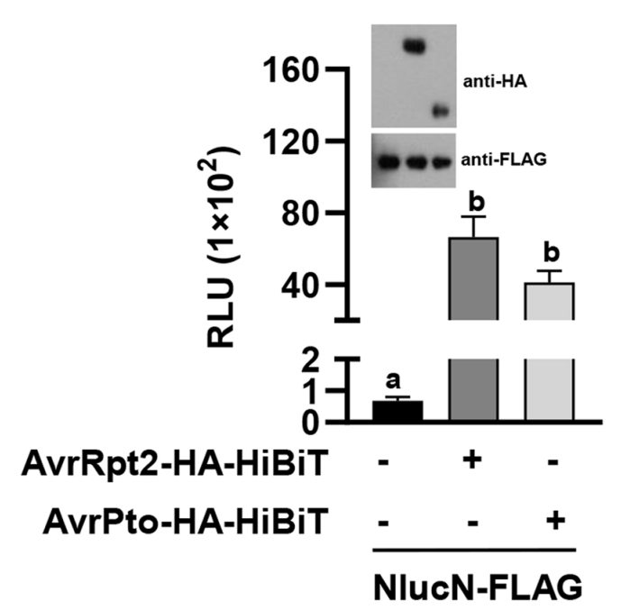
Coelenterazine h purchased from MedChemExpress. Usage Cited in: Stress Biol. 2024 Feb 16;4(1):14. [Abstract]
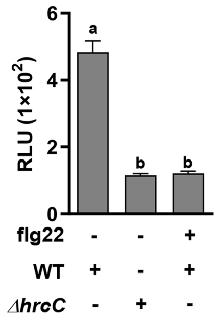
A. tumefaciens strains carrying NlucN-FLAG, along with avrRpt2-HA-HiBiT or avrPto-HA-HiBiT, were infiltrated into the leaves. Thirty hours after infiltration, leaves discs were collected and incubated with Coelenterazine h (10 µM; 10 min) in a 96-well plate, then the luminescence was monitored using EnSpire Multimode Plate Reader.
Coelenterazine h purchased from MedChemExpress. Usage Cited in: Stress Biol. 2024 Feb 16;4(1):14. [Abstract]
Leaves of transgenic A. thaliana plants expressing NlucN were infiltrated with H2O or 1 μM flg22. 24 hours after first infiltration, indicated bacteria (OD600 = 0.1) was infiltrated to the same leaves. Leaf discs were harvested 9 h later, and incubated with Coelenterazine h (10 µM; 10 min) in a 96-well plate, then the luminescence was monitored using EnSpire Multimode Plate Reader.
Coelenterazine h purchased from MedChemExpress. Usage Cited in: Stress Biol. 2024 Feb 16;4(1):14. [Abstract]
Effector secretion was induced in vitro. Culture supernatants were collected, incubated with Coelenterazine h (10 μM; 10 min) and NlucN-His, and measured for luminescence. Total protein was analyzed by immunoblot using anti-AvrPto and anti-HA antibodies to detect the accumulation of AvrPto-HiBiT and AvrRpt2-HA-HiBiT. Signal-to-noise ratio was shown.
-
J Biol Chem
Eyeless razor clam Sinonovacula constricta discriminates light spectra through opsins to guide Ca2+ and cAMP signaling pathways. [Abstract]2024 Jan;300(1):105527. PMID: 38043801
Coelenterazine h purchased from MedChemExpress. Usage Cited in: J Biol Chem. 2024 Jan;300(1):105527. [Abstract]
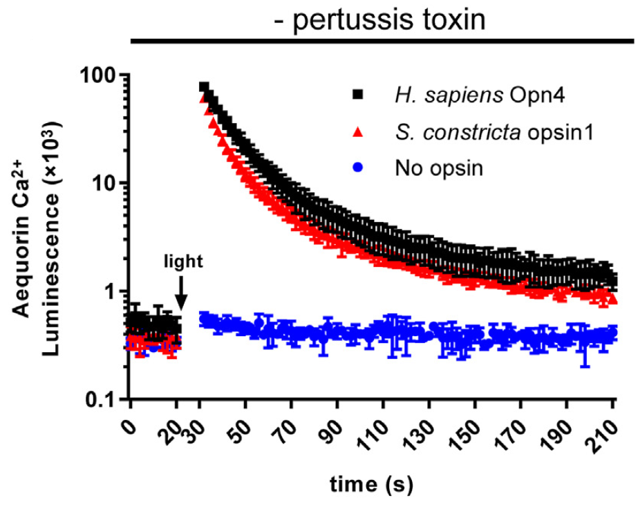
Coelenterazine h (10 μM; 2 h). HEK293T cells transfected with the empty vector (no opsin) served as the negative control, while those transfected with H. sapiens Opn4 were used as the positive control to measure the luminescence of Gαq-mediated Ca2+ increase.
-
ACS Pharmacol Transl Sci
2025 Apr 8;8(7):1996-2008. PMID: 40672676
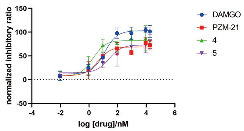
Coelenterazine h purchased from MedChemExpress. Usage Cited in: ACS Pharmacol Transl Sci. 2025 Apr 8;8(7):1996-2008. [Abstract]
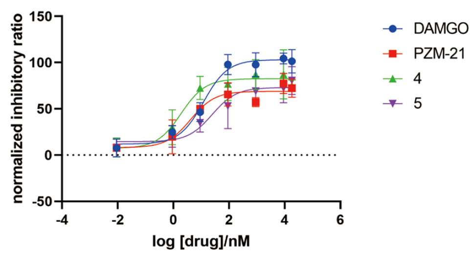
Coelenterazine h (5 μM; 5 min). Concentration-dependent inhibition of forskolin-induced (5 μM) cAMP accumulation using a BRET-Epac biosensor.


Pureza y Documentación
-
Ficha de datos (275 KB)
-
SDS (394 KB)
- English - EN (394 KB)
- Français - FR (394 KB)
- Deutsch - DE (394 KB)
- Norwegian - NO (394 KB)
- Español - ES (394 KB)
- Swedish - SV (394 KB)
- Italian - IT (394 KB)
- Korean - KR (394 KB)
- Portuguese - PT (394 KB)
-
Instrucciones de manejo (2659 KB)
Referencias
[1]. Jiang T, et al. New bioluminescent coelenterazine derivatives with various C-6 substitutions. Org Biomol Chem. 2017 Aug 23;15(33):7008-7018. [Content Brief]
[2]. M R Knight, et al. Imaging calcium dynamics in living plants using semi-synthetic recombinant aequorins. J Cell Biol. 1993 Apr;121(1):83-90. [Content Brief]
[3]. Kazushi Suzuki, et al. Five colour variants of bright luminescent protein for real-time multicolour bioimaging. Nat Commun. 2016 Dec 14:7:13718. [Content Brief]
[4]. Kenta Saito, et al. Auto-luminescent genetically-encoded ratiometric indicator for real-time Ca2+ imaging at the single cell level. PLoS One. 2010 Apr 1;5(4):e9935. [Content Brief]

Calculators
Concentration (start) × Volume (start) = Concentration (final) × Volume (final)


Coelenterazine h Related Classifications
HY-D1024 Related Classifications
 Powered by Bioz