



Alkaline phosphatase Antibody (YA3654)
(Synonyms: HOPS; TNAP; APTNAP; TNSALP; AP-TNAP)Based on 1 Customer Validation
Alkaline phosphatase Antibody (YA3654) is a Mouse-derived and non-conjugated IgG1 monoclonal antibody, targeting to Alkaline phosphatase.
-
Host:
Mouse
-
Isotype:
IgG1
-
Application:
WB, IHC-P, FC, ELISA
-
Reactivity :
Human, Mouse, Rat, Rabbit
-
Formulation:
Supplied in PBS with 0.05% sodium azide
-
Conjugation:
Non-conjugated


To place orders, for customer services and technical support, please contact: MedChemExpress USA
Tel: 609-228-6898 E-mail: [email protected] [email protected]





Applications
| Application |
WB

WB: Western Blot
|
IHC-P
IHC-P: Immunohistochemistry-Paraffin
|
FC
FC: Flow Cytometry
|
ELISA
ELISA: Enzyme Linked Immunosorbent Assay
|
|---|---|---|---|---|
| Dilution Ratio | 1:500-1:2000 | 1:200-1:1000 | 1:200-1:400 | 1:10000 |


Product Details
Alkaline phosphatase Antibody (YA3654) is a Mouse-derived and non-conjugated IgG1 monoclonal antibody, targeting to Alkaline phosphatase.
-
Host Mouse

-
Clonality Monoclonal

-
Species ReactivityHuman, Mouse, Rat, Rabbit
-
Observed Molecular WeightObserved band size: 75 kDa
Note: Due to possible protein modifications or aggregation, the molecular weight should be confirmed by actual measurement, and the predicted value is for reference only.

-
Calculated Molecular Weight Predicted band size: 57 kDa

Purified recombinant fragment of human ALPL aa 18-502.
affinity purified.
Non-conjugated
Unmodified
IgG1


Product Properties
-
Appearance
Solution
-
Formulation
Supplied in PBS with 0.05% sodium azide
-
Concentration
Batch-dependent, Please check the COA for the concentration of each lot. Check Lot Concentration
-
Storage & Stability
Stored at -20°C for 1 year. Avoid repeated freeze / thaw cycles.
-
Shipping
Shipping with blue ice.


Verification Images
-
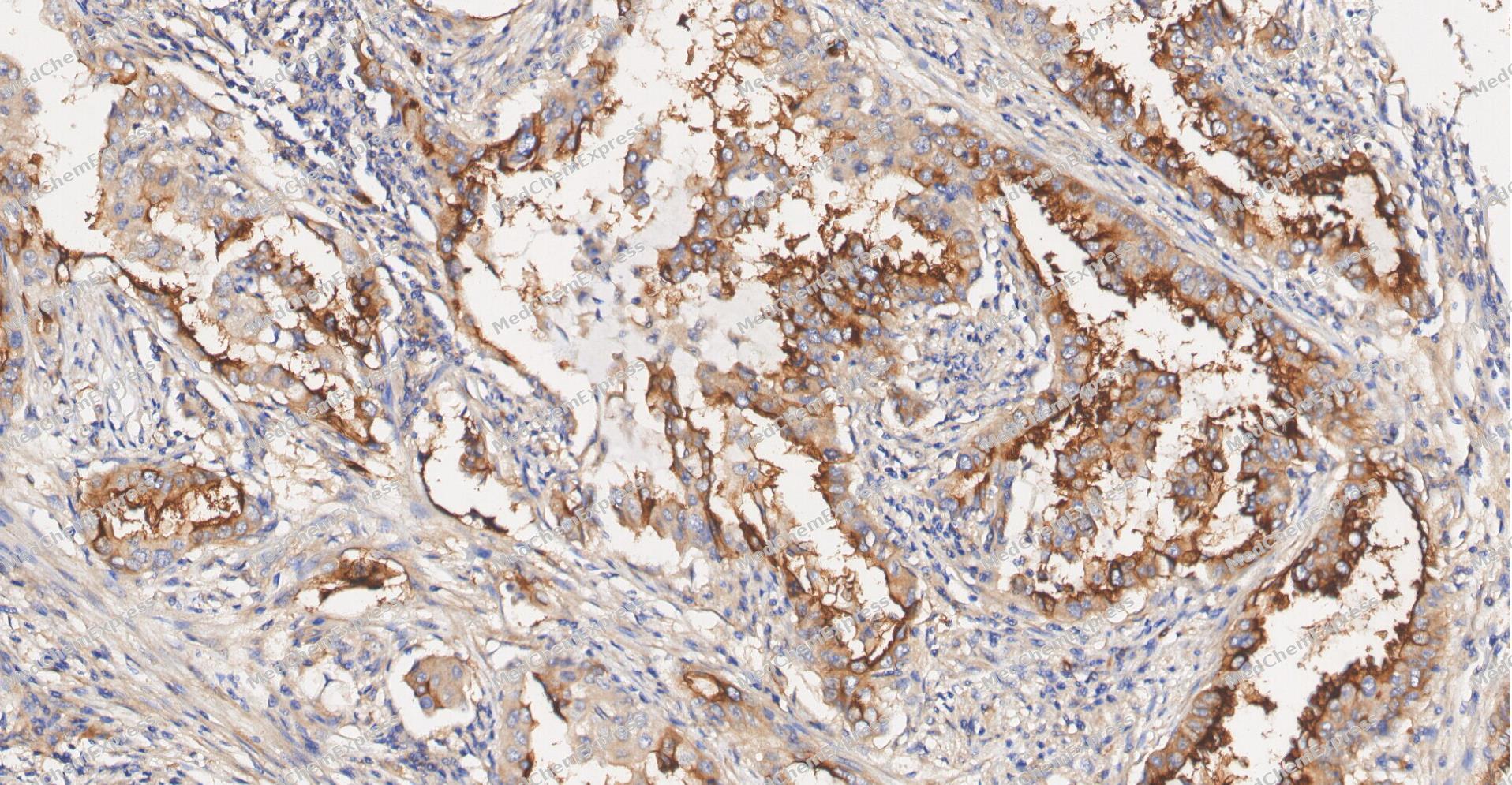 Immunohistochemical analysis of paraffin-embedded human Lung Adenocarcinom tissue using Alkaline phosphatase antibody was performed. The section was pretreated using high-temperature and high-pressure mediated EDTA antigen retrieval buffer (pH 9.0), for 5 minutes. The tissues were incubated with primary antibody (HY-P83957, 1:200 dilution) at room temperature for 60 minutes. Detection was performed using an HRP conjugated compact polymer system. DAB was used as the chromogen. The tissues were counterstained with hematoxylin and mounted with neutral balsam mounting medium.
 -
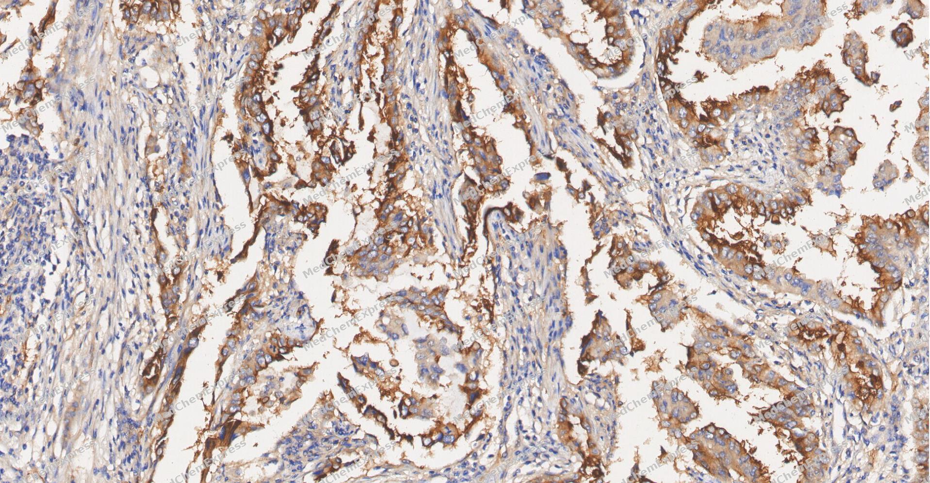 Immunohistochemical analysis of paraffin-embedded human Lung Adenocarcinom tissue using Alkaline phosphatase antibody was performed. The section was pretreated using high-temperature and high-pressure mediated EDTA antigen retrieval buffer (pH 9.0), for 5 minutes. The tissues were incubated with primary antibody (HY-P83957, 1:200 dilution) at room temperature for 60 minutes. Detection was performed using an HRP conjugated compact polymer system. DAB was used as the chromogen. The tissues were counterstained with hematoxylin and mounted with neutral balsam mounting medium.
 -
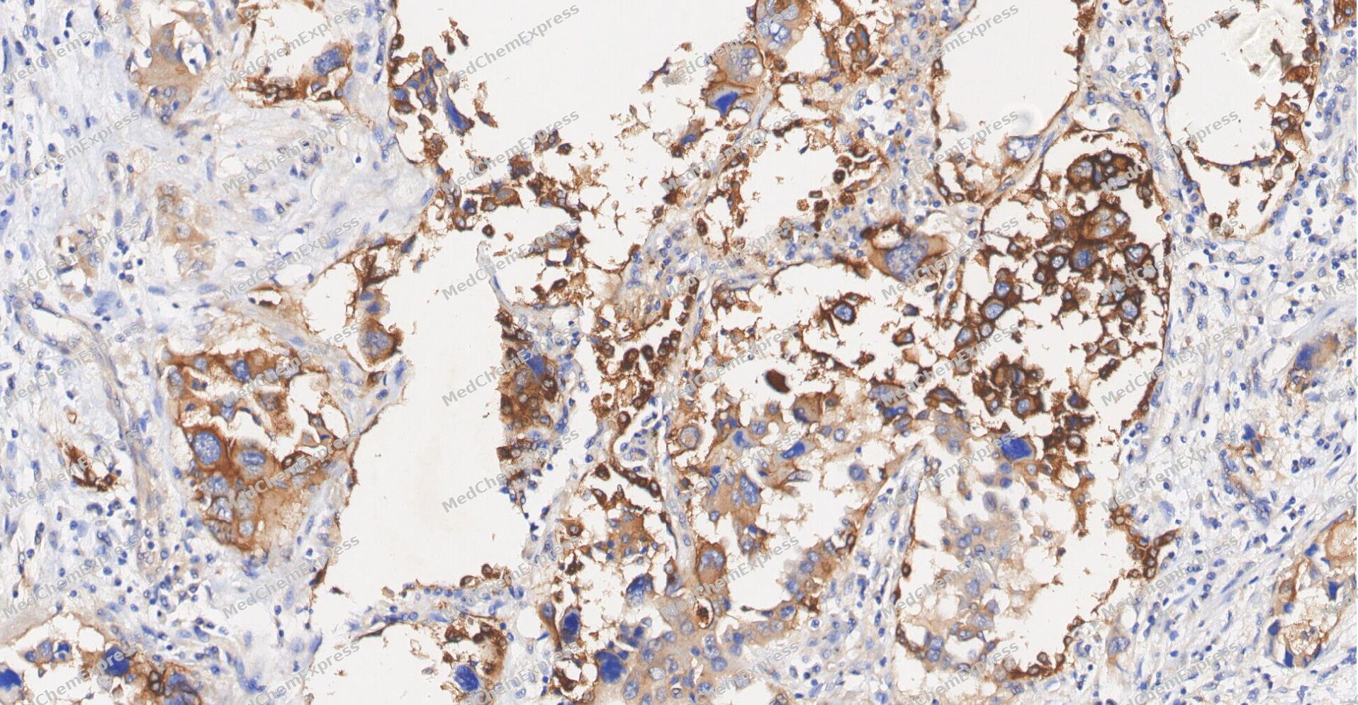 Immunohistochemical analysis of paraffin-embedded human Lung Adenocarcinom tissue using Alkaline phosphatase antibody was performed. The section was pretreated using high-temperature and high-pressure mediated EDTA antigen retrieval buffer (pH 9.0), for 5 minutes. The tissues were incubated with primary antibody (HY-P83957, 1:200 dilution) at room temperature for 60 minutes. Detection was performed using an HRP conjugated compact polymer system. DAB was used as the chromogen. The tissues were counterstained with hematoxylin and mounted with neutral balsam mounting medium.
 -
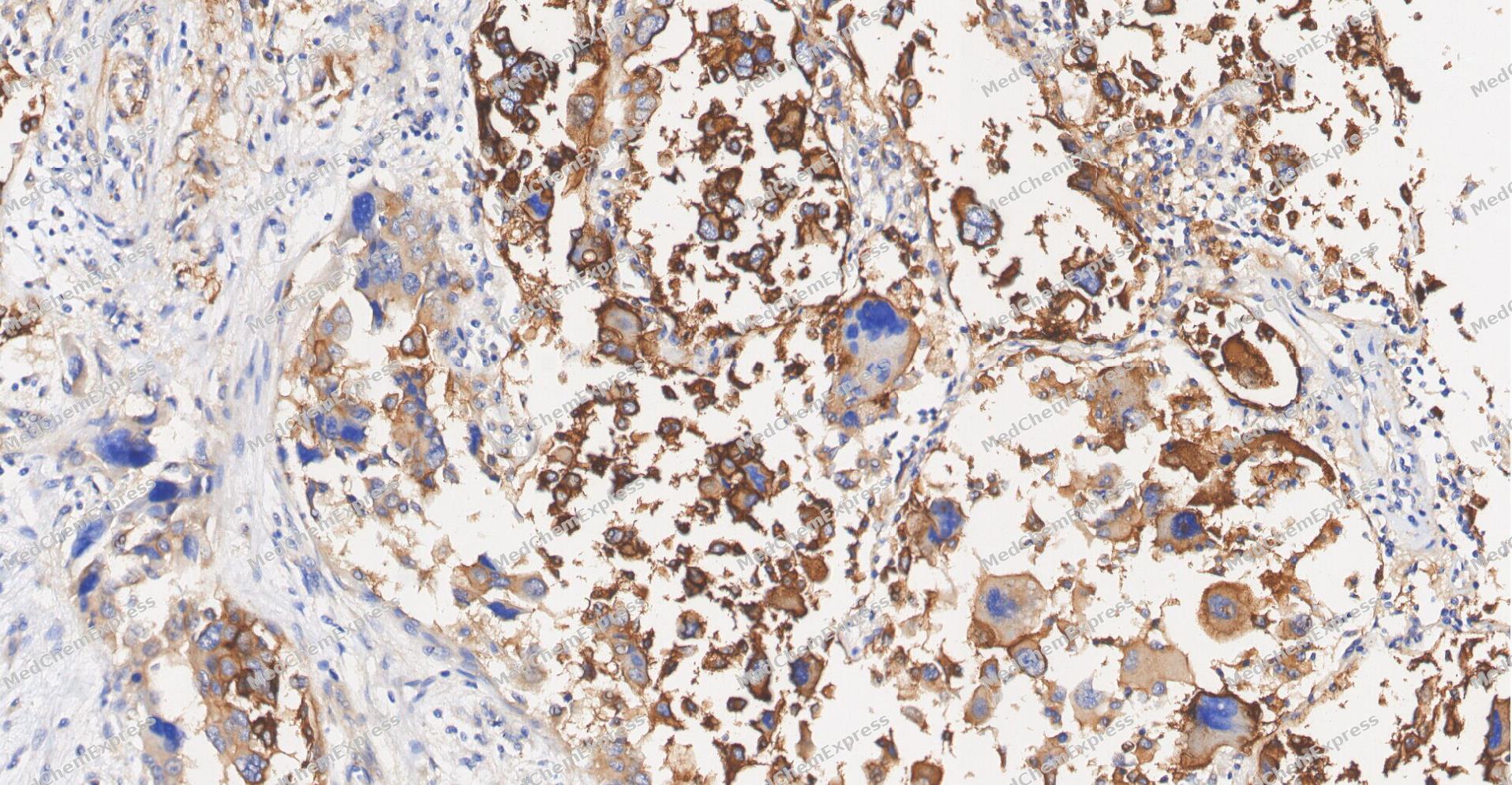 Immunohistochemical analysis of paraffin-embedded human Lung Adenocarcinom tissue using Alkaline phosphatase antibody was performed. The section was pretreated using high-temperature and high-pressure mediated EDTA antigen retrieval buffer (pH 9.0), for 5 minutes. The tissues were incubated with primary antibody (HY-P83957, 1:200 dilution) at room temperature for 60 minutes. Detection was performed using an HRP conjugated compact polymer system. DAB was used as the chromogen. The tissues were counterstained with hematoxylin and mounted with neutral balsam mounting medium.
 -
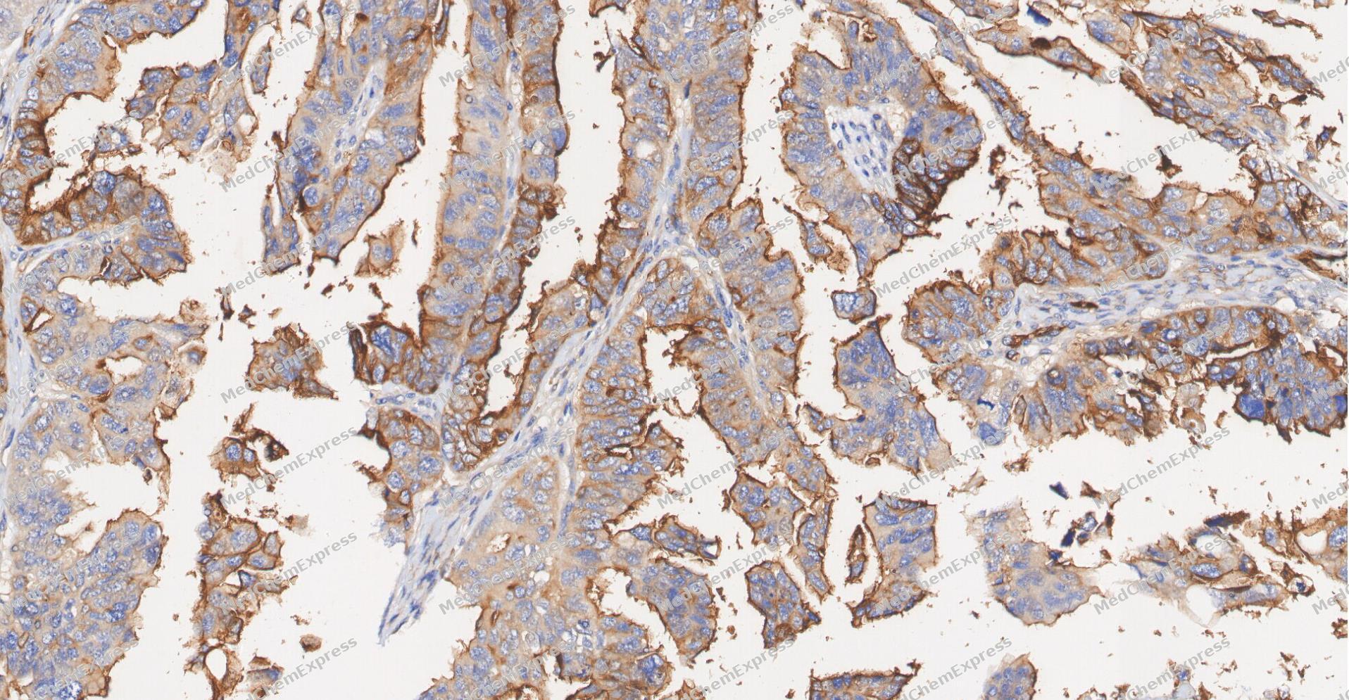 Immunohistochemical analysis of paraffin-embedded human ovarian carcinoma tissue using Alkaline phosphatase antibody was performed. The section was pretreated using high-temperature and high-pressure mediated EDTA antigen retrieval buffer (pH 9.0), for 5 minutes. The tissues were incubated with primary antibody (HY-P83957, 1:200 dilution) at room temperature for 60 minutes. Detection was performed using an HRP conjugated compact polymer system. DAB was used as the chromogen. The tissues were counterstained with hematoxylin and mounted with neutral balsam mounting medium.
 -
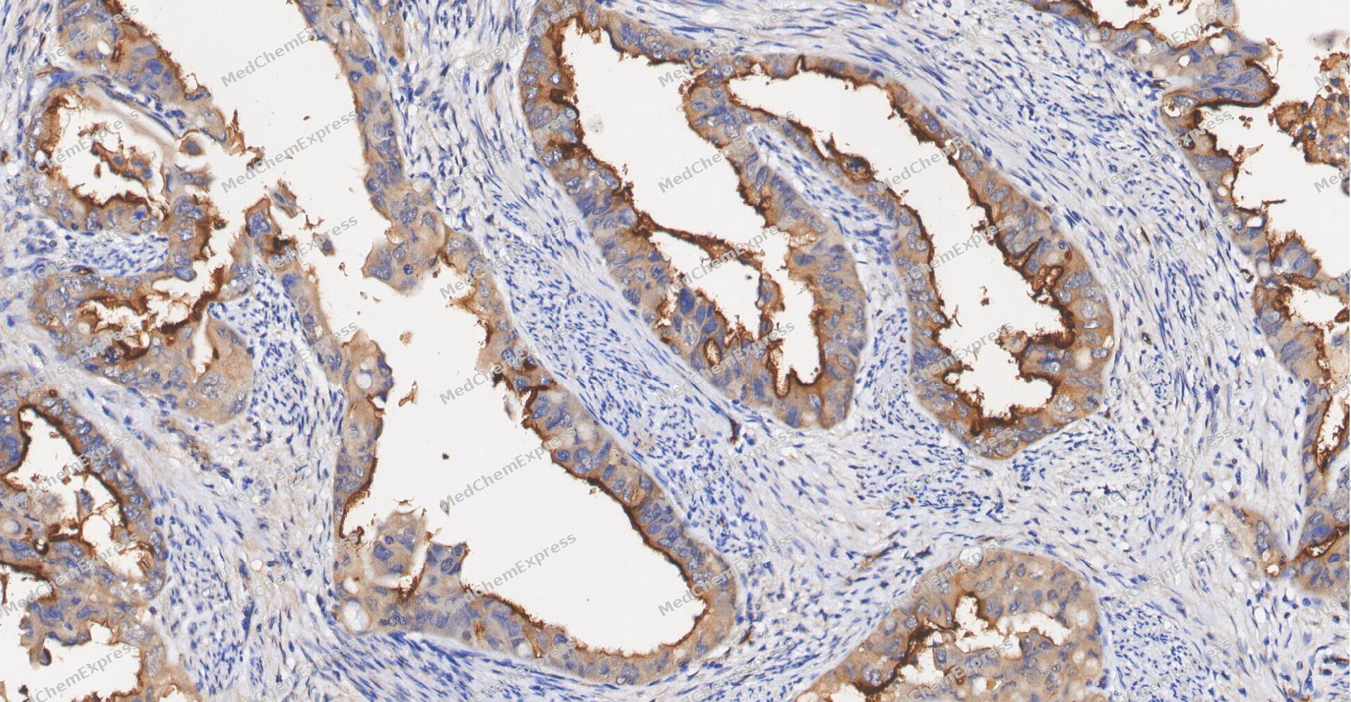 Immunohistochemical analysis of paraffin-embedded human Endometrial carcinoma tissue using Alkaline phosphatase antibody was performed. The section was pretreated using high-temperature and high-pressure mediated EDTA antigen retrieval buffer (pH 9.0), for 5 minutes. The tissues were incubated with primary antibody (HY-P83957, 1:200 dilution) at room temperature for 60 minutes. Detection was performed using an HRP conjugated compact polymer system. DAB was used as the chromogen. The tissues were counterstained with hematoxylin and mounted with neutral balsam mounting medium.
 -
 Immunohistochemical analysis of paraffin-embedded mouse stomach tissue using Alkaline phosphatase Antibody (HY-P83957, 1/1000). The section was pretreated using heat mediated antigen retrieval with sodium citrate buffer (pH 6.0) for 8 minutes. The tissues were blocked with quick block buffer for 0.5 hours at room temperature, washed with PBS and PBST, and then incubated with the primary antibody overnight at 4℃. The detection was performed using an HRP conjugated compact polymer system. DAB was used as the chromogen. Tissues were counterstained with hematoxylin and mounted with DPX.
 -
 Immunohistochemical analysis of paraffin-embedded mouse small intestine tissue using Alkaline phosphatase Antibody (HY-P83957, 1/1000). The section was pretreated using heat mediated antigen retrieval with sodium citrate buffer (pH 6.0) for 8 minutes. The tissues were blocked with quick block buffer for 0.5 hours at room temperature, washed with PBS and PBST, and then incubated with the primary antibody overnight at 4℃. The detection was performed using an HRP conjugated compact polymer system. DAB was used as the chromogen. Tissues were counterstained with hematoxylin and mounted with DPX.
 -
 Immunohistochemical analysis of paraffin-embedded mouse colon tissue using Alkaline phosphatase Antibody (HY-P83957, 1/1000). The section was pretreated using heat mediated antigen retrieval with sodium citrate buffer (pH 6.0) for 8 minutes. The tissues were blocked with quick block buffer for 0.5 hours at room temperature, washed with PBS and PBST, and then incubated with the primary antibody overnight at 4℃. The detection was performed using an HRP conjugated compact polymer system. DAB was used as the chromogen. Tissues were counterstained with hematoxylin and mounted with DPX.
 -
 Immunohistochemical analysis of paraffin-embedded mouse duodenum tissue using Alkaline phosphatase Antibody (HY-P83957, 1/1000). The section was pretreated using heat mediated antigen retrieval with sodium citrate buffer (pH 6.0) for 8 minutes. The tissues were blocked with quick block buffer for 0.5 hours at room temperature, washed with PBS and PBST, and then incubated with the primary antibody overnight at 4℃. The detection was performed using an HRP conjugated compact polymer system. DAB was used as the chromogen. Tissues were counterstained with hematoxylin and mounted with DPX.
 -
 Immunohistochemical analysis of paraffin-embedded human adrenal gland tissue using Alkaline phosphatase Antibody (HY-P83957, 1/1000). The section was pretreated using heat mediated antigen retrieval with sodium citrate buffer (pH 6.0) for 8 minutes. The tissues were blocked with quick block buffer for 0.5 hours at room temperature, washed with PBS and PBST, and then incubated with the primary antibody overnight at 4℃. The detection was performed using an HRP conjugated compact polymer system. DAB was used as the chromogen. Tissues were counterstained with hematoxylin and mounted with DPX.
 -
 Immunohistochemical analysis of paraffin-embedded human breast tissue using Alkaline phosphatase Antibody (HY-P83957, 1/1000). The section was pretreated using heat mediated antigen retrieval with sodium citrate buffer (pH 6.0) for 8 minutes. The tissues were blocked with quick block buffer for 0.5 hours at room temperature, washed with PBS and PBST, and then incubated with the primary antibody overnight at 4℃. The detection was performed using an HRP conjugated compact polymer system. DAB was used as the chromogen. Tissues were counterstained with hematoxylin and mounted with DPX.


Background
-
Function
Alkaline phosphatase is an Alkaline phosphatase that metabolizes various phosphate compounds and plays a key role in skeletal mineralization and adaptive thermogenesis. Has broad substrate specificity and can hydrolyze a considerable variety of compounds: however, only a few substrates, such as diphosphate (inorganic pyrophosphate; PPi), pyridoxal 5'-phosphate (PLP) and N-phosphocreatine are natural substrates. Plays an essential role in skeletal and dental mineralization via its ability to hydrolyze extracellular diphosphate, a potent mineralization inhibitor, to phosphate: it thereby promotes hydroxyapatite crystal formation and increases inorganic phosphate concentration. Acts in a non-redundant manner with PHOSPHO1 in skeletal mineralization: while PHOSPHO1 mediates the initiation of hydroxyapatite crystallization in the matrix vesicles (MVs), ALPL/TNAP catalyzes the spread of hydroxyapatite crystallization in the extracellular matrix. Also promotes dephosphorylation of osteopontin (SSP1), an inhibitor of hydroxyapatite crystallization in its phosphorylated state; it is however unclear whether ALPL/TNAP mediates SSP1 dephosphorylation via a direct or indirect manner. Catalyzes dephosphorylation of PLP to pyridoxal (PL), the transportable form of vitamin B6, in order to provide a sufficient amount of PLP in the brain, an essential cofactor for enzymes catalyzing the synthesis of diverse neurotransmitters. Additionally, also able to mediate ATP degradation in a stepwise manner to adenosine, thereby regulating the availability of ligands for purinergic receptors. Also capable of dephosphorylating microbial products, such as lipopolysaccharides (LPS) as well as other phosphorylated small-molecules, such as poly-inosine:cytosine (poly I:C). Acts as a key regulator of adaptive thermogenesis as part of the futile creatine cycle: localizes to the mitochondria of thermogenic fat cells and acts by mediating hydrolysis of N-phosphocreatine to initiate a futile cycle of creatine dephosphorylation and phosphorylation. During the futile creatine cycle, creatine and N-phosphocreatine are in a futile cycle, which dissipates the high energy charge of N-phosphocreatine as heat without performing any mechanical or chemical work[1][2][3][4][5][6].
-
Subcellular Localization
Cell membrane; Lipid-anchor, GPI-anchor; Extracellular vesicle membrane; Lipid-anchor, GPI-anchor; Mitochondrion membrane; Lipid-anchor, GPI-anchor; Mitochondrion intermembrane space
-
Isoforms & Post-Translational Modification
P05186 has 3 isomers: P05186-1: 57305 Da (predicted); P05186-2: 48909 Da (predicted); P05186-3: 51045 Da (predicted).
N-glycosylated -
Subunit
Homodimer
-
SwissProt ID
-
Synonyms
HOPS; TNAP; APTNAP; TNSALP; AP-TNAP


Documentation
-
Data Sheet (235 KB)
-
SDS (251 KB)
- English - EN (251 KB)
- Français - FR (251 KB)
- Deutsch - DE (251 KB)
- Norwegian - NO (251 KB)
- Español - ES (251 KB)
- Swedish - SV (251 KB)
- Italian - IT (251 KB)
- Korean - KR (251 KB)
- Portuguese - PT (251 KB)
-
User Guide for Antibodies (1077 KB)

 Powered by Bioz
