



Annexin A1/ANXA1 Antibody (YA832)
(Synonyms: ANXA1; ANX1; LPC1; Annexin A1; Annexin I; Annexin-1; Calpactin II; Calpactin-2; Chromobindin-9; Lipocortin I; Phospholipase A2 inhibitory protein; p35)Based on 1 Customer Validation
Annexin A1/ANXA1 Antibody (YA832) is a Mouse-derived and non-conjugated IgG1 monoclonal antibody, targeting to Annexin A1/ANXA1.
-
Host:
Mouse
-
Isotype:
IgG
-
Application:
WB
-
Reactivity :
Human, Mouse, Rat, Monkey
-
Formulation:
Supplied in 1*PBS (pH 7.3), 50% glycerol and 0.5% BSA. Preservative: 0.02% sodium azide.
-
Conjugation:
Non-conjugated


To place orders, for customer services and technical support, please contact: MedChemExpress USA
Tel: 609-228-6898 E-mail: [email protected] [email protected]





Applications
| Application |
WB

WB: Western Blot
|
|---|---|
| Dilution Ratio | 1:500-1:1000 |


Product Details
Annexin A1/ANXA1 Antibody (YA832) is a Mouse-derived and non-conjugated IgG1 monoclonal antibody, targeting to Annexin A1/ANXA1.
-
Host Mouse

-
Clonality Monoclonal

-
Species ReactivityHuman, Mouse, Rat, Monkey
-
Observed Molecular WeightObserved band size: 39 kDa
Note: Due to possible protein modifications or aggregation, the molecular weight should be confirmed by actual measurement, and the predicted value is for reference only.

-
Calculated Molecular Weight Predicted band size: 39 kDa

Entrez Gene: 301 Human ; 16952 Mouse ; 25380 Rat
SwissProt: P04083 Human ; P10107 Mouse ; P07150 Rat
OMIM: 151690 Human
Synthetic peptide corresponding to Human Annexin A1 beta.The exact sequence is proprietary to MCE.
Endogenous
affinity purified
Non-conjugated
Unmodified
IgG


Product Properties
-
Appearance
Solution
-
Formulation
Supplied in 1*PBS (pH 7.3), 50% glycerol and 0.5% BSA. Preservative: 0.02% sodium azide.
-
Concentration
Batch-dependent, Please check the COA for the concentration of each lot. Check Lot Concentration
-
Storage & Stability
Stored at -20°C for 1 year. Avoid repeated freeze / thaw cycles.
-
Shipping
Shipping with blue ice.


Verification Images
-
 Western blot analysis was performed on protein extracts (25 μg) from A549 (lane 2), HCT 116 (lane 3), NIH/3T3 (lane 4), K562 (lane 5) and Rat kidney (lane 6) using Annexin A1/ANXA1 antibody. Proteins were transferred onto a 0.45 μm PVDF membrane using the Trans-Blot® Turbo™ system for 13 min. The membrane was then blocked with 5% nonfat milk in TBST (HY-K1025) for 1 h at room temperature. The primary antibody (1:1000) and loading control antibody GAPDH Antibody (HRP) (HY-P80954A) (1:5000) were diluted in 5% nonfat milk in TBST and incubated with the membrane overnight at 4°C. After washing, the membrane of primary antibody was incubated with HRP-conjugated goat anti-rabbit/mouse IgG secondary antibody (HY-P8001/HY-P8004) (1:5000) diluted in 5% nonfat milk in TBST for 1 h at room temperature. Protein bands were visualized using an Ultra High Sensitivity ECL detection kit (HY-K1005).
 -
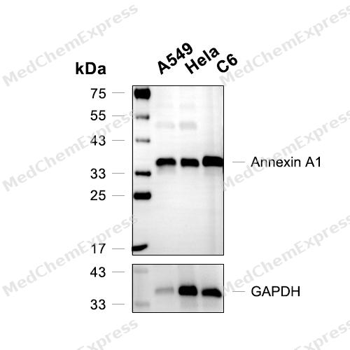 Western blot analysis of extracts from A549(lane 2(20μg) 、Hela (lane 3(20μg) and C6(lane 4(20μg)), using Annexin A1 (HY-P80544) Mouse mAb. Proteins were transferred to a PVDF membrane and blocked with 5% BSA in TBST for 2 hour at room temperature. The primary antibody (HY-P80544, 1/1000) and Loading control antibody (GAPDH, HY-P80954, 1/3000) was used in 5% BSA in TBST at 4°C overnight. Goat Anti-Mouse/Rabbit IgG-HRP Secondary Antibody (HY-P8004/HY-P8001, 1/10,000) was used for 1 hour at room temperature.


Background
-
Function
Annexin A1/ANXA1 plays important roles in the innate immune response as effector of glucocorticoid-mediated responses and regulator of the inflammatory process. Has anti-inflammatory activity. Plays a role in glucocorticoid-mediated down-regulation of the early phase of the inflammatory response. Contributes to the adaptive immune response by enhancing signaling cascades that are triggered by T-cell activation, regulates differentiation and proliferation of activated T-cells. Promotes the differentiation of T-cells into Th1 cells and negatively regulates differentiation into Th2 cells. Has no effect on unstimulated T cells. Negatively regulates hormone exocytosis via activation of the formyl peptide receptors and reorganization of the actin cytoskeleton. Has high affinity for Ca(2+) and can bind up to eight Ca(2+) ions. Displays Ca(2+)-dependent binding to phospholipid membranes. Plays a role in the formation of phagocytic cups and phagosomes. Plays a role in phagocytosis by mediating the Ca(2+)-dependent interaction between phagosomes and the actin cytoskeleton; Functions at least in part by activating the formyl peptide receptors and downstream signaling cascades. Promotes chemotaxis of granulocytes and monocytes via activation of the formyl peptide receptors. Promotes rearrangement of the actin cytoskeleton, cell polarization and cell migration. Promotes resolution of inflammation and wound healing. Acts via neutrophil N-formyl peptide receptors to enhance the release of CXCL2[1][2][3][4][5][6][7][8].
-
Subcellular Localization
Nucleus; Cytoplasm; Cell projection, cilium; Cell membrane; Membrane; Peripheral membrane protein; Endosome membrane; Peripheral membrane protein; Basolateral cell membrane; Apical cell membrane; Lateral cell membrane; Secreted; Secreted, extracellular space; Cell membrane; Peripheral membrane protein; Extracellular side; Secreted, extracellular exosome; Cytoplasmic vesicle, secretory vesicle lumen; Cell projection, phagocytic cup; Early endosome; Cytoplasmic vesicle membrane; Peripheral membrane protein
-
Expression
Tissue_specificity:Detected in resting neutrophils (PubMed: 10772777) . Detected in peripheral blood T cells (PubMed: 17008549) . Detected in extracellular vesicles of inflammatory bowel disease serum, but not detected in serum of healthy donors (PubMed: 25664854) . Detected in placenta (protein level) (PubMed: 2532504) . Detected in liver. -
Subunit
Homodimer; non-covalently linked (By similarity). Homodimer; linked by transglutamylation (PubMed:2532504).
-
SwissProt ID
-
Synonyms
ANXA1; ANX1; LPC1; Annexin A1; Annexin I; Annexin-1; Calpactin II; Calpactin-2; Chromobindin-9; Lipocortin I; Phospholipase A2 inhibitory protein; p35
-
Research Field
Signal Transduction


Documentation
-
Data Sheet (262 KB)
-
SDS (252 KB)
- English - EN (252 KB)
- Français - FR (252 KB)
- Deutsch - DE (252 KB)
- Norwegian - NO (252 KB)
- Español - ES (252 KB)
- Swedish - SV (252 KB)
- Italian - IT (252 KB)
- Korean - KR (252 KB)
- Portuguese - PT (252 KB)
-
User Guide for Antibodies (1077 KB)
References
[1]. Arcone R, et al. Structural characterization of a biologically active human lipocortin 1 expressed in Escherichia coli. Eur J Biochem. 1993 Jan 15;211(1-2):347-55. [Content Brief]
[2]. D'Acquisto F, et al. Annexin-1 modulates T-cell activation and differentiation. Blood. 2007 Feb 1;109(3):1095-102. [Content Brief]
[3]. McArthur S, et al. Annexin A1 regulates hormone exocytosis through a mechanism involving actin reorganization. FASEB J. 2009 Nov;23(11):4000-10. [Content Brief]
[4]. Pepinsky RB, et al. A dimeric form of lipocortin-1 in human placenta. Biochem J. 1989 Oct 1;263(1):97-103. [Content Brief]
[5]. Mailliard WS, et al. Calcium-dependent binding of S100C to the N-terminal domain of annexin I. J Biol Chem. 1996 Jan 12;271(2):719-25. [Content Brief]
[6]. Ernst S, et al. An annexin 1 N-terminal peptide activates leukocytes by triggering different members of the formyl peptide receptor family. J Immunol. 2004 Jun 15;172(12):7669-76. [Content Brief]
[7]. Woloszynek JC, et al. Cathepsin G-regulated release of formyl peptide receptor agonists modulate neutrophil effector functions. J Biol Chem. 2012 Oct 5;287(41):34101-9. [Content Brief]
[8]. Leoni G, et al. Annexin A1-containing extracellular vesicles and polymeric nanoparticles promote epithelial wound repair. J Clin Invest. 2015 Mar 2;125(3):1215-27. [Content Brief]

 Powered by Bioz
