



Aurora B Antibody (YA6131)
(Synonyms: AURKB; AIK2; AIM1; AIRK2; ARK2; STK1; STK12; STK5; Aurora kinase B; Aurora 1; Aurora- and IPL1-like midbody-associated protein 1; AIM-1; Aurora/IPL1-related kinase 2; ARK-2; Aurora-related kinase 2; STK-1; Serine/threonine-protein kinase 12)Based on 1 Customer Validation
Aurora B Antibody (YA6131) is a Rabbit-derived and non-conjugated IgG monoclonal antibody, targeting to Aurora B.
-
Host:
Rabbit
-
Isotype:
IgG
-
Application:
WB, IHC-P, ICC/IF, IP, ELISA
-
Reactivity :
Human
-
Formulation:
Supplied in PBS, 50% glycerol, 0.05% Proclin 300, 0.05%BSA
-
Conjugation:
Non-conjugated


To place orders, for customer services and technical support, please contact: MedChemExpress USA
Tel: 609-228-6898 E-mail: [email protected] [email protected]





Applications
| Application |
IHC-P

IHC-P: Immunohistochemistry-Paraffin
|
WB
WB: Western Blot
|
ICC/IF
ICC/IF: Immunocytochemistry/
Immunofluorescence |
ELISA
ELISA: Enzyme Linked Immunosorbent Assay
|
IP
IP: Immunoprecipitation
|
|---|---|---|---|---|---|
| Dilution Ratio | 1:500-1:1000 | 1:1000-1:5000 | 1:200-1:1000 | 1:5000-1:20000 | 1:50-1:200 |


Product Details
Aurora B Antibody (YA6131) is a Rabbit-derived and non-conjugated IgG monoclonal antibody, targeting to Aurora B.
-
Host Rabbit

-
Clonality Monoclonal

-
Species ReactivityHuman
-
Observed Molecular WeightObserved band size: 39 kDa
Note: Due to possible protein modifications or aggregation, the molecular weight should be confirmed by actual measurement, and the predicted value is for reference only.

-
Calculated Molecular Weight Predicted band size: 39 kDa

Protein A
Non-conjugated
Unmodified
IgG


Product Properties
-
Appearance
Solution
-
Formulation
Supplied in PBS, 50% glycerol, 0.05% Proclin 300, 0.05%BSA
-
Concentration
Batch-dependent, Please check the COA for the concentration of each lot. Check Lot Concentration
-
Storage & Stability
Stored at -20°C for 1 year. Avoid repeated freeze / thaw cycles.
-
Shipping
Shipping with blue ice.


Verification Images
-
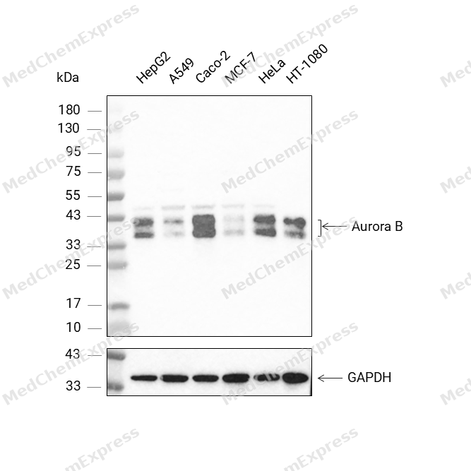 Western blot analysis was performed on protein extracts (25 μg) from HepG2 (lane 2), A549 (lane 3), Caco-2 (lane 4), MCF-7 (lane 5), HeLa (lane 6), and HT-1080 (lane 7) using Aurora B antibody. Proteins were transferred onto a 0.45 μm PVDF membrane using the Trans-Blot® Turbo™ system for 13 min. The membrane was then blocked with 5% nonfat milk in TBST (HY-K1025) for 1 h at room temperature. Thhe primary antibody (1:1000) and loading control antibody GAPDH Antibody (HRP) (HY-P80954A) (1:2500) were diluted in 5% nonfat milk in TBST and incubated with the membrane overnight at 4°C. After washing, the membrane of primary antibody was incubated with HRP-conjugated goat anti-rabbit IgG (H&L) secondary antibody (HY-P8001) (1:5000) diluted in 5% nonfat milk in TBST for 1 h at room temperature. Protein bands were visualized using an Ultra High Sensitivity ECL detection kit (HY-K1005).
 -
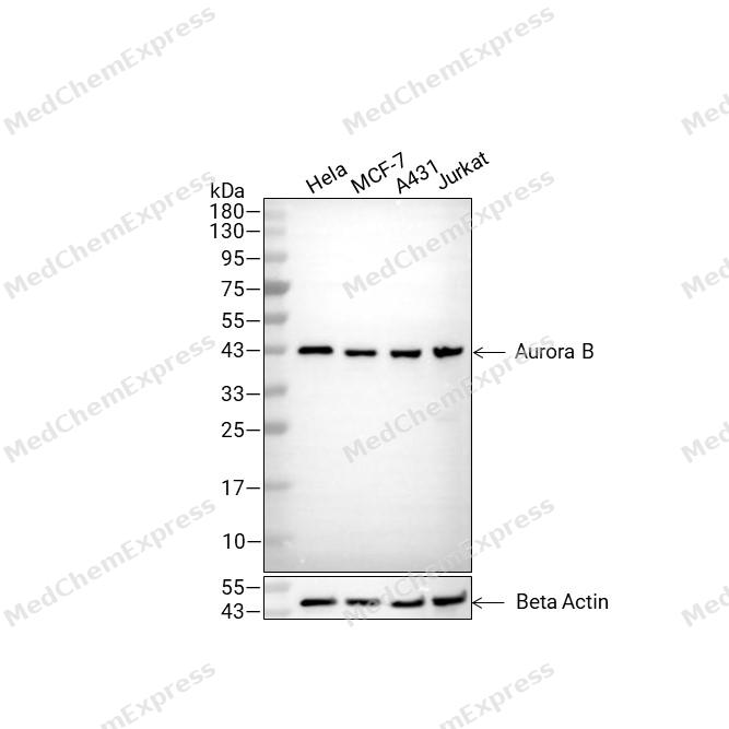 Western blot analysis of extracts from Hela (lane2(20μg), MCF-7 (lane3(20μg), A431 (lane4(20μg) and Jurkat (lane5(20μg) using Aurora B Antibody (HY-P86439). Proteins were transferred to a PVDF membrane and blocked with 5% non-fat milk in TBST for 2 hour at room temperature. The primary antibody (1/4000) and Loading control antibody (Beta Actin, HY-P80993, 1/10,000) was used in 5% non-fat milk in TBST at 4°C overnight. Goat Anti-Rabbit IgG-HRP Secondary Antibody (HY-P8001 ,1/10,000) was used for 1 hour at room temperature.


Background
-
Function
Aurora B (AURKB) is a mitotic serine/threonine kinase and the enzymatic core of the chromosomal passenger complex (CPC), which includes INCENP, Survivin, and Borealin[1]. Mechanistically, the CPC guides Aurora B through mitosis to control chromosome alignment, histone modification, kinetochore-microtubule error correction, spindle checkpoint signaling, and cytokinesis[1][2]. Therefore, Aurora B activity supports faithful chromosome segregation by releasing incorrect kinetochore-microtubule attachments, while reduced activity increases chromosome missegregation and aneuploid-cell generation[2]. In disease models, Aurora B deregulation or overexpression appears in several tumors, and studies in hepatocellular carcinoma, oral squamous cell carcinoma, prostate carcinoma, and glioblastoma connect Aurora B activity with recurrence, prognosis, tumor growth, or mitotic disruption[3][4][5][6]. Compared with related isoforms, Aurora A localizes to spindle poles and regulates mitotic entry, centrosome maturation, and spindle assembly, whereas Aurora B localizes with the CPC from inner centromeres to the spindle midzone and midbody[7]. Aurora C can act as a chromosomal passenger protein and complement Aurora B in mitotic cells, but Aurora B shows preferential INCENP binding and earlier M-phase expression[8]. For experimental applications, Aurora B or pan-Aurora inhibitors such as VX-680, AMG900, and danusertib help dissect metaphase-anaphase transition, cytokinetic initiation, mitotic progression, and cancer-cell proliferation[6][9][10].
-
Subcellular Localization
Nucleus; Chromosome; Chromosome, centromere; Chromosome, centromere, kinetochore; Cytoplasm, cytoskeleton, spindle; Midbody
-
Expression
Tissue_specificity:It is highly expressed in the thymus. It is also expressed in the spleen, lungs, testes, colon, placenta, and fetal liver. It is expressed in the S phase and G2/M phase, and its expression is upregulated in M phase cancer cells; it is not expressed in normal liver, but is highly expressed in metastatic liver.
Induction:Expression is cell cycle-regulated, with a low in G1/S, an increase during G2 and M. Expression decreases again after M phase -
Isoforms & Post-Translational Modification
Q96GD4 has 5 isomers: Q96GD4-1: 39311 Da (predicted); Q96GD4-2: 35302 Da (predicted); Q96GD4-3: 16211 Da (predicted); Q96GD4-4: 34760 Da (predicted); Q96GD4-5: 39467 Da (predicted).
The phosphorylation of Thr-232 requires the binding to INCENP and occurs by means of an autophosphorylation mechanism (PubMed:14722118). Thr-232 phosphorylation is indispensable for the AURKB kinase activity (PubMed:14722118, PubMed:26829474);Acetylated at Lys-215 by KAT5 at kinetochores, increasing AURKB activity and promoting accurate chromosome segregation in mitosis;Ubiquitinated by different BCR (BTB-CUL3-RBX1) E3 ubiquitin ligase complexes (PubMed:17543862, PubMed:19995937). Ubiquitinated by the BCR(KLHL9-KLHL13) E3 ubiquitin ligase complex, ubiquitination leads to removal from mitotic chromosomes and is required for cytokinesis (PubMed:17543862). During anaphase, the BCR(KLHL21) E3 ubiquitin ligase complex recruits the CPC complex from chromosomes to the spindle midzone and mediates the ubiquitination of AURKB (PubMed:17543862). Ubiquitination of AURKB by BCR(KLHL21) E3 ubiquitin ligase complex may not lead to its degradation by the proteasome (PubMed:19995937). Deubiquitinated by USP35; inhibiting CDH1-mediated degradation of AURKB (PubMed:29449677) -
Subunit
Component of the chromosomal passenger complex (CPC) composed of at least BIRC5/survivin, CDCA8/borealin, INCENP, AURKB or AURKC; predominantly independent AURKB- and AURKC-containing complexes exist (PubMed:11516652, PubMed:12925766, PubMed:14722118, PubMed:15249581, PubMed:18591255, PubMed:20562864, PubMed:27332895).
-
SwissProt ID
-
Synonyms
AURKB; AIK2; AIM1; AIRK2; ARK2; STK1; STK12; STK5; Aurora kinase B; Aurora 1; Aurora- and IPL1-like midbody-associated protein 1; AIM-1; Aurora/IPL1-related kinase 2; ARK-2; Aurora-related kinase 2; STK-1; Serine/threonine-protein kinase 12


Documentation
-
Data Sheet (263 KB)
-
SDS (252 KB)
- English - EN (252 KB)
- Français - FR (252 KB)
- Deutsch - DE (252 KB)
- Norwegian - NO (252 KB)
- Español - ES (252 KB)
- Swedish - SV (252 KB)
- Italian - IT (252 KB)
- Korean - KR (252 KB)
- Portuguese - PT (252 KB)
-
User Guide for Antibodies (1077 KB)
References
[1]. Vader G, et al. The chromosomal passenger complex: guiding Aurora-B through mitosis. J Cell Biol. 2006 Jun 19;173(6):833-7. [Content Brief]
[2]. Abe Y, et al. System-level deficiencies in Aurora B control in cancers. Cell Cycle. 2016 Aug 17;15(16):2091-2092. [Content Brief]
[3]. Tanaka S, et al. Aurora kinase B is a predictive factor for the aggressive recurrence of hepatocellular carcinoma after curative hepatectomy. Br J Surg. 2008 May;95(5):611-9. [Content Brief]
[4]. Pannone G, et al. Aurora B expression as a prognostic indicator and possible therapeutic target in oral squamous cell carcinoma. Int J Immunopathol Pharmacol. 2011 Jan-Mar;24(1):79-88. [Content Brief]
[5]. Cao M, et al. A Vector-Based Short Hairpin RNA Targeting Aurora B Suppresses Human Prostatic Carcinoma Growth. Technol Cancer Res Treat. 2017 Feb;16(1):112-119. [Content Brief]
[6]. Ryu J, et al. An Aurora kinase inhibitor, AMG900, inhibits glioblastoma cell proliferation by disrupting mitotic progression. Cancer Med. 2018 Nov;7(11):5589-5603. [Content Brief]
[7]. Carmena M, et al. Making the Auroras glow: regulation of Aurora A and B kinase function by interacting proteins. Curr Opin Cell Biol. 2009 Dec;21(6):796-805. [Content Brief]
[8]. Sasai K, et al. Aurora-C kinase is a novel chromosomal passenger protein that can complement Aurora-B kinase function in mitotic cells. Cell Motil Cytoskeleton. 2004 Dec;59(4):249-63. [Content Brief]
[9]. Li Z, et al. The Aurora Kinase in Trypanosoma brucei plays distinctive roles in metaphase-anaphase transition and cytokinetic initiation. PLoS Pathog. 2009 Sep;5(9):e1000575. [Content Brief]
[10]. Meulenbeld HJ, et al. Danusertib, an aurora kinase inhibitor. Expert Opin Investig Drugs. 2012 Mar;21(3):383-93. [Content Brief]

 Powered by Bioz
