



Axl Antibody
(Synonyms: Adhesion related kinase; AI323647; Ark; Axl; AXL oncogene; AXL receptor tyrosine kinase; AXL transforming gene; AXL transforming sequence/gene; EC 2.7.10.1; JTK11; Oncogene AXL; Tyro7; Tyrosine protein kinase receptor UFO; Tyrosine-protein kinase receptor UFO; UFO; UFO_HUMAN.)Based on 1 Customer Validation
Axl Antibody is a Rabbit-derived and non-conjugated IgG polyclonal antibody, targeting to Axl.
-
Host:
Rabbit
-
Isotype:
IgG
-
Application:
WB, ELISA, IHC-P, IHC-F, ICC/IF
-
Reactivity :
Human, Mouse
-
Formulation:
Supplied in 0.01M TBS(pH7.4) with 1% BSA, 0.03% Proclin300 and 50% Glycerol.
-
Conjugation:
Non-conjugated


To place orders, for customer services and technical support, please contact: MedChemExpress USA
Tel: 609-228-6898 E-mail: [email protected] [email protected]





Applications
| Application |
WB

WB: Western Blot
|
ELISA
ELISA: Enzyme Linked Immunosorbent Assay
|
IHC-P
IHC-P: Immunohistochemistry-Paraffin
|
IHC-F
IHC-F: Immunohistochemistry-Frozen
|
ICC/IF
ICC/IF: Immunocytochemistry/
Immunofluorescence |
|---|---|---|---|---|---|
| Dilution Ratio | 1:500-2000 | 1:5000-10000 | 1:100-500 | 1:100-500 | 1:100-500 |


Product Details
Axl Antibody is a Rabbit-derived and non-conjugated IgG polyclonal antibody, targeting to Axl.
-
Host Rabbit

-
Clonality Polyclonal

-
Species ReactivityHuman, Mouse Predicted Reactivity: Rat,Dog,Horse,Rabbit
Note: The predicted reactivity is for reference only and should not be considered a guarantee of product performance.
-
Observed Molecular WeightObserved band size: 130 kDa
Note: Due to possible protein modifications or aggregation, the molecular weight should be confirmed by actual measurement, and the predicted value is for reference only.

-
Calculated Molecular Weight Predicted band size: 95 kDa

Entrez Gene: 558 Human ; 26362 Mouse ; 308444 Rat
SwissProt: P30530 Human ; Q00993 Mouse ;
OMIM: 109135 Human
KLH conjugated synthetic peptide derived from human AXL: 151-250/894
Endogenous
affinity purified
Non-conjugated
Unmodified
IgG


Product Properties
-
Appearance
Solution
-
Formulation
Supplied in 0.01M TBS(pH7.4) with 1% BSA, 0.03% Proclin300 and 50% Glycerol.
-
Concentration
Batch-dependent, Please check the COA for the concentration of each lot. Check Lot Concentration
-
Storage & Stability
Stored at -20°C for 1 year. Avoid repeated freeze / thaw cycles.
-
Shipping
Shipping with blue ice.


Verification Images
-
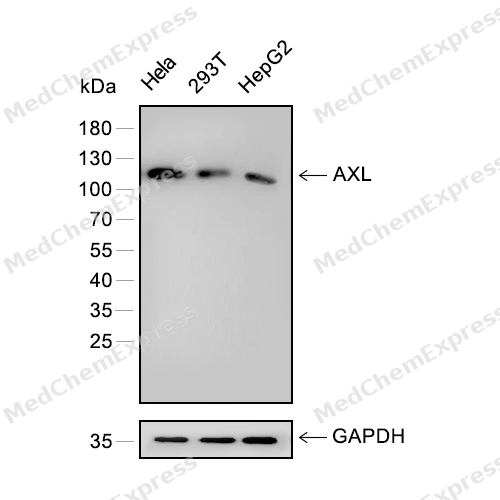 Western blot analysis of extracts from Hela (lane 1) and 293T (lane 2) and HepG2 (lane 3) using AXL antibody. Proteins were transferred to a PVDF membrane and blocked with 5% nonfat powdered milk in PBST for 2 hour at room temperature. The primary antibody (1/2000) and Loading control antibody (GAPDH, 1/3000) was diluted with 5% nonfat powdered milk in PBST at 4°C overnight. Goat Anti-Rabbit IgG-HRP Secondary Antibody (1/8,000) was incubated for 45min at room temperature.
 -
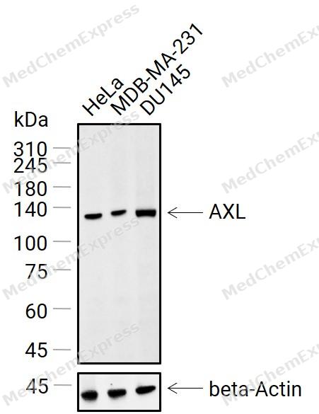 Western blot analysis of extracts from HeLa(lane 1(30μg)), MDA-MB-231(lane 2(30μg)) and DU145(lane 3(30μg)) using AXL antibody (HY-P81198). Proteins were transferred to a NC membrane and blocked with 5% Non-fat milk powder in TBST for 1 hour at room temperature. The primary antibody (1/1000) and Loading control antibody (Beta actin, HY-P80438,1/10000) was used in 5% Non-fat milk powder in TBST at 4°C overnight. Goat Anti-Rabbit IgG-HRP Secondary Antibody (HY-P8001, 1/20,000) was used for 1 hour at room temperature.
 -
 Immunohistochemical analysis of paraffin-embedded human skin tissue using Axl Antibody (HY-P81198, 1/200). The section was pretreated using heat mediated antigen retrieval with sodium citrate buffer (pH 6.0) for 8 minutes. The tissues were blocked with quick block buffer for 0.5 hours at room temperature, washed with PBS and PBST, and then incubated with the primary antibody overnight at 4℃. The detection was performed using an HRP conjugated compact polymer system. DAB was used as the chromogen. Tissues were counterstained with hematoxylin and mounted with DPX.
 -
 Immunohistochemical analysis of paraffin-embedded human liver tissue using Axl Antibody (HY-P81198, 1/200). The section was pretreated using heat mediated antigen retrieval with sodium citrate buffer (pH 6.0) for 8 minutes. The tissues were blocked with quick block buffer for 0.5 hours at room temperature, washed with PBS and PBST, and then incubated with the primary antibody overnight at 4℃. The detection was performed using an HRP conjugated compact polymer system. DAB was used as the chromogen. Tissues were counterstained with hematoxylin and mounted with DPX.
 -
 Immunohistochemical analysis of paraffin-embedded human heart muscle tissue using Axl Antibody (HY-P81198, 1/200). The section was pretreated using heat mediated antigen retrieval with sodium citrate buffer (pH 6.0) for 8 minutes. The tissues were blocked with quick block buffer for 0.5 hours at room temperature, washed with PBS and PBST, and then incubated with the primary antibody overnight at 4℃. The detection was performed using an HRP conjugated compact polymer system. DAB was used as the chromogen. Tissues were counterstained with hematoxylin and mounted with DPX.
 -
 Immunohistochemical analysis of paraffin-embedded human salivary gland tissue using Axl Antibody (HY-P81198, 1/200). The section was pretreated using heat mediated antigen retrieval with sodium citrate buffer (pH 6.0) for 8 minutes. The tissues were blocked with quick block buffer for 0.5 hours at room temperature, washed with PBS and PBST, and then incubated with the primary antibody overnight at 4℃. The detection was performed using an HRP conjugated compact polymer system. DAB was used as the chromogen. Tissues were counterstained with hematoxylin and mounted with DPX.
 -
 Immunohistochemical analysis of paraffin-embedded human testis tissue using Axl Antibody (HY-P81198, 1/200). The section was pretreated using heat mediated antigen retrieval with sodium citrate buffer (pH 6.0) for 8 minutes. The tissues were blocked with quick block buffer for 0.5 hours at room temperature, washed with PBS and PBST, and then incubated with the primary antibody overnight at 4℃. The detection was performed using an HRP conjugated compact polymer system. DAB was used as the chromogen. Tissues were counterstained with hematoxylin and mounted with DPX.
 -
 Immunohistochemical analysis of paraffin-embedded human lung tissue using Axl Antibody (HY-P81198, 1/200). The section was pretreated using heat mediated antigen retrieval with sodium citrate buffer (pH 6.0) for 8 minutes. The tissues were blocked with quick block buffer for 0.5 hours at room temperature, washed with PBS and PBST, and then incubated with the primary antibody overnight at 4℃. The detection was performed using an HRP conjugated compact polymer system. DAB was used as the chromogen. Tissues were counterstained with hematoxylin and mounted with DPX.
 -
 Immunohistochemical analysis of paraffin-embedded human cerebral cortex tissue using Axl Antibody (HY-P81198, 1/200). The section was pretreated using heat mediated antigen retrieval with sodium citrate buffer (pH 6.0) for 8 minutes. The tissues were blocked with quick block buffer for 0.5 hours at room temperature, washed with PBS and PBST, and then incubated with the primary antibody overnight at 4℃. The detection was performed using an HRP conjugated compact polymer system. DAB was used as the chromogen. Tissues were counterstained with hematoxylin and mounted with DPX.
 -
 Immunocytochemistry analysis of MCF-7 cells labeling Axl with Axl Antibody (HY-P81198) at 1/100 dilution. Cells were fixed in 4% paraformaldehyde for 15 minutes at room temperature, permeabilized with 0.1% Triton X-100 in PBS for 15 minutes at room temperature, then blocked with quick block buffer for 10 minutes at room temperature. Cells were then incubated with Axl Antibody (HY-P81198) at 1/100 dilution in quick block buffer overnight at 4 ℃. AF488-conjugated Goat Anti-Rabbit IgG H&L (HY-P8002, Green) was used as the secondary antibody at 1/1,000 dilution. PBS instead of the primary antibody was used as the secondary antibody only control. The Nuclear counterstain was DAPI (Blue).
 -
 Immunocytochemistry analysis of PC-3 cells labeling Axl with Axl Antibody (HY-P81198) at 1/100 dilution. Cells were fixed in 4% paraformaldehyde for 15 minutes at room temperature, permeabilized with 0.1% Triton X-100 in PBS for 15 minutes at room temperature, then blocked with quick block buffer for 10 minutes at room temperature. Cells were then incubated with Axl Antibody (HY-P81198) at 1/100 dilution in quick block buffer overnight at 4 ℃. AF488-conjugated Goat Anti-Rabbit IgG H&L (HY-P8002, Green) was used as the secondary antibody at 1/1,000 dilution. PBS instead of the primary antibody was used as the secondary antibody only control. The Nuclear counterstain was DAPI (Blue).
 -
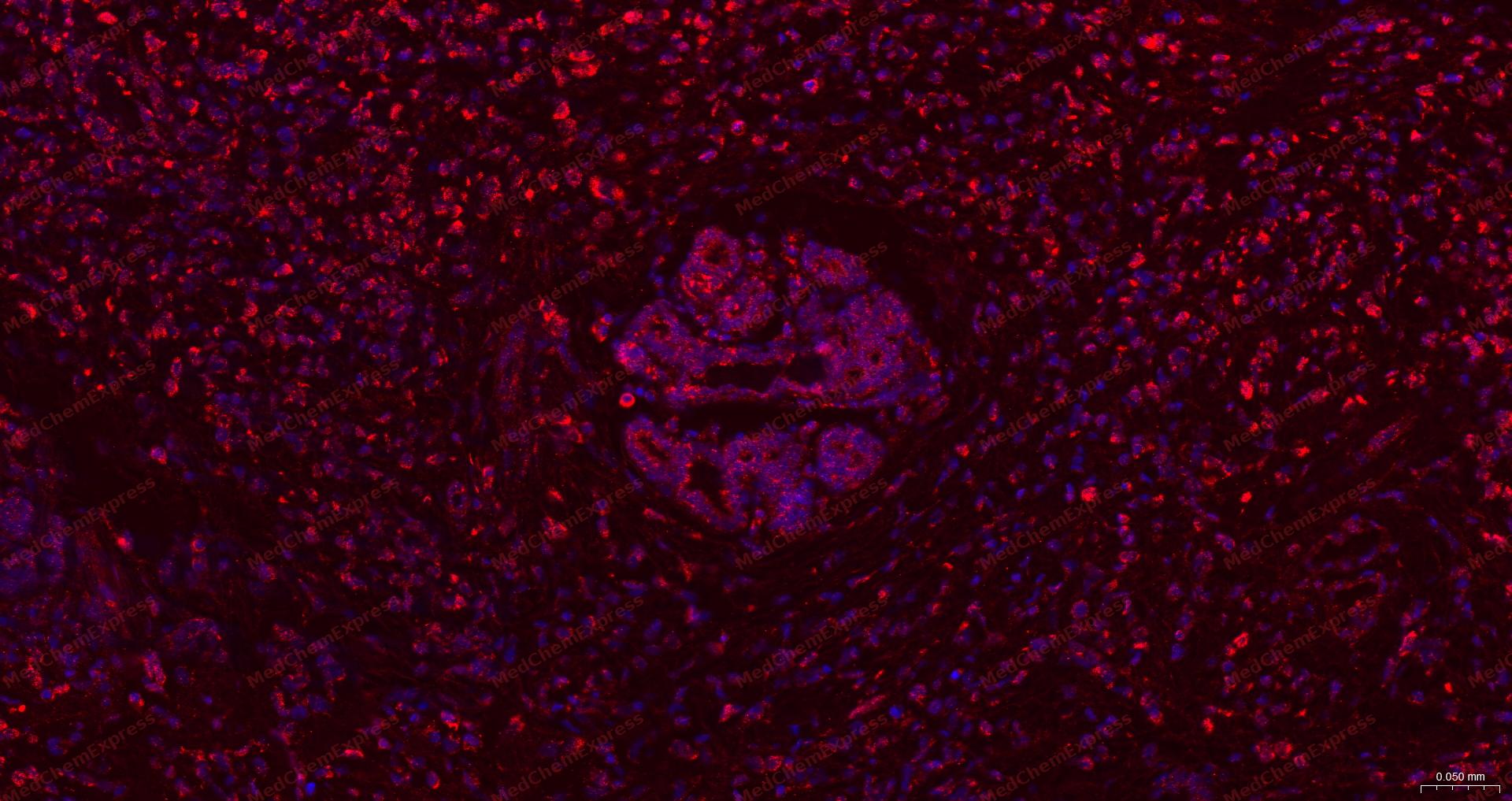 Immunofluorescence analysis of paraffin-embedded human breast cancer using AXL antibody. The section was pre-treated using heat mediated antigen retrieval with sodium citrate buffer (pH 6.0) for 15 minutes. The tissues were blocked in 10% normal goat serum for 30 minutes at room temperature, then incubated with AXL antibody (HY-P81198) at 1/200 dilution overnight at 4 ℃. Alexa Fluor® 594-conjugated AffiniPure Goat Anti-Rabbit IgG H&L(HY-P8003 , Red) was used as the secondary antibody at 1/200 dilution. The Nuclear counterstain was DAPI (Blue).
 -
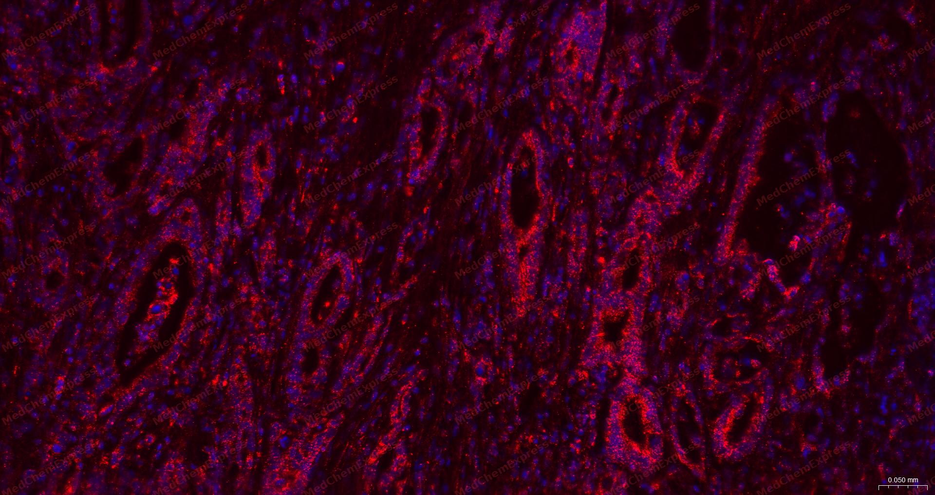 Immunofluorescence analysis of paraffin-embedded human pancreatic cancer using AXL antibody. The section was pre-treated using heat mediated antigen retrieval with sodium citrate buffer (pH 6.0) for 15 minutes. The tissues were blocked in 10% normal goat serum for 30 minutes at room temperature, then incubated with AXL antibody (HY-P81198) at 1/200 dilution overnight at 4 ℃. Alexa Fluor® 594-conjugated AffiniPure Goat Anti-Rabbit IgG H&L(HY-P8003 , Red) was used as the secondary antibody at 1/200 dilution. The Nuclear counterstain was DAPI (Blue).


Background
-
Function
Axl is a TAM receptor tyrosine kinase activated by GAS6, and TAM receptors support apoptotic-cell recognition, efferocytosis, immune regulation, tissue homeostasis, and cancer-related signaling[1][2]. Mechanistically, Axl participates in ligand-induced TAM activation with TYRO3 and MERTK, but each receptor shows distinct activation patterns with GAS6, PROS1, apoptotic cells, phosphatidylserine vesicles, and enveloped virus[3]. In cancer models, Axl activation stimulates MAPK, AKT, and FAK pathways, while Axl inhibition or knockdown reduces tumor growth, migration, colony formation, and chemoresistance in NSCLC and neuroblastoma[2][4]. In inflammatory disease models, AXL/MERTK inhibition protected against pancreatic necrosis by limiting CXCL2-related neutrophil infiltration, whereas soluble Axl increased in lupus nephritis and multiple sclerosis lesions, indicating disease-linked dysregulation of GAS6-TAM signaling[5][6][7]. Compared with MERTK, Axl showed stronger effects on chemosensitivity in NSCLC, distinct ligand-response behavior among TAM isoforms, and critical roles with TYRO3 in GPVI-mediated platelet activation[3][2][8]. For experimental applications, Axl inhibitors, soluble TAM domains, and antagonist or agonist antibodies provide tools to test TAM signaling, immune regulation, apoptosis, and therapeutic sensitization[3][5][9].
-
Subcellular Localization
Cell membrane; Single-pass type I membrane protein
-
Expression
Tissue_specificity:Highly expressed in metastatic colon tumors. Expressed in primary colon tumors. Weakly expressed in normal colon tissue.
Induction: (Microbial infection) Up-regulated by Aedes aegypti lymphotoxin beta receptor inhibitor during Zika virus infection -
Isoforms & Post-Translational Modification
P30530 has 2 isomers: P30530-1: 98337 Da (predicted); P30530-2: 97378 Da (predicted).
Monoubiquitinated upon GAS6-binding. A very small proportion of the receptor could be subjected to polyubiquitination in a very transient fashion;Phosphorylated at tyrosine residues by autocatalysis, which activates kinase activity -
Subunit
Heterodimer and heterotetramer with ligand GAS6. Interacts with CBL, GRB2, LCK, NCK2, PIK3R1, PIK3R2, PIK3R3, PLCG1, SOCS1 and TNS2. Part of a complex including AXL, TNK2 and GRB2, in which GRB2 promotes AXL recruitment by TNK2
-
SwissProt ID
-
Synonyms
Adhesion related kinase; AI323647; Ark; Axl; AXL oncogene; AXL receptor tyrosine kinase; AXL transforming gene; AXL transforming sequence/gene; EC 2.7.10.1; JTK11; Oncogene AXL; Tyro7; Tyrosine protein kinase receptor UFO; Tyrosine-protein kinase receptor UFO; UFO; UFO_HUMAN.


Documentation
-
Data Sheet (263 KB)
-
SDS (252 KB)
- English - EN (252 KB)
- Français - FR (252 KB)
- Deutsch - DE (252 KB)
- Norwegian - NO (252 KB)
- Español - ES (252 KB)
- Swedish - SV (252 KB)
- Italian - IT (252 KB)
- Korean - KR (252 KB)
- Portuguese - PT (252 KB)
-
User Guide for Antibodies (1077 KB)
References
[1]. Zhou L, et al. Tyro3, Axl, Mertk receptor-mediated efferocytosis and immune regulation in the tumor environment. Int Rev Cell Mol Biol. 2021;361:165-210. [Content Brief]
[2]. Linger RM, et al. Mer or Axl receptor tyrosine kinase inhibition promotes apoptosis, blocks growth and enhances chemosensitivity of human non-small cell lung cancer. Oncogene. 2013 Jul 18;32(29):3420-31. [Content Brief]
[3]. Tsou WI, et al. Receptor tyrosine kinases, TYRO3, AXL, and MER, demonstrate distinct patterns and complex regulation of ligand-induced activation. J Biol Chem. 2014 Sep 12;289(37):25750-63. [Content Brief]
[4]. Li Y, et al. Inhibition of Mer and Axl receptor tyrosine kinases leads to increased apoptosis and improved chemosensitivity in human neuroblastoma. Biochem Biophys Res Commun. 2015 Feb 13;457(3):461-6. [Content Brief]
[5]. Bao J, et al. AXL and MERTK receptor tyrosine kinases inhibition protects against pancreatic necrosis via selectively limiting CXCL2-related neutrophil infiltration. Biochim Biophys Acta Mol Basis Dis. 2022 Dec 1;1868(12):166490. [Content Brief]
[6]. Bellan M, et al. Increased plasma levels of Gas6 and its soluble tyrosine kinase receptors Mer and Axl are associated with immunological activity and severity of lupus nephritis. Clin Exp Rheumatol. 2021 Jan-Feb;39(1):132-138. [Content Brief]
[7]. Weinger JG, et al. Up-regulation of soluble Axl and Mer receptor tyrosine kinases negatively correlates with Gas6 in established multiple sclerosis lesions. Am J Pathol. 2009 Jul;175(1):283-93. [Content Brief]
[9]. Ali SR, et al. Nerve Density and Neuronal Biomarkers in Cancer. Cancers (Basel). 2022 Oct 1;14(19):4817. [Content Brief]

 Powered by Bioz
