



Caspase-6 Antibody (YA556)
(Synonyms: MCH2, CASP6, Caspase-6, CASP-6, CSP-6, Apoptotic protease Mch-2)
Based on 1 Customer Validation
Caspase-6 Antibody (YA556) is a Rabbit-derived and non-conjugated IgG monoclonal antibody, targeting to Caspase-6.
-
Host:
Rabbit
-
Isotype:
IgG
-
Application:
WB, ICC/IF, IHC-P, IP
-
Reactivity :
Human, Mouse, Rat
-
Formulation:
Supplied in 1*TBS (pH7.4), 0.05% BSA and 40% Glycerol. Preservative: 0.05% Sodium Azide.
-
Conjugation:
Non-conjugated


To place orders, for customer services and technical support, please contact: MedChemExpress USA
Tel: 609-228-6898 E-mail: [email protected] [email protected]





Applications
| Application |
WB

WB: Western Blot
|
ICC/IF
ICC/IF: Immunocytochemistry/
Immunofluorescence |
IHC-P
IHC-P: Immunohistochemistry-Paraffin
|
|---|---|---|---|
| Dilution Ratio | 1:1000-1:2000 | 1:50-1:200 | 1:50-1:400 |


Product Details
Caspase-6 Antibody (YA556) is a Rabbit-derived and non-conjugated IgG monoclonal antibody, targeting to Caspase-6.
-
Host Rabbit

-
Clonality Recombinant,Monoclonal

-
Species ReactivityHuman, Mouse, Rat
-
Observed Molecular WeightObserved band size: 33/11 kDa
Note: Due to possible protein modifications or aggregation, the molecular weight should be confirmed by actual measurement, and the predicted value is for reference only.

-
Calculated Molecular Weight Predicted band size: 33 kDa

Entrez Gene: 839 Human ; 12368 Mouse ;
SwissProt: P55212 Human ; O08738 Mouse ; O35397 Rat
OMIM: 601532 Human
Synthetic peptide corresponding to Human Apolipoprotein E.AA range:210-249.
Endogenous
Protein A affinity purified.
Non-conjugated
Unmodified
IgG


Product Properties
-
Appearance
Solution
-
Formulation
Supplied in 1*TBS (pH7.4), 0.05% BSA and 40% Glycerol. Preservative: 0.05% Sodium Azide.
-
Concentration
Batch-dependent, Please check the COA for the concentration of each lot. Check Lot Concentration
-
Storage & Stability
Stored at -20°C for 1 year. Avoid repeated freeze / thaw cycles.
-
Shipping
Shipping with blue ice.


Verification Images
-
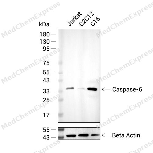 Western blot analysis of extracts from Jurkat (lane 2(20μg), C2C12 (lane 3(20μg), C6 (lane 4(20μg) using Caspase-6 Antibody (HY-P80047) Rabbit mAb. Proteins were transferred to a PVDF membrane and blocked with 5% BSA in TBST for 2 hour at room temperature. The primary antibody (HY-P80416, 1/1000) and Loading control antibody (Beta Actin, HY-P80438, 1/3000) was used in 5% BSA in TBST at 4°C overnight. Goat Anti-Mouse/Rabbit IgG-HRP Secondary Antibody (HY-P8004/HY-P8001, 1/10,000) was used for 1 hour at room temperature.
 -
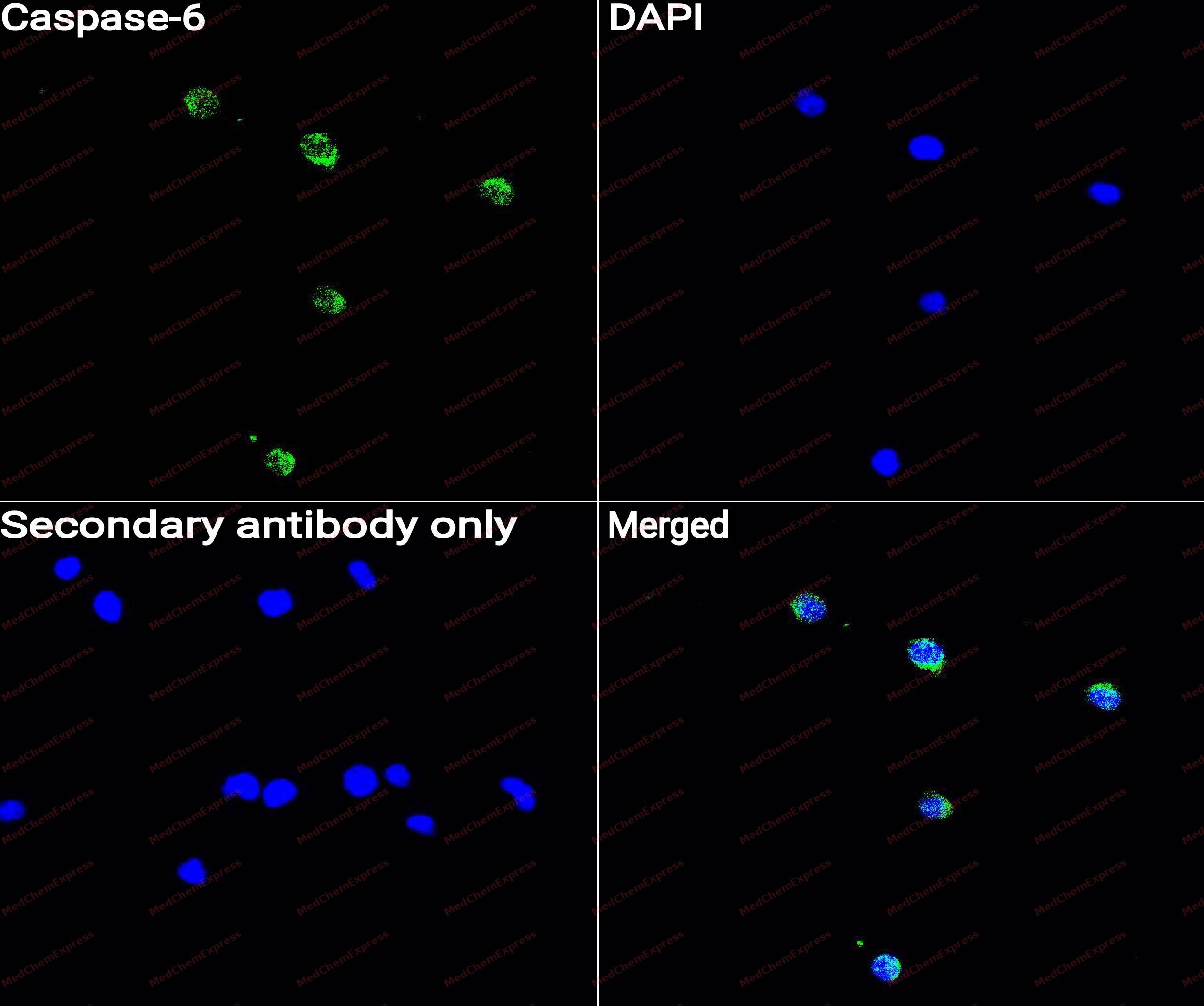 Immunocytochemistry analysis of Jurkat cells labeling Caspase-6 with Caspase-6 Antibody (HY-P80047) at 1/50 dilution. Cells were fixed in 4% paraformaldehyde for 15 minutes at room temperature, permeabilized with 0.1% Triton X-100 for 10 minutes at room temperature, then blocked with QuickBlock™ Blocking Buffer for Immunol Staining for 10 min at room temperature. Cells were then incubated with Caspase-6 Antibody (HY-P80047)at 1/50 dilution in QuickBlock™ Blocking Buffer for Immunol Staining at 4 ℃. Alexa Fluor® 488-conjugated AffiniPure Goat Anti-Rabbit IgG H&L(HY-P8002, Green) was used as the secondary antibody at 1/1,000 dilution. PBS instead of the primary antibody was used as the secondary antibody only control. The Nuclear counterstain was DAPI (Blue).
 -
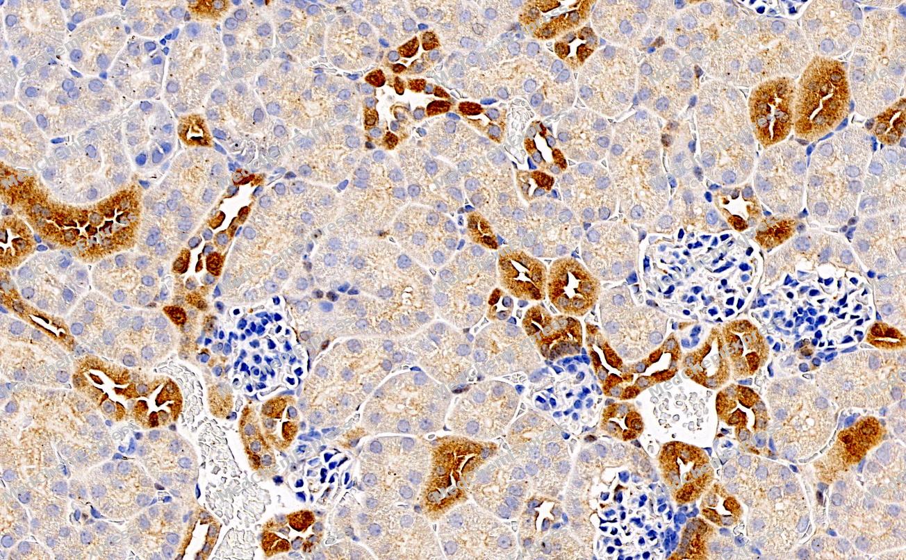 Immunohistochemical analysis of paraffin-embedded Mouse kidney tissue using Caspase-6 Antibody. The section was pre-treated using heat mediated antigen retrieval with sodium citrate buffer (pH 6.0) for 8 minutes. The tissues were blocked in QuickBlock for 20 minutes at room temperature, washed with ddH2O and PBS, and then probed with the primary antibody (HY-P80047, 1/500) in 4℃ overnight. The detection was performed using an HRP conjugated compact polymer system. DAB was used as the chromogen. Tissues were counterstained with hematoxylin and mounted with DPX.
 -
Immunohistochemical analysis of paraffin-embedded Mouse kidney tissue using Caspase-6 Antibody. The section was pre-treated using heat mediated antigen retrieval with sodium citrate buffer (pH 6.0) for 8 minutes. The tissues were blocked in QuickBlock for 20 minutes at room temperature, washed with ddH2O and PBS, and then probed with the primary antibody (HY-P80047, 1/500) in 4℃ overnight. The detection was performed using an HRP conjugated compact polymer system. DAB was used as the chromogen. Tissues were counterstained with hematoxylin and mounted with DPX.


Background
-
Function
Caspase-6 (CASP6) is a cysteine-aspartate protease traditionally classified as an executioner caspase that participates in apoptotic signaling and substrate cleavage during programmed cell death[1][2]. Mechanistically, caspase-6 functions within the broader caspase network that regulates apoptosis, inflammation, and cellular homeostasis, and accumulating evidence demonstrates that its biological activities extend beyond classical apoptotic execution[1][3]. In innate immune pathways, caspase-6 promotes inflammasome activation and facilitates the assembly of the ZBP1-PANoptosome, thereby supporting pyroptotic, apoptotic, and necroptotic signaling collectively termed PANoptosis[3]. During influenza A virus infection, caspase-6 enhances the interaction between RIPK3 and ZBP1, contributing to inflammatory cell death, cytokine production, and host defense responses[3]. Disease relevance is further supported by studies linking caspase-6 activity to neurodegenerative disorders, including Alzheimer’s disease and Huntington’s disease, where aberrant caspase-6 activation has been associated with pathological processes independent of overt apoptosis[2][4]. Compared with the closely related executioner caspases caspase-3 and caspase-7, caspase-6 displays distinct biological functions, including non-enzymatic regulation of innate immune signaling and PANoptosome-associated pathways, highlighting functional divergence within the executioner caspase subgroup[1][3]. For experimental applications, the development of isoform-selective caspase-6 inhibitors has attracted significant interest because selective modulation of caspase-6 may enable mechanistic studies while reducing off-target effects associated with broad-spectrum caspase inhibition[5].
-
Subcellular Localization
Cytoplasm; Nucleus
-
Subunit
Heterotetramer that consists of two anti-parallel arranged heterodimers, each one formed by a 18 kDa (p18) and a 11 kDa (p11) subunits (PubMed:19133298, PubMed:19694615, PubMed:20890311). Interacts with BIRC6/bruce (PubMed:15200957). Interacts with RIPK3 (PubMed:32298652)
-
SwissProt ID
-
Synonyms
MCH2, CASP6, Caspase-6, CASP-6, CSP-6, Apoptotic protease Mch-2
-
Research Field
Cell Biology


Documentation
-
Data Sheet (262 KB)
-
SDS (251 KB)
- English - EN (251 KB)
- Français - FR (251 KB)
- Deutsch - DE (251 KB)
- Norwegian - NO (251 KB)
- Español - ES (251 KB)
- Swedish - SV (251 KB)
- Italian - IT (251 KB)
- Korean - KR (251 KB)
- Portuguese - PT (251 KB)
-
User Guide for Antibodies (1077 KB)
References
[1]. Nadendla EK, et al. Caspases: structural and molecular mechanisms and functions in cell death, innate immunity, and disease. Cell Discov. 2025 May 5;11(1):42. [Content Brief]
[2]. Wang XJ, et al. Activation and regulation of caspase-6 and its role in neurodegenerative diseases. Annu Rev Pharmacol Toxicol. 2015;55:553-72. [Content Brief]
[3]. Zheng M, et al. Caspase-6 Is a Key Regulator of Innate Immunity, Inflammasome Activation, and Host Defense. Cell. 2020 Apr 30;181(3):674-687.e13. [Content Brief]
[4]. Yang L, et al. ncRNA- and Pc2 methylation-dependent gene relocation between nuclear structures mediates gene activation programs. Cell. 2011 Nov 11;147(4):773-88. [Content Brief]
[5]. Van Horn KS, et al. Engaging a Non-catalytic Cysteine Residue Drives Potent and Selective Inhibition of Caspase-6. J Am Chem Soc. 2023 May 10;145(18):10015-10021. [Content Brief]

 Powered by Bioz
