



CPT2 Antibody (YA2715)
(Synonyms: CPT1; CPT2; IIAE4; CPTASE)Based on 1 Customer Validation
CPT2 Antibody (YA2715) is a Rabbit-derived and non-conjugated IgG monoclonal antibody, targeting to CPT2.
-
Host:
Rabbit
-
Isotype:
IgG
-
Application:
WB, IHC-P, ICC/IF
-
Reactivity :
Human, Mouse, Rat
-
Formulation:
Supplied in Rabbit IgG in 10mM phosphate buffered saline , pH 7.4, 150mM sodium chloride, 0.05% BSA, 0.02% sodium azide and 50% glycerol.
-
Conjugation:
Non-conjugated


To place orders, for customer services and technical support, please contact: MedChemExpress USA
Tel: 609-228-6898 E-mail: [email protected] [email protected]





Applications
| Application |
WB

WB: Western Blot
|
IHC-P
IHC-P: Immunohistochemistry-Paraffin
|
ICC/IF
ICC/IF: Immunocytochemistry/
Immunofluorescence |
|---|---|---|---|
| Dilution Ratio | 1:500-1:1000 | 1:50-1:100 | 1:50-1:200 |


Product Details
CPT2 Antibody (YA2715) is a Rabbit-derived and non-conjugated IgG monoclonal antibody, targeting to CPT2.
-
Host Rabbit

-
Clonality Recombinant,Monoclonal

-
Species ReactivityHuman, Mouse, Rat
-
Observed Molecular WeightObserved band size: 74 kDa
Note: Due to possible protein modifications or aggregation, the molecular weight should be confirmed by actual measurement, and the predicted value is for reference only.

-
Calculated Molecular Weight Predicted band size: 74 kDa

Entrez Gene: 1376 Human ; 12896 Mouse ; 25413 Rat
SwissProt: P23786 Human ; P52825 Mouse ; P18886 Rat
OMIM: 255110 Human
A synthesized peptide derived from human CPT2
Endogenous
Affinity Chromatography
Non-conjugated
Unmodified
IgG


Product Properties
-
Appearance
Solution
-
Formulation
Supplied in Rabbit IgG in 10mM phosphate buffered saline , pH 7.4, 150mM sodium chloride, 0.05% BSA, 0.02% sodium azide and 50% glycerol.
-
Concentration
Batch-dependent, Please check the COA for the concentration of each lot. Check Lot Concentration
-
Storage & Stability
Stored at -20°C for 1 year. Avoid repeated freeze / thaw cycles.
-
Shipping
Shipping with blue ice.


Verification Images
-
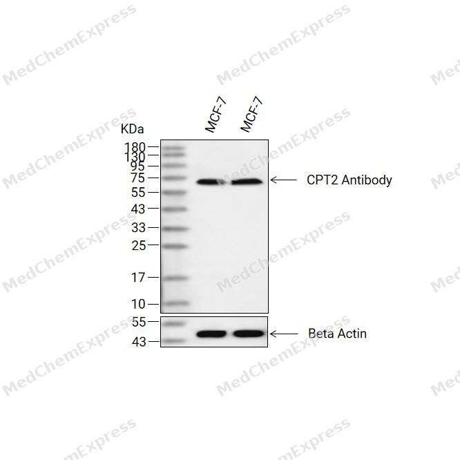 Western blot analysis of extracts from MCF-7(lane 2(20ug) and MCF-7(lane 3(40ug) using CPT2 Antibody (HY-P82970) Rabbit mAb. Proteins were transferred to a PVDF membrane and blocked with 5% non-fat milk in TBST for 2 hour at room temperature. The primary antibody (1/1000) and Loading control antibody (Beta Actin, HY-P83730, 1/10000) was used in 5% non-fat milk in TBST at 4°C overnight. Goat Anti-Mouse/Rabbit IgG-HRP Secondary Antibody (1/10000) was used for 1 hour at room temperature.
 -
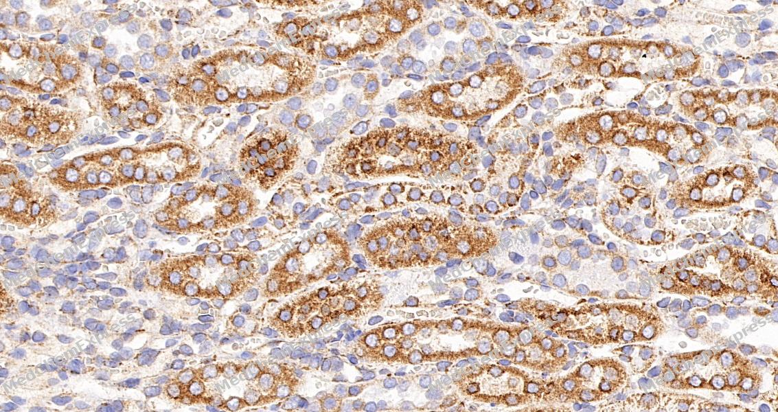 Immunohistochemical analysis of paraffin-embedded mouse kidney tissue using CPT2 Antibody. The section was pre-treated using heat mediated antigen retrieval with sodium citrate buffer (pH 6.0) for 8 minutes. The tissues were blocked in QuickBlock for 20 minutes at room temperature, washed with ddH2O and PBS, and then probed with the primary antibody at 1/100 dilution in 4℃ overnight. The detection was performed using an HRP conjugated compact polymer system. DAB was used as the chromogen. Tissues were counterstained with hematoxylin and mounted with DPX.
 -
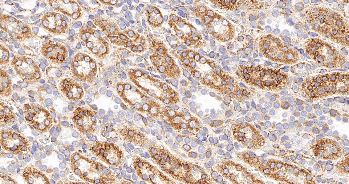 Immunohistochemical analysis of paraffin-embedded mouse kidney tissue using CPT2 Antibody. The section was pre-treated using heat mediated antigen retrieval with sodium citrate buffer (pH 6.0) for 8 minutes. The tissues were blocked in QuickBlock for 20 minutes at room temperature, washed with ddH2O and PBS, and then probed with the primary antibody at 1/100 dilution in 4℃ overnight. The detection was performed using an HRP conjugated compact polymer system. DAB was used as the chromogen. Tissues were counterstained with hematoxylin and mounted with DPX.
 -
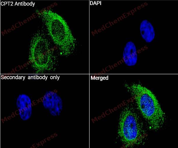 Immunocytochemistry analysis of MCF-7 cells labeling CPT2 with CPT2 Antibody (YA2715) (HY-P82970) at 1/50 dilution. Cells were fixed in 4% paraformaldehyde for 15 minutes at room temperature, permeabilized with 0.1% Triton X-100 for 10 minutes at room temperature, then blocked with BSA for Immunol Staining for 10 min at room temperature. Cells were then incubated with CPT2 Antibody (YA2715) (HY-P82970) at 1/50 dilution in BSA for Immunol Staining at 4 ℃,stay overnight. AF488-conjugated AffiniPure Goat Anti-Rabbit IgG H&L(HY-P8002, Green) was used as the secondary antibody at 1/1,000 dilution. PBS instead of the primary antibody was used as the secondary antibody only control. The Nuclear counterstain was DAPI (Blue).
 -
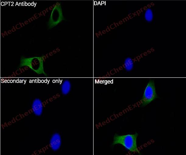 Immunocytochemistry analysis of SK-BR-3 cells labeling CPT2 with CPT2 Antibody (YA2715) (HY-P82970) at 1/100 dilution. Cells were fixed in 4% paraformaldehyde for 15 minutes at room temperature, permeabilized with 0.1% Triton X-100 for 10 minutes at room temperature, then blocked with BSA for Immunol Staining for 10 min at room temperature. Cells were then incubated with CPT2 Antibody (YA2715) (HY-P82970) at 1/100 dilution in BSA for Immunol Staining at 4 ℃,stay overnight. AF488-conjugated AffiniPure Goat Anti-Rabbit IgG H&L(HY-P8002, Green) was used as the secondary antibody at 1/1,000 dilution. PBS instead of the primary antibody was used as the secondary antibody only control. The Nuclear counterstain was DAPI (Blue).


Background
-
Function
Carnitine palmitoyltransferase 2 (CPT-2) is a ubiquitously expressed mitochondrial enzyme that catalyzes the conversion of long-chain acylcarnitines to acyl-CoA within mitochondria, thereby enabling mitochondrial β-oxidation of long-chain fatty acids and cellular energy production[1][2]. CPT-2 functions as a core component of the carnitine shuttle, acting downstream of CPT1 to utilize acylcarnitines generated at the outer mitochondrial membrane and support fatty acid flux into oxidative pathways[1][3]. Mechanistically, mitochondrial fatty acid oxidation depends on the coordinated activities of CPT1 and CPT-2, with CPT1 generating acylcarnitines and CPT-2 converting these intermediates back to metabolically active acyl-CoA species[3]. Deficiency of CPT-2 disrupts long-chain fatty acid oxidation and causes CPT II deficiency, a metabolic disorder characterized by impaired utilization of fatty acids as an energy source, particularly during fasting or increased energy demand[2][4]. Disease manifestations range from severe neonatal and infantile forms to the more common myopathic phenotype associated with recurrent muscle symptoms and metabolic crises[4][5]. Compared with related CPT1 isoforms, which display tissue-specific expression patterns including liver, muscle, and brain variants, CPT-2 is ubiquitously expressed and serves as the common downstream mitochondrial enzyme of the carnitine transport system[1][5]. Experimental studies using Cpt2-deficient models demonstrate that loss of CPT-2 abolishes mitochondrial oxidation of non-esterified fatty acids, making CPT-2 a valuable target for investigating fatty acid metabolism and mitochondrial dysfunction[3].
-
Subcellular Localization
Mitochondrion inner membrane; Peripheral membrane protein; Matrix side
-
SwissProt ID
-
Synonyms
CPT1; CPT2; IIAE4; CPTASE
-
Research Field
Tags & Cell Markers


Documentation
-
Data Sheet (261 KB)
-
SDS (251 KB)
- English - EN (251 KB)
- Français - FR (251 KB)
- Deutsch - DE (251 KB)
- Norwegian - NO (251 KB)
- Español - ES (251 KB)
- Swedish - SV (251 KB)
- Italian - IT (251 KB)
- Korean - KR (251 KB)
- Portuguese - PT (251 KB)
-
User Guide for Antibodies (1077 KB)
References
[1]. Rubio-Gozalbo ME, et al. Carnitine-acylcarnitine translocase deficiency, clinical, biochemical and genetic aspects. Mol Aspects Med. 2004 Oct-Dec;25(5-6):521-32. [Content Brief]
[2]. Choi J, et al. Carnitine palmitoyltransferase 1 facilitates fatty acid oxidation in a non-cell-autonomous manner. Cell Rep. 2024 Dec 24;43(12):115006. [Content Brief]
[3]. Seferi S, et al. Carnitine Palmitoyltransferase II (CPT2) Deficiency: An Overlooked and Elusive Cause of Acute Kidney Injury. Cureus. 2024 Sep 29;16(9):e70442. [Content Brief]

 Powered by Bioz
