



EGFR Antibody (YA775)
(Synonyms: EGFR; ERBB; ERBB1; HER1; Epidermal growth factor receptor; Proto-oncogene c-ErbB-1; Receptor tyrosine-protein kinase erbB-1)
Based on 1 Customer Validation
EGFR Antibody (YA775) is a Mouse-derived and non-conjugated IgG1 monoclonal antibody, targeting to EGFR.
-
Host:
Mouse
-
Isotype:
IgG
-
Application:
WB, ICC/IF, IP
-
Reactivity :
Human, Monkey
-
Formulation:
Supplied in 1*PBS (pH 7.3), 50% glycerol and 0.5% BSA. Preservative: 0.02% sodium azide.
-
Conjugation:
Non-conjugated


To place orders, for customer services and technical support, please contact: MedChemExpress USA
Tel: 609-228-6898 E-mail: [email protected] [email protected]





Applications
| Application |
WB

WB: Western Blot
|
ICC/IF
ICC/IF: Immunocytochemistry/
Immunofluorescence |
IP
IP: Immunoprecipitation
|
|---|---|---|---|
| Dilution Ratio | 1:500-1:1000 | 1:50-1:200 | 1:20 |


Product Details
EGFR Antibody (YA775) is a Mouse-derived and non-conjugated IgG1 monoclonal antibody, targeting to EGFR.
-
Host Mouse

-
Clonality Monoclonal

-
Species ReactivityHuman, Monkey
-
Observed Molecular WeightObserved band size: 175 kDa
Note: Due to possible protein modifications or aggregation, the molecular weight should be confirmed by actual measurement, and the predicted value is for reference only.

-
Calculated Molecular Weight Predicted band size: 134 kDa

Synthetic peptide corresponding to Human EGFR.The exact sequence is proprietary to MCE.
Endogenous
affinity purified
Non-conjugated
Unmodified
IgG


Product Properties
-
Appearance
Solution
-
Formulation
Supplied in 1*PBS (pH 7.3), 50% glycerol and 0.5% BSA. Preservative: 0.02% sodium azide.
-
Concentration
Batch-dependent, Please check the COA for the concentration of each lot. Check Lot Concentration
-
Storage & Stability
Stored at -20°C for 1 year. Avoid repeated freeze / thaw cycles.
-
Shipping
Shipping with blue ice.


Verification Images
-
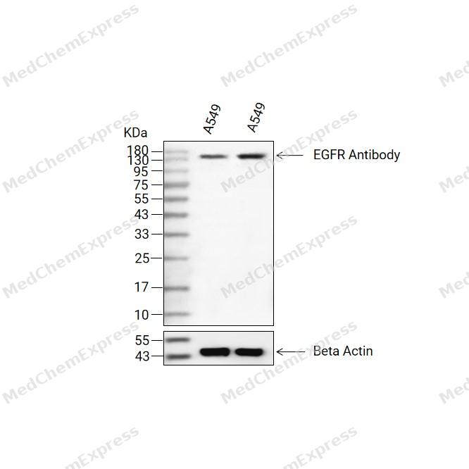 Western blot analysis of extracts from A549(lane 2(20ug) and A549(lane 3(40ug) using EGFR Antibody (HY-P80648) Rabbit mAb. Proteins were transferred to a PVDF membrane and blocked with 5% non-fat milk in TBST for 2 hour at room temperature. The primary antibody (1/1000) and Loading control antibody (Beta Actin, HY-P83730, 1/10000) was used in 5% non-fat milk in TBST at 4°C overnight. Goat Anti-Mouse/Rabbit IgG-HRP Secondary Antibody (1/10000) was used for 1 hour at room temperature.
 -
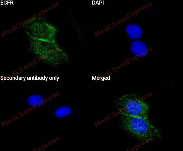 Immunocytochemistry analysis of Hela cells labeling EGFR with EGFR Antibody (HY-P80648) at 1/50 dilution. Cells were fixed in 4% paraformaldehyde for 15 minutes at room temperature, permeabilized with 0.1% Triton X-100 for 10 minutes at room temperature, then blocked with QuickBlock™ Blocking Buffer for Immunol Staining for 10 min at room temperature. Cells were then incubated with EGFR Antibody (HY-P80648) at 1/50 dilution in QuickBlock™ Blocking Buffer for Immunol Staining at 4 ℃. Alexa Fluor® 488-conjugated AffiniPure Goat Anti-Mouse IgG H&L(HY-P8005, Green) was used as the secondary antibody at 1/1,000 dilution. PBS instead of the primary antibody was used as the secondary antibody only control. The Nuclear counterstain was DAPI (Blue).
 -
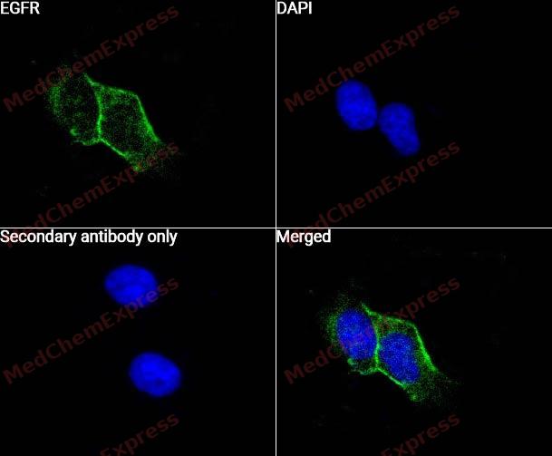 Immunocytochemistry analysis of Hela cells labeling EGFR with EGFR Antibody (HY-P80648) at 1/100 dilution. Cells were fixed in 4% paraformaldehyde for 15 minutes at room temperature, permeabilized with 0.1% Triton X-100 for 10 minutes at room temperature, then blocked with QuickBlock™ Blocking Buffer for Immunol Staining for 10 min at room temperature. Cells were then incubated with EGFR Antibody (HY-P80648) at 1/100 dilution in QuickBlock™ Blocking Buffer for Immunol Staining at 4 ℃. Alexa Fluor® 488-conjugated AffiniPure Goat Anti-Mouse IgG H&L(HY-P8005, Green) was used as the secondary antibody at 1/1,000 dilution. PBS instead of the primary antibody was used as the secondary antibody only control. The Nuclear counterstain was DAPI (Blue).


Background
-
Function
The EGFR family of receptor tyrosine kinases (RTK) comprises four distinct receptors: the EGFR (also known as ErbB-1/HER1), ErbB-2 (neu, HER2), ErbB-3 (HER3) and ErbB-4 (HER4) . All EGFR family members are characterized by a modular structure consisting of an extracellular ligand-binding domain, a single hydrophobic transmembrane region, and the intracellular part harbouring the highly conserved tyrosine kinase domain. The ErbB family of receptor tyrosine kinases (RTKs) couples binding of extracellular growth factor ligands to intracellular signaling pathways regulating diverse biologic responses, including proliferation, differentiation, cell motility, and survival. Ten growth factors and their ErbB specificities are: EGF, amphiregulin (AR), and TGF bind ErbB-1; betacellulin, and epiregulin bind both ErbB-1 and ErbB-4; the neuregulins (also called heregulins and Neu differentiation factors) NRG-1 and NRG-2 bind ErbB-3 and ErbB-4; and NRG-3 and NRG-4 bind ErbB-4. No known ligand binds ErbB-2. The three best characterized signaling pathways induced through ErbBs are Ras-mitogen-activated protein kinase (Ras-MAPK), phosphatidylinositol 3 kinase-protein kinase B (PI3K-PKB/Akt), and phospholipase C-protein kinase C (PLC-PKC) pathways.
-
Subcellular Localization
Cell membrane; Single-pass type I membrane protein; Endoplasmic reticulum membrane; Single-pass type I membrane protein; Golgi apparatus membrane; Single-pass type I membrane protein; Nucleus membrane; Single-pass type I membrane protein; Endosome; Endosome membrane; Nucleus; Secreted
-
Expression
Tissue_specificity:Widely expressed. Isomer 2 is also expressed in ovarian cancer. -
Isoforms & Post-Translational Modification
P00533 has 4 isomers: P00533-1: 134277 Da (predicted); P00533-2: 44664 Da (predicted); P00533-3: 77312 Da (predicted); P00533-4: 69228 Da (predicted).
Phosphorylated on Tyr residues in response to EGF (PubMed:20462955, PubMed:27153536). Phosphorylation at Ser-695 is partial and occurs only if Thr-693 is phosphorylated. Phosphorylation at Thr-678 and Thr-693 by PRKD1 inhibits EGF-induced MAPK8/JNK1 activation. Dephosphorylation by PTPRJ prevents endocytosis and stabilizes the receptor at the plasma membrane. Autophosphorylation at Tyr-1197 is stimulated by methylation at Arg-1199 and enhances interaction with PTPN6. Autophosphorylation at Tyr-1092 and/or Tyr-1110 recruits STAT3. Dephosphorylated by PTPN1 and PTPN2;Monoubiquitinated and polyubiquitinated upon EGF stimulation; which does not affect tyrosine kinase activity or signaling capacity but may play a role in lysosomal targeting (PubMed:27153536). Polyubiquitin linkage is mainly through 'Lys-63', but linkage through 'Lys-48', 'Lys-11' and 'Lys-29' also occurs. Deubiquitination by OTUD7B prevents degradation. Ubiquitinated by RNF115 and RNF126 (By similarity). Ubiquitinated by ZNRF1 or CBL at different lysines in response to EGF stimulation; leading to recruitment of the ESCRT machinery and subsequent degradation in the lysosomes (PubMed:33996800). Deubiquitinated by UCHL1 leading to the inhibition of its degradation (By similarity);Palmitoylated on Cys residues by ZDHHC20. Palmitoylation inhibits internalization after ligand binding, and increases the persistence of tyrosine-phosphorylated EGFR at the cell membrane. Palmitoylation increases the amplitude and duration of EGFR signaling;Methylated. Methylation at Arg-1199 by PRMT5 stimulates phosphorylation at Tyr-1197 -
Subunit
Binding of the ligand triggers homo- and/or heterodimerization of the receptor triggering its autophosphorylation. Heterodimer with ERBB2 (PubMed:10805725). Forms a complex with CCDC88A/GIV (via SH2-like regions) and GNAI3 which leads to enhanced EGFR signaling and triggering of cell migration; binding to CCDC88A requires autophosphorylation of the EGFR C-terminal region, and ligand stimulation is required for recruitment of GNAI3 to the complex (PubMed:20462955, PubMed:25187647). Interacts with ERRFI1; inhibits dimerization of the kinase domain and autophosphorylation (PubMed:18046415). Part of a complex with ERBB2 and either PIK3C2A or PIK3C2B (PubMed:10805725). Interacts with GRB2; an adapter protein coupling the receptor to downstream signaling pathways. Interacts with GAB2; involved in signaling downstream of EGFR. Interacts with STAT3; mediates EGFR downstream signaling in cell proliferation. Interacts with RIPK1; involved in NF-kappa-B activation. Interacts (autophosphorylated) with CBL, CBLB and CBLC; involved in EGFR ubiquitination and regulation; interaction with CBL is reduced in the presence of tensin TNS4 (PubMed:23774213). Interacts with SOCS5; regulates EGFR degradation through ELOC- and ELOB-mediated ubiquitination and proteasomal degradation. Interacts with PRMT5; methylates EGFR and enhances interaction with PTPN6. Interacts (phosphorylated) with PTPN6; inhibits EGFR-dependent activation of MAPK/ERK. Interacts with COPG1; essential for regulation of EGF-dependent nuclear transport of EGFR by retrograde trafficking from the Golgi to the ER. Interacts with TNK2; this interaction is dependent on EGF stimulation and kinase activity of EGFR. Interacts with PCNA; positively regulates PCNA (PubMed:17115032). Interacts with PELP1. Interacts with MUC1. Interacts with AP2M1. Interacts with FER. May interact with EPS8; mediates EPS8 phosphorylation. Interacts (via SH2 domains) with GRB2, NCK1 and NCK2 (PubMed:10026169). Interacts with ATXN2. Interacts with GAREM1. Interacts (ubiquitinated) with ANKRD13A/B/D; the interaction is direct and may regulate EGFR internalization after EGF stimulation. Interacts with GPER1; the interaction occurs in an estrogen-dependent manner. Interacts (via C-terminal cytoplasmic kinase domain) with ZPR1 (via zinc fingers). Interacts with RNF115 and RNF126 (PubMed:23418353). Interacts with GPRC5A (via its transmembrane domain) (PubMed:25311788). Interacts with FAM83B; positively regulates EGFR inducing its autophosphorylation in absence of stimulation by EGF (PubMed:23912460). Interacts with LAPTM4B; positively correlates with EGFR activation (PubMed:28479384). Interacts with STX19 (PubMed:16420529). Interacts with CD44 (PubMed:23589287). Interacts with PGRMC1; the interaction requires PGRMC1 homodimerization (PubMed:26988023). Interacts with PIKFYVE (PubMed:17909029). Interacts with NEU3. Interacts with TRAF4 (PubMed:30352854). Interacts with the ant venom OMEGA-myrmeciitoxin(02)-Mg1a (By similarity). Interacts with CD82; this interaction facilitates ligand-induced endocytosis of the receptor and its subsequent desensitization (PubMed:35538033)
-
SwissProt ID
-
Synonyms
EGFR; ERBB; ERBB1; HER1; Epidermal growth factor receptor; Proto-oncogene c-ErbB-1; Receptor tyrosine-protein kinase erbB-1
-
Research Field
Signal Transduction


Documentation
-
Data Sheet (234 KB)
-
SDS (252 KB)
- English - EN (252 KB)
- Français - FR (252 KB)
- Deutsch - DE (252 KB)
- Norwegian - NO (252 KB)
- Español - ES (252 KB)
- Swedish - SV (252 KB)
- Italian - IT (252 KB)
- Korean - KR (252 KB)
- Portuguese - PT (252 KB)
-
User Guide for Antibodies (1077 KB)

 Powered by Bioz
