



EIF5 Antibody (YA4096)
(Synonyms: EIF-5; EIF-5A)
Based on 1 Customer Validation
EIF5 Antibody (YA4096) is a Mouse-derived and non-conjugated IgG2a monoclonal antibody, targeting to EIF5.
-
Host:
Mouse
-
Isotype:
IgG
-
Application:
IHC-P, ICC/IF, FC, ELISA
-
Reactivity :
Human
-
Formulation:
Supplied in PBS with 0.05% sodium azide
-
Conjugation:
Non-conjugated


To place orders, for customer services and technical support, please contact: MedChemExpress USA
Tel: 609-228-6898 E-mail: [email protected] [email protected]





Applications
| Application |
IHC-P

IHC-P: Immunohistochemistry-Paraffin
|
ICC/IF
ICC/IF: Immunocytochemistry/
Immunofluorescence |
FC
FC: Flow Cytometry
|
ELISA
ELISA: Enzyme Linked Immunosorbent Assay
|
|---|---|---|---|---|
| Dilution Ratio | 1:200-1:1000 | 1:50-1:250 | 1:200-1:400 | 1:10000 |


Product Details
EIF5 Antibody (YA4096) is a Mouse-derived and non-conjugated IgG2a monoclonal antibody, targeting to EIF5.
-
Host Mouse

-
Clonality Monoclonal

-
Species ReactivityHuman
-
Observed Molecular WeightObserved band size: 50-60 kDa
Note: Due to possible protein modifications or aggregation, the molecular weight should be confirmed by actual measurement, and the predicted value is for reference only.

-
Calculated Molecular Weight Predicted band size: 50 kDa

Purified recombinant fragment of human EIF5 (AA: 1-300) expressed in E. Coli.
affinity purified.
Non-conjugated
Unmodified
IgG


Product Properties
-
Appearance
Solution
-
Formulation
Supplied in PBS with 0.05% sodium azide
-
Concentration
Batch-dependent, Please check the COA for the concentration of each lot. Check Lot Concentration
-
Storage & Stability
Stored at -20°C for 1 year. Avoid repeated freeze / thaw cycles.
-
Shipping
Shipping with blue ice.


Verification Images
-
 Immunohistochemical analysis of paraffin-embedded human liver tissue using EIF5 Antibody (HY-P84399, 1/500). The section was pretreated using heat mediated antigen retrieval with sodium citrate buffer (pH 6.0) for 8 minutes. The tissues were blocked with quick block buffer for 0.5 hours at room temperature, washed with PBS and PBST, and then incubated with the primary antibody overnight at 4℃. The detection was performed using an HRP conjugated compact polymer system. DAB was used as the chromogen. Tissues were counterstained with hematoxylin and mounted with DPX.
 -
 Immunohistochemical analysis of paraffin-embedded human adrenal gland tissue using EIF5 Antibody (HY-P84399, 1/500). The section was pretreated using heat mediated antigen retrieval with sodium citrate buffer (pH 6.0) for 8 minutes. The tissues were blocked with quick block buffer for 0.5 hours at room temperature, washed with PBS and PBST, and then incubated with the primary antibody overnight at 4℃. The detection was performed using an HRP conjugated compact polymer system. DAB was used as the chromogen. Tissues were counterstained with hematoxylin and mounted with DPX.
 -
 Immunohistochemical analysis of paraffin-embedded human salivary gland tissue using EIF5 Antibody (HY-P84399, 1/800). The section was pretreated using heat mediated antigen retrieval with sodium citrate buffer (pH 6.0) for 8 minutes. The tissues were blocked with quick block buffer for 0.5 hours at room temperature, washed with PBS and PBST, and then incubated with the primary antibody overnight at 4℃. The detection was performed using an HRP conjugated compact polymer system. DAB was used as the chromogen. Tissues were counterstained with hematoxylin and mounted with DPX.
 -
 Immunohistochemical analysis of paraffin-embedded human testis tissue using EIF5 Antibody (HY-P84399, 1/800). The section was pretreated using heat mediated antigen retrieval with sodium citrate buffer (pH 6.0) for 8 minutes. The tissues were blocked with quick block buffer for 0.5 hours at room temperature, washed with PBS and PBST, and then incubated with the primary antibody overnight at 4℃. The detection was performed using an HRP conjugated compact polymer system. DAB was used as the chromogen. Tissues were counterstained with hematoxylin and mounted with DPX.
 -
 Immunohistochemical analysis of paraffin-embedded human epididymis tissue using EIF5 Antibody (HY-P84399, 1/800). The section was pretreated using heat mediated antigen retrieval with sodium citrate buffer (pH 6.0) for 8 minutes. The tissues were blocked with quick block buffer for 0.5 hours at room temperature, washed with PBS and PBST, and then incubated with the primary antibody overnight at 4℃. The detection was performed using an HRP conjugated compact polymer system. DAB was used as the chromogen. Tissues were counterstained with hematoxylin and mounted with DPX.
 -
 Immunohistochemical analysis of paraffin-embedded human fallopian tube tissue using EIF5 Antibody (HY-P84399, 1/800). The section was pretreated using heat mediated antigen retrieval with sodium citrate buffer (pH 6.0) for 8 minutes. The tissues were blocked with quick block buffer for 0.5 hours at room temperature, washed with PBS and PBST, and then incubated with the primary antibody overnight at 4℃. The detection was performed using an HRP conjugated compact polymer system. DAB was used as the chromogen. Tissues were counterstained with hematoxylin and mounted with DPX.
 -
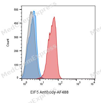 Flow cytometric analysis of 1X10^6 PC-3 cells labeling EIF5 Antibody(red). Cells were fixed with 4% paraformaldehyde and permeabilised with 0.2% Triton X-100. Then stained with the primary antibody at 1/400 dilution overnight at 4℃. AF 488 Goat Anti-mouse IgG H&L was used as the secondary antibody at 1/1,000 dilution for 45 minutes at room temperature. Mouse IgG Isotype Control (blue) was used as the isotype control, cells without incubation with primary antibody were used as the unlabeled control (black).
 -
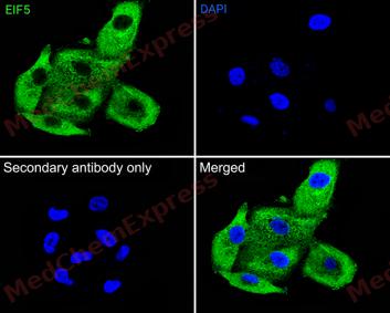 Immunofluorescence analysis of A549 cells labeling EIF5 antibody at 1/50 dilution. Cells were fixed in 4% paraformaldehyde for 15 minutes at room temperature, permeabilized with 0.1% Triton X-100 in PBS for 15 minutes at room temperature, then blocked with 1% BSA in 10% negative goat serum for 1 hour at room temperature.Cells were then incubated with EIF5 antibody at 1/50 dilution in 1% BSA in PBST overnight at 4 ℃. AF 488 Goat Anti-mouse IgG H&L (green) was used as the secondary antibody at 1/500 dilution. PBS instead of the primary antibody was used as the secondary antibody only control. The Nuclear counterstain was DAPI (Blue)


Background
-
Function
eIF5 (eukaryotic translation initiation factor 5) is a central regulator of eukaryotic translation initiation that functions as a GTPase-activating protein (GAP) for eIF2 and promotes GTP hydrolysis during start-codon recognition[1][2]. Mechanistically, eIF5 participates in assembly and progression of the translation pre-initiation complex, interacts with multiple initiation factors, and contributes to accurate AUG selection during ribosomal scanning[2][3]. Beyond its canonical GAP activity, eIF5 also exhibits GDP dissociation inhibitor (GDI) activity toward eIF2, linking GTP hydrolysis to recycling and control of translation initiation complexes[4]. Therefore, eIF5 occupies a critical position in the molecular pathway that couples start-codon recognition with productive protein synthesis[2][4]. Experimental studies in yeast and reconstituted eukaryotic translation systems established that eIF5 is required for efficient eIF2 GTPase activation and proper initiation complex function[1][4]. Structural and biochemical analyses further showed that eIF5 can replace eIF1 on the 40S initiation complex and thereby contribute to stringent start-site selection[3]. Compared with the related factor eIF5A, which primarily functions during translation elongation and termination rather than initiation, eIF5 acts directly within the translation initiation pathway and regulates eIF2-dependent start-codon selection[3][4]. Consequently, eIF5 serves as an important experimental target for mechanistic studies of translational control, initiation fidelity, and ribosome-associated regulatory processes[1][3][4].
-
Subcellular Localization
Cytoplasm
-
Subunit
Component of the 43S pre-initiation complex (43S PIC), which is composed of the 40S ribosomal subunit, EIF1, eIF1A (EIF1AX), eIF3 complex, EIF5 and eIF2-GTP-initiator tRNA complex (eIF2 ternary complex). Interacts with eIF1A (EIF1AX) during scanning (PubMed:24319994). Interacts through its C-terminal domain (CTD) with EIF1 or with eIF2-beta (EIF2S2) (mutually exclusive) through a common binding site (PubMed:21745818, PubMed:22813744). Interacts through its C-terminal domain (CTD) with the CTD of EIF5B (PubMed:30211544). Interacts with FMR1 isoform 6; this interaction occurs in a RNA-dependent manner (PubMed:24658146)
-
SwissProt ID
-
Synonyms
EIF-5; EIF-5A


Documentation
-
Data Sheet (261 KB)
-
SDS (251 KB)
- English - EN (251 KB)
- Français - FR (251 KB)
- Deutsch - DE (251 KB)
- Norwegian - NO (251 KB)
- Español - ES (251 KB)
- Swedish - SV (251 KB)
- Italian - IT (251 KB)
- Korean - KR (251 KB)
- Portuguese - PT (251 KB)
-
User Guide for Antibodies (1077 KB)
References
[1]. Kimura H, et al. Identification of hypoxia-inducible factor 1 ancillary sequence and its function in vascular endothelial growth factor gene induction by hypoxia and nitric oxide. J Biol Chem. 2001 Jan 19;276(3):2292-8. [Content Brief]
[2]. Llácer JL, et al. Translational initiation factor eIF5 replaces eIF1 on the 40S ribosomal subunit to promote start-codon recognition. Elife. 2018 Nov 30;7:e39273. [Content Brief]
[3]. Jennings MD, et al. eIF5 is a dual function GAP and GDI for eukaryotic translational control. Small GTPases. 2010 Sep;1(2):118-123. [Content Brief]

 Powered by Bioz
