



EpCAM Antibody (YA458)
(Synonyms: EPCAM; GA733-2; M1S2; M4S1; MIC18; TACSTD1; TROP1; Epithelial cell adhesion molecule; Ep-CAM; Adenocarcinoma-associated antigen; Cell surface glycoprotein Trop-1; Epithelial cell surface antigen; Epithelial glycoprotein; EGP; Epithelial glycoprotein 314; )
Based on 1 Customer Validation
EpCAM Antibody (YA458) is a Rabbit-derived and non-conjugated IgG monoclonal antibody, targeting to EpCAM.
-
Host:
Rabbit
-
Isotype:
IgG
-
Application:
WB, IHC-F, IHC-P, ICC/IF, IP
-
Reactivity :
Human
-
Formulation:
Supplied in 50 mM Tris-Glycine (pH 7.4), 0.15 M NaCl, 40% Glycerol and 0.05% BSA. Preservative: 0.01% Sodium azide
-
Conjugation:
Non-conjugated


To place orders, for customer services and technical support, please contact: MedChemExpress USA
Tel: 609-228-6898 E-mail: [email protected] [email protected]





Applications
| Application |
WB

WB: Western Blot
|
IHC-P
IHC-P: Immunohistochemistry-Paraffin
|
IHC-F
IHC-F: Immunohistochemistry-Frozen
|
ICC/IF
ICC/IF: Immunocytochemistry/
Immunofluorescence |
IP
IP: Immunoprecipitation
|
|---|---|---|---|---|---|
| Dilution Ratio | 1:500-1:1000 | 1:50-1:100 | 1:50-1:100 | 1:50-1:200 | 1:20 |


Product Details
EpCAM Antibody (YA458) is a Rabbit-derived and non-conjugated IgG monoclonal antibody, targeting to EpCAM.
-
Host Rabbit

-
Clonality Recombinant,Monoclonal

-
Species ReactivityHuman
-
Observed Molecular WeightObserved band size: 42 kDa
Note: Due to possible protein modifications or aggregation, the molecular weight should be confirmed by actual measurement, and the predicted value is for reference only.

-
Calculated Molecular Weight Predicted band size: 35 kDa

Synthetic peptide corresponding to Human EpCAM aa24-265.
Endogenous
affinity purified
Non-conjugated
Unmodified
IgG


Product Properties
-
Appearance
Solution
-
Formulation
Supplied in 50 mM Tris-Glycine (pH 7.4), 0.15 M NaCl, 40% Glycerol and 0.05% BSA. Preservative: 0.01% Sodium azide
-
Concentration
Batch-dependent, Please check the COA for the concentration of each lot. Check Lot Concentration
-
Storage & Stability
Stored at -20°C for 1 year. Avoid repeated freeze / thaw cycles.
-
Shipping
Shipping with blue ice.


Verification Images
-
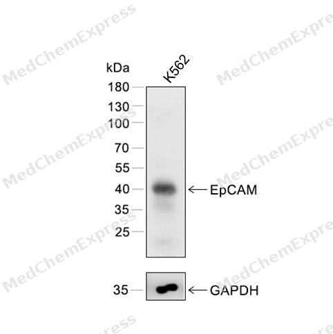 Western blot analysis of extracts from K562 using EpCAM Antibody (YA458) antibody. Proteins were transferred to a PVDF membrane and blocked with 5% nonfat powdered milk in PBST for 2 hour at room temperature. The primary antibody (1/1000) and loading control antibody (GAPDH, 1/3000) was diluted with 5% nonfat powdered milk in PBST at 4°C overnight. Goat Anti-Rabbit IgG-HRP Secondary Antibody (1/8,000) was incubated for 45min at room temperature.
 -
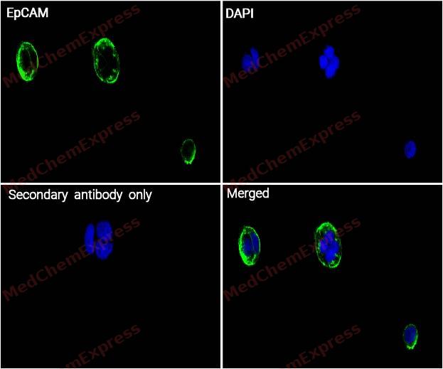 Immunocytochemistry analysis of CACO-2 cells labeling EpCAM with EpCAM Antibody (HY-P80657) at 1/100 dilution. Cells were fixed in 4% paraformaldehyde for 15 minutes at room temperature, permeabilized with 0.1% Triton X-100 in PBS for 15 minutes at room temperature, then blocked with quick block buffer for 10 minutes at room temperature. Cells were then incubated with EpCAM Antibody (HY-P80657) at 1/100 dilution in quick block buffer overnight at 4 ℃. AF488-conjugated Goat Anti-Rabbit IgG H&L(HY-P8002, Green) was used as the secondary antibody at 1/1,000 dilution. PBS instead of the primary antibody was used as the secondary antibody only control. The Nuclear counterstain was DAPI (Blue).
 -
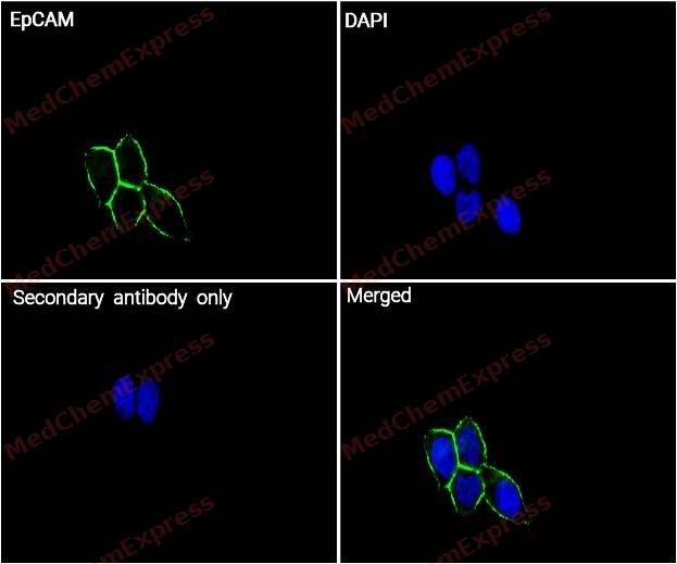 Immunocytochemistry analysis of MCF-7 cells labeling EpCAM with EpCAM Antibody (HY-P80657) at 1/100 dilution. Cells were fixed in 4% paraformaldehyde for 15 minutes at room temperature, permeabilized with 0.1% Triton X-100 in PBS for 15 minutes at room temperature, then blocked with quick block buffer for 10 minutes at room temperature. Cells were then incubated with EpCAM Antibody (HY-P80657) at 1/100 dilution in quick block buffer overnight at 4 ℃. AF488-conjugated Goat Anti-Rabbit IgG H&L(HY-P8002, Green) was used as the secondary antibody at 1/1,000 dilution. PBS instead of the primary antibody was used as the secondary antibody only control. The Nuclear counterstain was DAPI (Blue).


Background
-
Function
EpCAM (CD326) is a transmembrane glycoprotein expressed on the surface of epithelial cells and serves as a classic marker for epithelial cells and epithelial-derived tumors. It plays a role in cell adhesion, the regulation of proliferation, and the maintenance of tumor stemness, holding significant value in tumor diagnosis, circulating tumor cell detection, cancer stem cell research, and the development of targeted therapies.
-
Subcellular Localization
Lateral cell membrane; Single-pass type I membrane protein; Cell junction, tight junction
-
Expression
Tissue_specificity:This gene is highly and selectively expressed in undifferentiated embryonic stem cells (ESCs) , but not in differentiated ESCs. Once ESCs differentiate, its protein level rapidly declines. This gene is expressed on almost all epithelial cell membranes, but not on mesoderm or neural cell membranes. It has also been found on the surface of adenocarcinomas. -
Subunit
Monomer. Interacts with phosphorylated CLDN7
-
SwissProt ID
-
Synonyms
EPCAM; GA733-2; M1S2; M4S1; MIC18; TACSTD1; TROP1; Epithelial cell adhesion molecule; Ep-CAM; Adenocarcinoma-associated antigen; Cell surface glycoprotein Trop-1; Epithelial cell surface antigen; Epithelial glycoprotein; EGP; Epithelial glycoprotein 314;
-
Research Field
Tags & Cell Markers


Documentation
-
Data Sheet (234 KB)
-
SDS (252 KB)
- English - EN (252 KB)
- Français - FR (252 KB)
- Deutsch - DE (252 KB)
- Norwegian - NO (252 KB)
- Español - ES (252 KB)
- Swedish - SV (252 KB)
- Italian - IT (252 KB)
- Korean - KR (252 KB)
- Portuguese - PT (252 KB)
-
User Guide for Antibodies (1077 KB)

 Powered by Bioz
