



FMRP Antibody (YA850)
(Synonyms: CREB1; Cyclic AMP-responsive element-binding protein 1; CREB-1; cAMP-responsive element-binding protein 1)
Based on 1 Customer Validation
FMRP Antibody (YA850) is a Rabbit-derived and non-conjugated IgG monoclonal antibody, targeting to FMRP.
-
Host:
Rabbit
-
Isotype:
IgG
-
Application:
WB, ICC/IF, IHC-P
-
Reactivity :
Human, Mouse, Rat
-
Formulation:
Supplied in 1*TBS (pH7.4), 0.05% BSA, 40% Glycerol. Preservative: 0.05% Sodium Azide.
-
Conjugation:
Non-conjugated


To place orders, for customer services and technical support, please contact: MedChemExpress USA
Tel: 609-228-6898 E-mail: [email protected] [email protected]





Applications
| Application |
WB

WB: Western Blot
|
ICC/IF
ICC/IF: Immunocytochemistry/
Immunofluorescence |
IF-Tissue
IF-Tissue: Immunofluorescence-Tissue
|
IHC-P
IHC-P: Immunohistochemistry-Paraffin
|
|---|---|---|---|---|
| Dilution Ratio | 1:1000-1:5000 | 1:100-1:500 | 1:100-1:500 | 1:50-1:1000 |


Product Details
FMRP Antibody (YA850) is a Rabbit-derived and non-conjugated IgG monoclonal antibody, targeting to FMRP.
-
Host Rabbit

-
Clonality Recombinant,Monoclonal

-
Species ReactivityHuman, Mouse, Rat
-
Observed Molecular WeightObserved band size: 71-75 kDa
Note: Due to possible protein modifications or aggregation, the molecular weight should be confirmed by actual measurement, and the predicted value is for reference only.

-
Calculated Molecular Weight Predicted band size: 71 kDa

Entrez Gene: 2332 Human ; 24948 Rat
SwissProt: Q06787 Human ; P35922 Mouse ; Q80WE1 Rat
OMIM: 300624 Human
Synthetic peptide within human FMRP aa 20-60.
Endogenous
Protein A affinity purified.
Non-conjugated
Unmodified
IgG


Product Properties
-
Appearance
Solution
-
Formulation
Supplied in 1*TBS (pH7.4), 0.05% BSA, 40% Glycerol. Preservative: 0.05% Sodium Azide.
-
Concentration
Batch-dependent, Please check the COA for the concentration of each lot. Check Lot Concentration
-
Storage & Stability
Stored at -20°C for 1 year. Avoid repeated freeze / thaw cycles.
-
Shipping
Shipping with blue ice


Verification Images
-
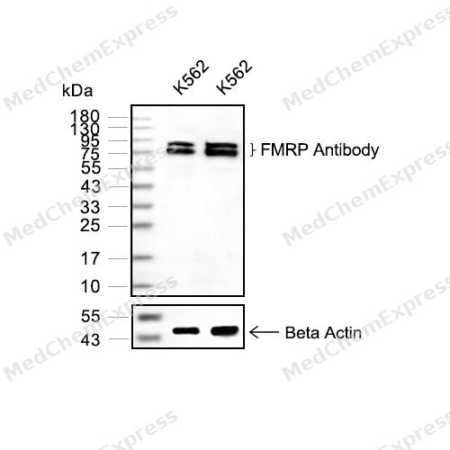 Western blot analysis of extracts from K562(lane 2(20μg) and K562(lane 3(40μg) using FMRP(HY-P80970 Rabbit mAb. Proteins were transferred to a PVDF membrane and blocked with 5% non-fat milk in TBST for 2 hour at room temperature. The primary antibody (1/1000) and Loading control antibody (Beta Actin, HY-P80438, 1/10000) was used in 5% non-fat milk in TBST at 4°C overnight. Goat Anti-Mouse/Rabbit IgG-HRP Secondary Antibody (1/10000) was used for 1 hour at room temperature.
 -
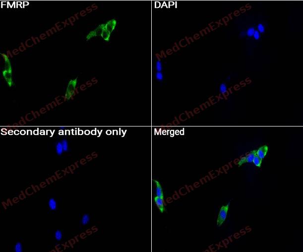 Immunocytochemistry analysis of NIH3T3 cells labeling FMRP with FMRP antibody (HY-P80970) at 1/100 dilution. Cells were fixed in 4% paraformaldehyde for 15 minutes at room temperature, permeabilized with 0.1% Triton X-100 for 10 minutes at room temperature, then blocked with QuickBlock™ Blocking Buffer for Immunol Staining for 10 min at room temperature. Cells were then incubated with FMRP antibody (HY-P80970) at 1/100 dilution in QuickBlock™ Blocking Buffer for Immunol Staining at 4 ℃. Alexa Fluor® 488-conjugated AffiniPure Goat Anti-Rabbit IgG H&L(HY-P8002, Green) was used as the secondary antibody at 1/1,000 dilution. PBS instead of the primary antibody was used as the secondary antibody only control. The Nuclear counterstain was DAPI (Blue).
 -
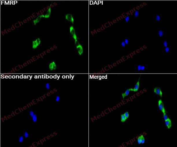 Immunocytochemistry analysis of NIH3T3 cells labeling FMRP with FMRP antibody (HY-P80970) at 1/250 dilution. Cells were fixed in 4% paraformaldehyde for 15 minutes at room temperature, permeabilized with 0.1% Triton X-100 for 10 minutes at room temperature, then blocked with QuickBlock™ Blocking Buffer for Immunol Staining for 10 min at room temperature. Cells were then incubated with FMRP antibody (HY-P80970) at 1/250 dilution in QuickBlock™ Blocking Buffer for Immunol Staining at 4 ℃. Alexa Fluor® 488-conjugated AffiniPure Goat Anti-Rabbit IgG H&L(HY-P8002, Green) was used as the secondary antibody at 1/1,000 dilution. PBS instead of the primary antibody was used as the secondary antibody only control. The Nuclear counterstain was DAPI (Blue).
 -
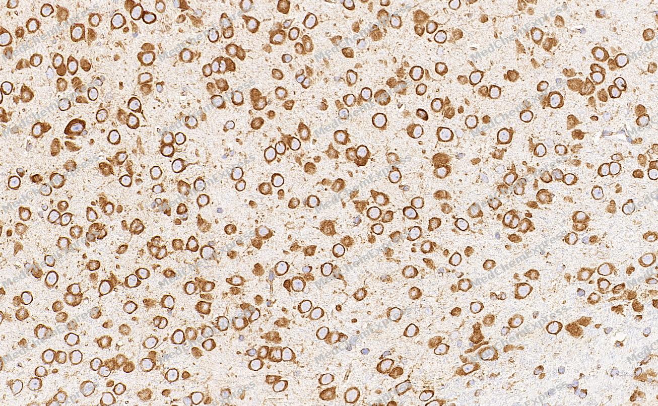 Immunohistochemical analysis of paraffin-embedded Mouse brain tissue using FMRP Antibody. The section was pre-treated using heat mediated antigen retrieval with Tris-EDTA buffer (pH 9.0) for 8 minutes. The tissues were blocked in QuickBlock for 20 minutes at room temperature, washed with ddH2O and PBS, and then probed with the primary antibody (HY-P80970, 1/400) in 4℃ overnight. The detection was performed using an HRP conjugated compact polymer system. DAB was used as the chromogen. Tissues were counterstained with hematoxylin and mounted with DPX.
 -
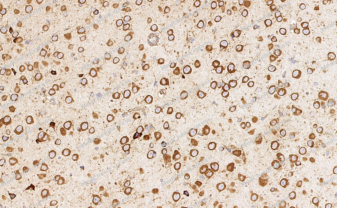 Immunohistochemical analysis of paraffin-embedded Mouse brain tissue using FMRP Antibody. The section was pre-treated using heat mediated antigen retrieval with Tris-EDTA buffer (pH 9.0) for 8 minutes. The tissues were blocked in QuickBlock for 20 minutes at room temperature, washed with ddH2O and PBS, and then probed with the primary antibody (HY-P80970, 1/400) in 4℃ overnight. The detection was performed using an HRP conjugated compact polymer system. DAB was used as the chromogen. Tissues were counterstained with hematoxylin and mounted with DPX.


Background
-
Function
FMRP multifunctional polyribosome-associated RNA-binding protein that plays a central role in neuronal development and synaptic plasticity through the regulation of alternative mRNA splicing, mRNA stability, mRNA dendritic transport and postsynaptic local protein synthesis of target mRNAs. Acts as an mRNA regulator by mediating formation of some phase-separated membraneless compartment: undergoes liquid-liquid phase separation upon binding to target mRNAs, leading to assemble mRNAs into cytoplasmic ribonucleoprotein granules that concentrate mRNAs with associated regulatory factors. Plays a role in the alternative splicing of its own mRNA. Stabilizes the scaffolding postsynaptic density protein DLG4/PSD-95 and the myelin basic protein (MBP) mRNAs in hippocampal neurons and glial cells, respectively; this stabilization is further increased in response to metabotropic glutamate receptor (mGluR) stimulation. Plays a role in selective delivery of a subset of dendritic mRNAs to synaptic sites in response to mGluR activation in a kinesin-dependent manner. Undergoes liquid-liquid phase separation following phosphorylation and interaction with CAPRIN1, promoting formation of cytoplasmic ribonucleoprotein granules that concentrate mRNAs with factors that inhibit translation and mediate deadenylation of target mRNAs. Acts as a repressor of mRNA translation in synaptic regions by mediating formation of neuronal ribonucleoprotein granules and promoting recruitmtent of EIF4EBP2. Plays a role as a repressor of mRNA translation during the transport of dendritic mRNAs to postsynaptic dendritic spines. Component of the CYFIP1-EIF4E-FMR1 complex which blocks cap-dependent mRNA translation initiation. Represses mRNA translation by stalling ribosomal translocation during elongation. Reports are contradictory with regards to its ability to mediate translation inhibition of MBP mRNA in oligodendrocytes. Also involved in the recruitment of the RNA helicase MOV10 to a subset of mRNAs and hence regulates microRNA (miRNA)-mediated translational repression by AGO2. Facilitates the assembly of miRNAs on specific target mRNAs. Also plays a role as an activator of mRNA translation of a subset of dendritic mRNAs at synapses. In response to mGluR stimulation, FMR1-target mRNAs are rapidly derepressed, allowing for local translation at synapses. Binds to a large subset of dendritic mRNAs that encode a myriad of proteins involved in pre- and postsynaptic functions. Binds to 5'-ACU[GU]-3' and/or 5'-[AU]GGA-3' RNA consensus sequences within mRNA targets, mainly at coding sequence (CDS) and 3'-untranslated region (UTR) and less frequently at 5'-UTR. Binds to intramolecular G-quadruplex structures in the 5'- or 3'-UTRs of mRNA targets. Binds to G-quadruplex structures in the 3'-UTR of its own mRNA. Also binds to RNA ligands harboring a kissing complex (kc) structure; this binding may mediate the association of FMR1 with polyribosomes. Binds mRNAs containing U-rich target sequences. Binds to a triple stem-loop RNA structure, called Sod1 stem loop interacting with FMRP (SoSLIP), in the 5'-UTR region of superoxide dismutase SOD1 mRNA. Binds to the dendritic, small non-coding brain cytoplasmic RNA 1 (BC1); which may increase the association of the CYFIP1-EIF4E-FMR1 complex to FMR1 target mRNAs at synapses. Plays a role in mRNA nuclear export. Specifically recognizes and binds a subset of N6-methyladenosine (m6A)-containing mRNAs, promoting their nuclear export in a XPO1/CRM1-dependent manner. Together with export factor NXF2, is involved in the regulation of the NXF1 mRNA stability in neurons. Associates with export factor NXF1 mRNA-containing ribonucleoprotein particles (mRNPs) in a NXF2-dependent manner. Binds to a subset of miRNAs in the brain. May associate with nascent transcripts in a nuclear protein NXF1-dependent manner. In vitro, binds to RNA homomer; preferentially on poly(G) and to a lesser extent on poly(U), but not on poly(A) or poly(C). Moreover, plays a role in the modulation of the sodium-activated potassium channel KCNT1 gating activity. Negatively regulates the voltage-dependent calcium channel current density in soma and presynaptic terminals of dorsal root ganglion (DRG) neurons, and hence regulates synaptic vesicle exocytosis. Modulates the voltage-dependent calcium channel CACNA1B expression at the plasma membrane by targeting the channels for proteasomal degradation. Plays a role in regulation of MAP1B-dependent microtubule dynamics during neuronal development. Has been shown to play a translation-independent role in the modulation of presynaptic action potential (AP) duration and neurotransmitter release via large-conductance calcium-activated potassium (BK) channels in hippocampal and cortical excitatory neurons. May be involved in the control of DNA damage response (DDR) mechanisms through the regulation of ATR-dependent signaling pathways such as histone H2AX/H2A.x and BRCA1 phosphorylations. Forms a cytoplasmic messenger ribonucleoprotein (mRNP) network by packaging long mRNAs, serving as a scaffold that recruits proteins and signaling molecules. This network facilitates signaling reactions by maintaining proximity between kinases and substrates; Binds to RNA homomer; preferentially on poly(G) and to a lesser extent on poly(U), but not on poly(A) or poly(C). May bind to RNA in Cajal bodies; Binds to RNA homomer; preferentially on poly(G) and to a lesser extent on poly(U), but not on poly(A) or poly(C). May bind to RNA in Cajal bodies; (Microbial infection) Acts as a positive regulator of influenza A virus (IAV) replication. Required for the assembly and nuclear export of the viral ribonucleoprotein (vRNP) components[1][2][3][4][5][6][7][8][9][10][11][12][13][14][15][16][17][18][19][20][21][22][23][24][25][26][27][28][29][30][31][32][33][34][35][36].
-
Subcellular Localization
Cytoplasm, Cytoplasmic ribonucleoprotein granule; Cytoplasm, Stress granule; Cytoplasm; Perikaryon; Cytoplasm, perinuclear region; Cell projection, neuron projection; Cell projection, axon; Cell projection, dendrite; Cell projection, dendritic spine; Synapse, synaptosome; Cell projection, growth cone; Cell projection, filopodium tip; Synapse; Postsynaptic cell membrane; Presynaptic cell membrane; Nucleus; Nucleus, nucleolus; Chromosome, centromere; Chromosome; Cell membrane; Cytoplasm; Cytoplasm, perinuclear region; Cytoplasm; Nucleus; Nucleus, Cajal body; Nucleus; Nucleus, Cajal body
-
Expression
Tissue_specificity:Expressed in the brain, cerebellum and testis (PubMed:8401578, PubMed:9259278) . Also expressed in epithelial tissues (PubMed:8401578) . Expressed in mature oligodendrocytes (OLGs) (PubMed:23891804) . Expressed in fibroblast (PubMed:24204304) . Expressed in neurons, Purkinje cells and spermatogonias (at protein level) (PubMed:8401578, PubMed:9259278) . Expressed in brain, testis and placenta (PubMed:8504300, PubMed:9259278) . Expressed in neurons and lymphocytes (PubMed:8504300)
Induction: (Microbial infection) Up-regulated in response to infection by influenza A virus -
Subunit
Homodimer (PubMed:12950170, PubMed:16636078, PubMed:39106863, PubMed:7489725). Forms heterodimer with FXR1; heterodimerization occurs in a methylation-dependent manner (PubMed:11157796, PubMed:16636078, PubMed:7489725).
-
SwissProt ID
-
Synonyms
CREB1; Cyclic AMP-responsive element-binding protein 1; CREB-1; cAMP-responsive element-binding protein 1
-
Research Field
Neuroscience


Documentation
-
Data Sheet (268 KB)
-
SDS (252 KB)
- English - EN (252 KB)
- Français - FR (252 KB)
- Deutsch - DE (252 KB)
- Norwegian - NO (252 KB)
- Español - ES (252 KB)
- Swedish - SV (252 KB)
- Italian - IT (252 KB)
- Korean - KR (252 KB)
- Portuguese - PT (252 KB)
-
User Guide for Antibodies (1077 KB)
References
[1]. Mazroui R, et al. Trapping of messenger RNA by Fragile X Mental Retardation protein into cytoplasmic granules induces translation repression. Hum Mol Genet. 2002 Nov 15;11(24):3007-17. [Content Brief]
[2]. Antar LN, et al. Local functions for FMRP in axon growth cone motility and activity-dependent regulation of filopodia and spine synapses. Mol Cell Neurosci. 2006 May-Jun;32(1-2):37-48. [Content Brief]
[3]. Didiot MC, et al. The G-quartet containing FMRP binding site in FMR1 mRNA is a potent exonic splicing enhancer. Nucleic Acids Res. 2008 Sep;36(15):4902-12. [Content Brief]
[4]. Bechara EG, et al. A novel function for fragile X mental retardation protein in translational activation. PLoS Biol. 2009 Jan 20;7(1):e16. [Content Brief]
[5]. Ascano M Jr, et al. FMRP targets distinct mRNA sequence elements to regulate protein expression. Nature. 2012 Dec 20;492(7429):382-6. [Content Brief]
[6]. Kenny PJ, et al. MOV10 and FMRP regulate AGO2 association with microRNA recognition elements. Cell Rep. 2014 Dec 11;9(5):1729-1741. [Content Brief]
[7]. Tsang B, et al. Phosphoregulated FMRP phase separation models activity-dependent translation through bidirectional control of mRNA granule formation. Proc Natl Acad Sci U S A. 2019 Mar 5;116(10):4218-4227. [Content Brief]
[8]. Kim TH, et al. Phospho-dependent phase separation of FMRP and CAPRIN1 recapitulates regulation of translation and deadenylation. Science. 2019 Aug 23;365(6455):825-829. [Content Brief]
[9]. Laggerbauer B, et al. Evidence that fragile X mental retardation protein is a negative regulator of translation. Hum Mol Genet. 2001 Feb 15;10(4):329-38. [Content Brief]
[10]. Schaeffer C, et al. The fragile X mental retardation protein binds specifically to its mRNA via a purine quartet motif. EMBO J. 2001 Sep 3;20(17):4803-13. [Content Brief]
[11]. Sung YJ, et al. The fragile X mental retardation protein FMRP binds elongation factor 1A mRNA and negatively regulates its translation in vivo. J Biol Chem. 2003 May 2;278(18):15669-78. [Content Brief]
[12]. Giampetruzzi A, et al. FMRP and myelin protein expression in oligodendrocytes. Mol Cell Neurosci. 2013 Sep;56:333-41. [Content Brief]
[13]. Jin P, et al. Biochemical and genetic interaction between the fragile X mental retardation protein and the microRNA pathway. Nat Neurosci. 2004 Feb;7(2):113-7. [Content Brief]
[14]. Plante I, et al. Dicer-derived microRNAs are utilized by the fragile X mental retardation protein for assembly on target RNAs. J Biomed Biotechnol. 2006;2006(4):64347. [Content Brief]
[15]. Fähling M, et al. Translational regulation of the human achaete-scute homologue-1 by fragile X mental retardation protein. J Biol Chem. 2009 Feb 13;284(7):4255-66. [Content Brief]
[16]. Darnell JC, et al. Fragile X mental retardation protein targets G quartet mRNAs important for neuronal function. Cell. 2001 Nov 16;107(4):489-99. [Content Brief]
[17]. Zalfa F, et al. A new function for the fragile X mental retardation protein in regulation of PSD-95 mRNA stability. Nat Neurosci. 2007 May;10(5):578-87. [Content Brief]
[18]. Myrick LK, et al. Fragile X syndrome due to a missense mutation. Eur J Hum Genet. 2014 Oct;22(10):1185-9. [Content Brief]
[19]. Ashley CT Jr, et al. FMR1 protein: conserved RNP family domains and selective RNA binding. Science. 1993 Oct 22;262(5133):563-6. [Content Brief]
[20]. Menon L, et al. Fragile X mental retardation protein interactions with the microtubule associated protein 1B RNA. RNA. 2008 Aug;14(8):1644-55. [Content Brief]
[21]. Zhang Y, et al. FMRP interacts with G-quadruplex structures in the 3'-UTR of its dendritic target Shank1 mRNA. RNA Biol. 2014;11(11):1364-74. [Content Brief]
[22]. Rackham O, et al. Visualization of RNA-protein interactions in living cells: FMRP and IMP1 interact on mRNAs. EMBO J. 2004 Aug 18;23(16):3346-55. [Content Brief]
[23]. Darnell JC, et al. Kissing complex RNAs mediate interaction between the Fragile-X mental retardation protein KH2 domain and brain polyribosomes. Genes Dev. 2005 Apr 15;19(8):903-18. [Content Brief]
[24]. Chen L, et al. The fragile X mental retardation protein binds and regulates a novel class of mRNAs containing U rich target sequences. Neuroscience. 2003;120(4):1005-17. [Content Brief]
[25]. Hsu PJ, et al. The RNA-binding protein FMRP facilitates the nuclear export of N(6)-methyladenosine-containing mRNAs. J Biol Chem. 2019 Dec 27;294(52):19889-19895. [Content Brief]
[26]. Kim M, et al. Fragile X mental retardation protein FMRP binds mRNAs in the nucleus. Mol Cell Biol. 2009 Jan;29(1):214-28. [Content Brief]
[27]. Adinolfi S, et al. The N-terminus of the fragile X mental retardation protein contains a novel domain involved in dimerization and RNA binding. Biochemistry. 2003 Sep 9;42(35):10437-44. [Content Brief]
[28]. Menon RP, et al. The C terminus of fragile X mental retardation protein interacts with the multi-domain Ran-binding protein in the microtubule-organising centre. J Mol Biol. 2004 Oct 8;343(1):43-53. [Content Brief]
[29]. Siomi H, et al. The protein product of the fragile X gene, FMR1, has characteristics of an RNA-binding protein. Cell. 1993 Jul 30;74(2):291-8. [Content Brief]
[30]. Siomi MC, et al. FXR1, an autosomal homolog of the fragile X mental retardation gene. EMBO J. 1995 Jun 1;14(11):2401-8. [Content Brief]
[31]. Siomi H, et al. Essential role for KH domains in RNA binding: impaired RNA binding by a mutation in the KH domain of FMR1 that causes fragile X syndrome. Cell. 1994 Apr 8;77(1):33-9. [Content Brief]
[32]. Brown MR, et al. Fragile X mental retardation protein controls gating of the sodium-activated potassium channel Slack. Nat Neurosci. 2010 Jul;13(7):819-21. [Content Brief]
[33]. Myrick LK, et al. Independent role for presynaptic FMRP revealed by an FMR1 missense mutation associated with intellectual disability and seizures. Proc Natl Acad Sci U S A. 2015 Jan 27;112(4):949-56. [Content Brief]
[34]. Alpatov R, et al. A chromatin-dependent role of the fragile X mental retardation protein FMRP in the DNA damage response. Cell. 2014 May 8;157(4):869-81. [Content Brief]
[35]. Chen X, et al. The FXR1 network acts as a signaling scaffold for actomyosin remodeling. Cell. 2024 Sep 5;187(18):5048-5063.e25. [Content Brief]
[36]. Dury AY, et al. Nuclear Fragile X Mental Retardation Protein is localized to Cajal bodies. PLoS Genet. 2013 Oct;9(10):e1003890. [Content Brief]

 Powered by Bioz
