



Glutamate Receptor 1 (AMPA Subtype) Antibody (YA407)
(Synonyms: GLUA1, GLUH1, GLUR1, GRIA1, Glutamate receptor 1, GluR-1, AMPA-selective glutamate receptor 1, GluR-A, GluR-K1)
Based on 1 Customer Validation
Glutamate Receptor 1 (AMPA Subtype) Antibody (YA407) is a Rabbit-derived and non-conjugated IgG monoclonal antibody, targeting to Glutamate Receptor 1 (AMPA Subtype).
-
Host:
Rabbit
-
Isotype:
IgG
-
Application:
WB, IHC-P, IP
-
Reactivity :
Mouse, Rat
-
Formulation:
Supplied in 1*TBS (pH7.4), 0.05% BSA and 40% Glycerol. Preservative: 0.05% Sodium Azide.
-
Conjugation:
Non-conjugated


To place orders, for customer services and technical support, please contact: MedChemExpress USA
Tel: 609-228-6898 E-mail: [email protected] [email protected]





Applications
| Application |
WB

WB: Western Blot
|
IHC-P
IHC-P: Immunohistochemistry-Paraffin
|
IP
IP: Immunoprecipitation
|
|---|---|---|---|
| Dilution Ratio | 1:500-1:2000 | 1:50-1:200 | Use at an assay dependent concentration. |


Product Details
Glutamate Receptor 1 (AMPA Subtype) Antibody (YA407) is a Rabbit-derived and non-conjugated IgG monoclonal antibody, targeting to Glutamate Receptor 1 (AMPA Subtype).
-
Host Rabbit

-
Clonality Recombinant,Monoclonal

-
Species ReactivityMouse, Rat
-
Observed Molecular WeightObserved band size: 90 kDa
Note: Due to possible protein modifications or aggregation, the molecular weight should be confirmed by actual measurement, and the predicted value is for reference only.

-
Calculated Molecular Weight Predicted band size: 90 kDa

Entrez Gene: 2890 Human ; 14799 Mouse ; 50592 Rat
SwissProt: P42261 Human ; P23818 Mouse ; P19490 Rat
OMIM: 138248 Human
Synthetic peptide corresponding to Human Glutamate receptor 1.AA range:857-906.
Endogenous
Protein A affinity purified.
Non-conjugated
Unmodified
IgG


Product Properties
-
Appearance
Solution
-
Formulation
Supplied in 1*TBS (pH7.4), 0.05% BSA and 40% Glycerol. Preservative: 0.05% Sodium Azide.
-
Concentration
Batch-dependent, Please check the COA for the concentration of each lot. Check Lot Concentration
-
Storage & Stability
Stored at -20°C for 1 year. Avoid repeated freeze / thaw cycles.
-
Shipping
Shipping with blue ice.


Verification Images
-
 Western blot analysis of extracts from Rat brain tissue (lane2, 20μg), Mouse brain tissue (lane3, 20μg) using Glutamate Receptor 1 (AMPA Subtype) Antibody (HY-P80407). Proteins were transferred to a PVDF membrane and blocked with 5% non-fat milk in TBST for 2 hour at room temperature. The primary antibody (1/1000) and Loading control antibody (Beta Actin, HY-P80993, 1/10,000) was used in 5% non-fat milk in TBST at 4°C overnight. Goat Anti-Rabbit IgG-HRP Secondary Antibody (HY-P8001,1/10,000) was used for 1 hour at room temperature.
 -
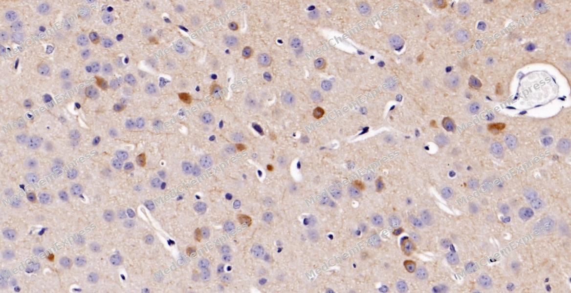 Immunohistochemical analysis of paraffin-embedded mouse brain tissue using Glutamate Receptor 1 (AMPA Subtype) Antibody. The section was pre-treated using heat mediated antigen retrieval with sodium citrate buffer (pH 6.0) for 8 minutes. The tissues were blocked in QuickBlock for 20 minutes at room temperature, washed with ddH2O and PBS, and then probed with the primary antibody at 1/100 dilution in 4℃ overnight. The detection was performed using an HRP conjugated compact polymer system. DAB was used as the chromogen. Tissues were counterstained with hematoxylin and mounted with DPX.
 -
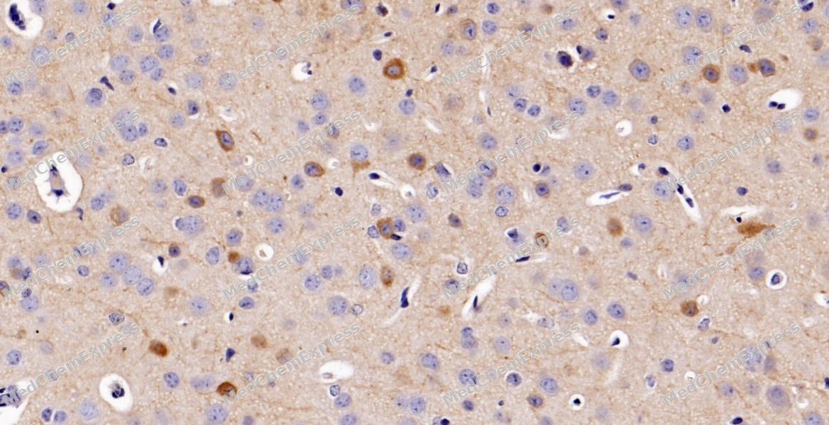 Immunohistochemical analysis of paraffin-embedded mouse brain tissue using Glutamate Receptor 1 (AMPA Subtype) Antibody. The section was pre-treated using heat mediated antigen retrieval with sodium citrate buffer (pH 6.0) for 8 minutes. The tissues were blocked in QuickBlock for 20 minutes at room temperature, washed with ddH2O and PBS, and then probed with the primary antibody at 1/100 dilution in 4℃ overnight. The detection was performed using an HRP conjugated compact polymer system. DAB was used as the chromogen. Tissues were counterstained with hematoxylin and mounted with DPX.


Background
-
Function
Glutamate Receptor 1 is an Ionotropic glutamate receptor that functions as a ligand-gated cation channel, gated by L-glutamate and glutamatergic agonists such as alpha-amino-3-hydroxy-5-methyl-4-isoxazolepropionic acid (AMPA), quisqualic acid, and kainic acid. L-glutamate acts as an excitatory neurotransmitter at many synapses in the central nervous system. Binding of the excitatory neurotransmitter L-glutamate induces a conformation change, leading to the opening of the cation channel, and thereby converts the chemical signal to an electrical impulse upon entry of monovalent and divalent cations such as sodium and calcium. The receptor then desensitizes rapidly and enters in a transient inactive state, characterized by the presence of bound agonist. In the presence of CACNG2 or CACNG4 or CACNG7 or CACNG8, shows resensitization which is characterized by a delayed accumulation of current flux upon continued application of L-glutamate. Resensitization is blocked by CNIH2 tH2O2gh interaction with CACNG8 in the CACNG8-containing AMPA receptors complex. Calcium (Ca(2+)) permeability depends on subunits composition and, heteromeric channels containing edited GRIA2 subunit are calcium-impermeable. Also permeable to other divalents cations such as strontium(2+) and magnesium(2+) and monovalent cations such as potassium(1+) and lithium(1+)[1][2][3][4][5].
-
Subcellular Localization
Cell membrane; Multi-pass membrane protein; Endoplasmic reticulum membrane; Multi-pass membrane protein; Postsynaptic cell membrane; Multi-pass membrane protein; Postsynaptic density membrane; Multi-pass membrane protein; Cell projection, dendrite; Cell projection, dendritic spine; Early endosome membrane; Multi-pass membrane protein; Recycling endosome membrane; Multi-pass membrane protein; Presynapse; Synapse
-
Expression
Tissue_specificity:Widely expressed in the brain -
Subunit
Homotetramer or heterotetramer of pore-forming glutamate receptor subunits; heteromeric assembly can be the result of both receptor subtype and flip or flop form and according the composition, one partner can be dominant with respect to the fast desensitizing current component, whereas the other can determine the steady-state component (By similarity). Tetramers may be formed by the dimerization of dimers (PubMed:23739980). Found in a complex with GRIA2, GRIA3, GRIA4, CNIH2, CNIH3, CACNG2, CACNG3, CACNG4, CACNG5, CACNG7 and CACNG8 (By similarity). Interacts with HIP1 and RASGRF2. Interacts with SYNDIG1 and GRIA2 (By similarity). Interacts with DLG1 (via C-terminus). Interacts with LRFN1. Interacts with PRKG2. Interacts with CNIH2 and CACNG2 (PubMed:20805473). Interacts with CACNG5; this interaction modulates the gating. Interacts (via C-terminus) with PDLIM4 (via LIM domain); this interaction as well as the interaction of PDLIM4 with alpha-actinin is required for their colocalization in early endosomes (By similarity). Interacts with SNX27 (via PDZ domain); the interaction is required for recycling to the plasma membrane when endocytosed and prevent degradation in lysosomes. Interacts (via PDZ-binding motif) with SHANK3 (via PDZ domain) (By similarity). Interacts with CACNG3; associates GRIA1 with the adapter protein complex 4 (AP-4) to target GRIA1 to the somatodendritic compartment of neurons (By similarity). Interacts with CACNG2; this interaction mediates traffick to the plasma membrane and modulation of desensitization. Interacts with CNIH2 and CNIH3; this interaction promotes expression at the plasma membrane and extensively modulates their gating properties by slowing deactivation and desensitization kinetics (By similarity). Found in a complex with GRIA2, GRIA3, GRIA4, DLG4, CACNG8 and CNIH2 (By similarity)
-
SwissProt ID
-
Synonyms
GLUA1, GLUH1, GLUR1, GRIA1, Glutamate receptor 1, GluR-1, AMPA-selective glutamate receptor 1, GluR-A, GluR-K1
-
Research Field
Neuroscience


Documentation
-
Data Sheet (262 KB)
-
SDS (252 KB)
- English - EN (252 KB)
- Français - FR (252 KB)
- Deutsch - DE (252 KB)
- Norwegian - NO (252 KB)
- Español - ES (252 KB)
- Swedish - SV (252 KB)
- Italian - IT (252 KB)
- Korean - KR (252 KB)
- Portuguese - PT (252 KB)
-
User Guide for Antibodies (1077 KB)
References
[1]. Sun W, et al. Molecular cloning, chromosomal mapping, and functional expression of human brain glutamate receptors. Proc Natl Acad Sci U S A. 1992 Feb 15;89(4):1443-7. [Content Brief]
[2]. Shi Y, et al. Functional comparison of the effects of TARPs and cornichons on AMPA receptor trafficking and gating. Proc Natl Acad Sci U S A. 2010 Sep 14;107(37):16315-9. [Content Brief]
[3]. Kato AS, et al. Hippocampal AMPA receptor gating controlled by both TARP and cornichon proteins. Neuron. 2010 Dec 22;68(6):1082-96. [Content Brief]
[4]. Geisheker MR, et al. Hotspots of missense mutation identify neurodevelopmental disorder genes and functional domains. Nat Neurosci. 2017 Aug;20(8):1043-1051. [Content Brief]
[5]. Ismail V, et al. Identification and functional evaluation of GRIA1 missense and truncation variants in individuals with ID: An emerging neurodevelopmental syndrome. Am J Hum Genet. 2022 Jul 7;109(7):1217-1241. [Content Brief]

 Powered by Bioz
