



Hamartin Antibody (YA743)
(Synonyms: KIAA0243, TSC, TSC1, Hamartin, Tuberous sclerosis 1 protein)
Based on 1 Customer Validation
Hamartin Antibody (YA743) is a Mouse-derived and non-conjugated IgG1 monoclonal antibody, targeting to Hamartin.
-
Host:
Mouse
-
Isotype:
IgG
-
Application:
WB, ICC/IF, IHC-P, FC
-
Reactivity :
Human, Mouse
-
Formulation:
Supplied in 1*PBS (pH7.4), 0.05% BSA and 40% Glycerol. Preservative: 0.05% Sodium Azide
-
Conjugation:
Non-conjugated


To place orders, for customer services and technical support, please contact: MedChemExpress USA
Tel: 609-228-6898 E-mail: [email protected] [email protected]





Applications
| Application |
WB

WB: Western Blot
|
ICC/IF
ICC/IF: Immunocytochemistry/
Immunofluorescence |
IHC-P
IHC-P: Immunohistochemistry-Paraffin
|
FC
FC: Flow Cytometry
|
|---|---|---|---|---|
| Dilution Ratio | 1:500 | 1:100 | 1:200-1:600 | 1:500-1:1000 |


Product Details
Hamartin Antibody (YA743) is a Mouse-derived and non-conjugated IgG1 monoclonal antibody, targeting to Hamartin.
-
Host Mouse

-
Clonality Monoclonal

-
Species ReactivityHuman, Mouse
-
Observed Molecular WeightObserved band size: 140 kDa
Note: Due to possible protein modifications or aggregation, the molecular weight should be confirmed by actual measurement, and the predicted value is for reference only.

-
Calculated Molecular Weight Predicted band size: 130 kDa

Entrez Gene: 7248 Human ; 64930 Mouse ; 60445 Rat
SwissProt: Q92574 Human ; Q9EP53 Mouse ; Q9Z136 Rat
OMIM: 191100 Human
Synthetic peptide corresponding to Human Hamartin.AA range:401-600.
Endogenous
Protein G affinity purified.
Non-conjugated
Unmodified
IgG


Product Properties
-
Appearance
Solution
-
Formulation
Supplied in 1*PBS (pH7.4), 0.05% BSA and 40% Glycerol. Preservative: 0.05% Sodium Azide
-
Concentration
Batch-dependent, Please check the COA for the concentration of each lot. Check Lot Concentration
-
Storage & Stability
Stored at -20°C for 1 year. Avoid repeated freeze / thaw cycles.
-
Shipping
Shipping with blue ice.


Verification Images
-
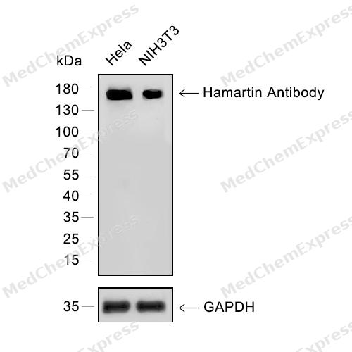 Western blot analysis of extracts from Hela (lane 1) and NIH3T3 (lane 2) using Hamartin antibody. Proteins were transferred to a PVDF membrane and blocked with 5% nonfat powdered milk in PBST for 2 hour at room temperature. The primary antibody (1/1000) and Loading control antibody (GAPDH, 1/3000) was diluted with 5% nonfat powdered milk in PBST at 4°C overnight. Goat Anti-Rabbit IgG-HRP Secondary Antibody (1/8,000) was incubated for 45min at room temperature.
 -
 Flow cytometric analysis of 1X10⁶ HeLa cells labeling Hamartin Antibody (HY-P80498, red). Cells were fixed with 4% paraformaldehyde and permeabilised with 90% methanol. Then stained with the primary antibody at 1/500 dilution for an hour at 4℃. AF488-conjugated Goat Anti-Mouse IgG H&L (HY-P8005) was used as the secondary antibody at 1/1,000 dilution for 30 minutes at 4℃. Mouse IgG Isotype Control (HY-P80757, blue) was used as the isotype control, cells without incubation with primary antibody were used as the unlabeled control (black).
 -
 Immunocytochemistry analysis of NIH/3T3 cells labeling Hamartin with Hamartin Antibody (HY-P80498) at 1/100 dilution. Cells were fixed in 4% paraformaldehyde for 15 minutes at room temperature, permeabilized with 0.1% Triton X-100 in PBS for 15 minutes at room temperature, then blocked with quick block buffer for 10 minutes at room temperature. Cells were then incubated with Hamartin Antibody (HY-P80498) at 1/100 dilution in quick block buffer overnight at 4 ℃. AF488-conjugated Goat Anti-Mouse IgG H&L(HY-P8005, Green) was used as the secondary antibody at 1/1,000 dilution. PBS instead of the primary antibody was used as the secondary antibody only control. The Nuclear counterstain was DAPI (Blue).
 -
 Immunocytochemistry analysis of SH-SY5Y cells labeling Hamartin with Hamartin Antibody (HY-P80498) at 1/100 dilution. Cells were fixed in 4% paraformaldehyde for 15 minutes at room temperature, permeabilized with 0.1% Triton X-100 in PBS for 15 minutes at room temperature, then blocked with quick block buffer for 10 minutes at room temperature. Cells were then incubated with Hamartin Antibody (HY-P80498) at 1/100 dilution in quick block buffer overnight at 4 ℃. AF488-conjugated Goat Anti-Mouse IgG H&L(HY-P8005, Green) was used as the secondary antibody at 1/1,000 dilution. PBS instead of the primary antibody was used as the secondary antibody only control. The Nuclear counterstain was DAPI (Blue).


Background
-
Function
Hamartin is a Non-catalytic component of the TSC-TBC complex, a multiprotein complex that acts as a negative regulator of the canonical mTORC1 complex, an evolutionarily conserved central nutrient sensor that stimulates anabolic reactions and macromolecule biosynthesis to promote cellular biomass generation and growth. The TSC-TBC complex acts as a GTPase-activating protein (GAP) for the small GTPase RHEB, a direct activator of the protein kinase activity of mTORC1. In absence of nutrients, the TSC-TBC complex inhibits mTORC1, thereby preventing phosphorylation of ribosomal protein S6 kinase (RPS6KB1 and RPS6KB2) and EIF4EBP1 (4E-BP1) by the mTORC1 signaling. The TSC-TBC complex is inactivated in response to nutrients, relieving inhibition of mTORC1. Within the TSC-TBC complex, TSC1 stabilizes TSC2 and prevents TSC2 self-aggregation. Involved in microtubule-mediated protein transport via its ability to regulate mTORC1 signaling. Also acts as a co-chaperone for HSP90AA1 facilitating HSP90AA1 chaperoning of protein clients such as kinases, TSC2 and glucocorticoid receptor NR3C1. Increases ATP binding to HSP90AA1 and inhibits HSP90AA1 ATPase activity. Competes with the activating co-chaperone AHSA1 for binding to HSP90AA1, thereby providing a reciprocal regulatory mechanism for chaperoning of client proteins. Recruits TSC2 to HSP90AA1 and stabilizes TSC2 by preventing the interaction between TSC2 and ubiquitin ligase HERC1[1][2][3].
-
Subcellular Localization
Lysosome membrane; Peripheral membrane protein; Cytoplasm, cytosol
-
Subunit
Interacts (via C-terminus) with the closed form of HSP90AA1 (via the middle domain and TPR repeat-binding motif) (PubMed:29127155).
-
SwissProt ID
-
Synonyms
KIAA0243, TSC, TSC1, Hamartin, Tuberous sclerosis 1 protein
-
Research Field
Cell Biology


Documentation
-
Data Sheet (262 KB)
-
SDS (251 KB)
- English - EN (251 KB)
- Français - FR (251 KB)
- Deutsch - DE (251 KB)
- Norwegian - NO (251 KB)
- Español - ES (251 KB)
- Swedish - SV (251 KB)
- Italian - IT (251 KB)
- Korean - KR (251 KB)
- Portuguese - PT (251 KB)
-
User Guide for Antibodies (1077 KB)
References
[1]. Garami A, et al. Insulin activation of Rheb, a mediator of mTOR/S6K/4E-BP signaling, is inhibited by TSC1 and 2. Mol Cell. 2003 Jun;11(6):1457-66. [Content Brief]
[2]. Jiang X, et al. Regulation of microtubule-dependent protein transport by the TSC2/mammalian target of rapamycin pathway. Cancer Res. 2006 May 15;66(10):5258-69. [Content Brief]
[3]. Woodford MR, et al. Tumor suppressor Tsc1 is a new Hsp90 co-chaperone that facilitates folding of kinase and non-kinase clients. EMBO J. 2017 Dec 15;36(24):3650-3665. [Content Brief]

 Powered by Bioz
