



Hsp27 Antibody (YA732)
(Synonyms: HSPB1; HSP27; HSP28; Heat shock protein beta-1; HspB1; 28 kDa heat shock protein; Estrogen-regulated 24 kDa protein; Heat shock 27 kDa protein; HSP 27; Stress-responsive protein 27; SRP27)
Based on 1 Customer Validation
Hsp27 Antibody (YA732) is a Mouse-derived and non-conjugated IgG1 monoclonal antibody, targeting to Hsp27.
-
Host:
Mouse
-
Isotype:
IgG
-
Application:
WB, ICC/IF
-
Reactivity :
Human, Monkey
-
Formulation:
Supplied in 1*PBS (pH 7.3), 50% glycerol and 0.5% BSA. Preservative: 0.02% sodium azide.
-
Conjugation:
Non-conjugated


To place orders, for customer services and technical support, please contact: MedChemExpress USA
Tel: 609-228-6898 E-mail: [email protected] [email protected]





Applications
| Application |
WB

WB: Western Blot
|
ICC/IF
ICC/IF: Immunocytochemistry/
Immunofluorescence |
|---|---|---|
| Dilution Ratio | 1:500-1:1000 | 1:50-1:200 |


Product Details
Hsp27 Antibody (YA732) is a Mouse-derived and non-conjugated IgG1 monoclonal antibody, targeting to Hsp27.
-
Host Mouse

-
Clonality Monoclonal

-
Species ReactivityHuman, Monkey
-
Observed Molecular WeightObserved band size: 27 kDa
Note: Due to possible protein modifications or aggregation, the molecular weight should be confirmed by actual measurement, and the predicted value is for reference only.

-
Calculated Molecular Weight Predicted band size: 23 kDa

Entrez Gene: 3315 Human ; 15507 Mouse ;
SwissProt: P04792 Human ; P14602 Mouse ; P42930 Rat
OMIM: 606595 Human
Synthetic peptide corresponding to Human Hsp27.The exact sequence is proprietary to MCE.
Endogenous
affinity purified
Non-conjugated
Unmodified
IgG


Product Properties
-
Appearance
Solution
-
Formulation
Supplied in 1*PBS (pH 7.3), 50% glycerol and 0.5% BSA. Preservative: 0.02% sodium azide.
-
Concentration
Batch-dependent, Please check the COA for the concentration of each lot. Check Lot Concentration
-
Storage & Stability
Stored at -20°C for 1 year. Avoid repeated freeze / thaw cycles.
-
Shipping
Shipping with blue ice.


Verification Images
-
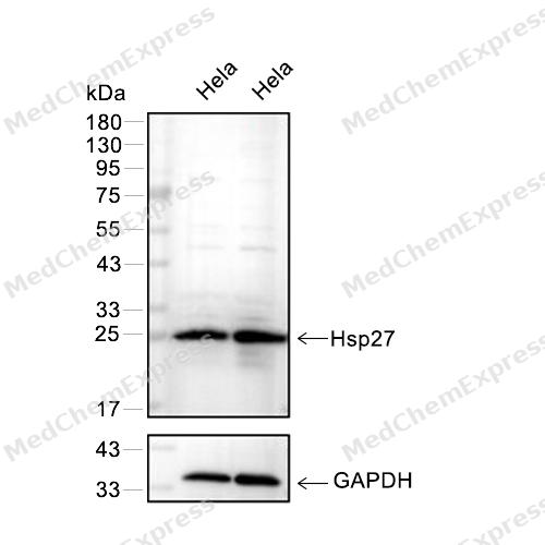 Western blot analysis of extracts from Hela(lane 2(20μg) or lane 3(40μg)), using Hsp27 (HY-P80709) Rabbit mAb. Proteins were transferred to a PVDF membrane and blocked with 5% BSA in TBST for 2 hour at room temperature. The primary antibody (HY-P80709, 1/1000) and Loading control antibody (GAPDH, HY-P80954, 1/3000) was used in 5% BSA in TBST at 4°C overnight. Goat Anti-Rabbit/Mouse IgG-HRP Secondary Antibody (HY-P8001/HY-P8004, 1/10,000) was used for 1 hour at room temperature.
 -
 Western blot analysis was performed on protein extracts (25 μg) from HeLa (lane 2), DU145 (lane 3), A549 (lane 4) and MCF-7 (lane 5) using Hsp27 antibody. Proteins were transferred onto a 0.45 μm PVDF membrane using the Trans-Blot® Turbo™ system for 13 min. The membrane was then blocked with 5% nonfat milk in TBST (HY-K1025) for 1 h at room temperature. The primary antibody (1:1000) and loading control antibody GAPDH Antibody (HRP) (HY-P80954A) (1:5000) were diluted in 5% nonfat milk in TBST and incubated with the membrane overnight at 4°C. After washing, the membrane of primary antibody was incubated with HRP-conjugated goat anti-rabbit/mouse IgG secondary antibody (HY-P8001/HY-P8004) (1:5000) diluted in 5% nonfat milk in TBST for 1 h at room temperature. Protein bands were visualized using an Ultra High Sensitivity ECL detection kit (HY-K1005).
 -
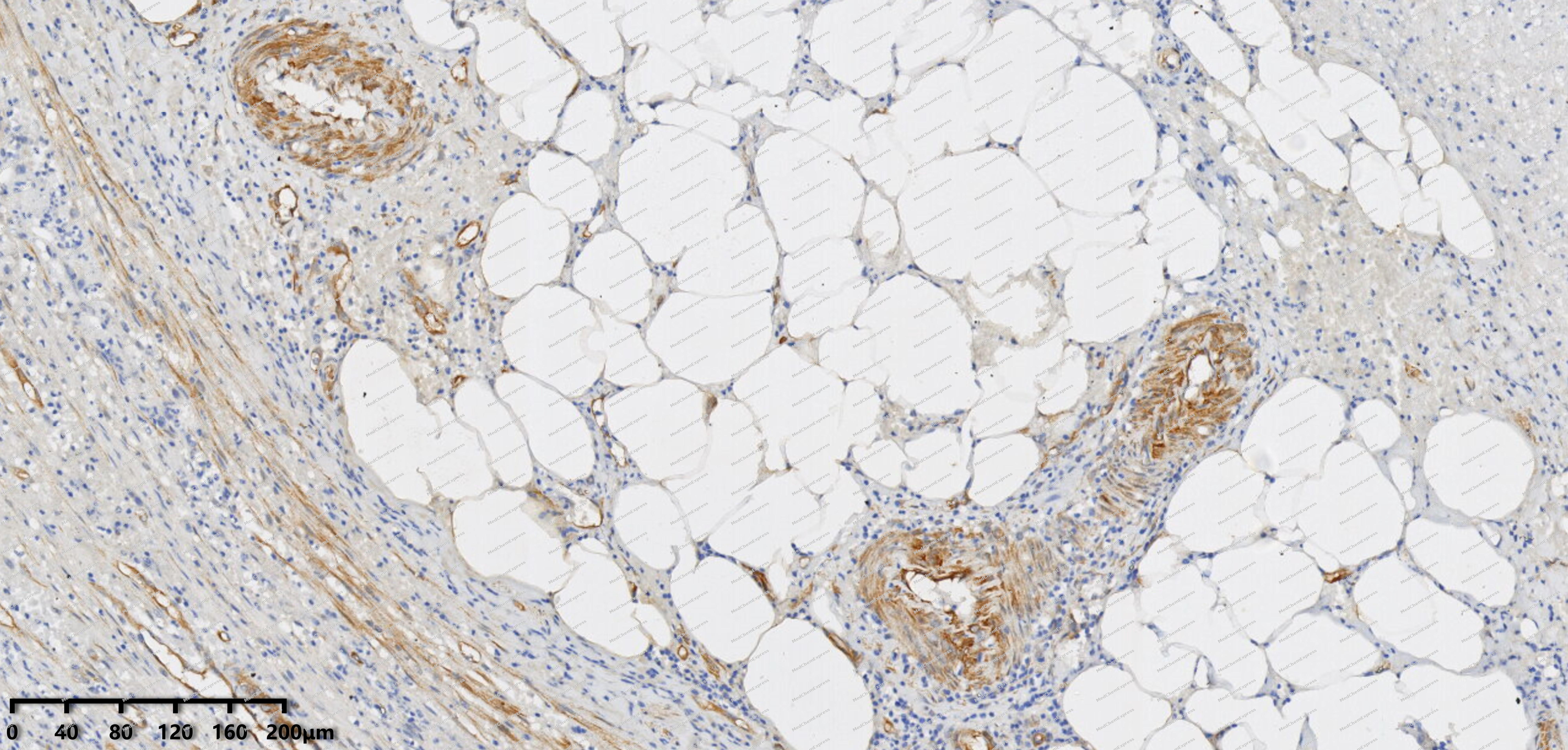 Immunohistochemical analysis of paraffin-embedded human appendix tissue using HSP27 antibody was performed. The section was pretreated using high-temperature mediated EDTA antigen retrieval buffer (pH 9.0), for 20 minutes. The tissues were incubated with primary antibody (HY-P80709, 1:100 dilution) at room temperature for 20 minutes. Detection was performed using an HRP conjugated compact polymer system. DAB was used as the chromogen. The tissues were counterstained with hematoxylin and mounted with neutral balsam mounting medium.
 -
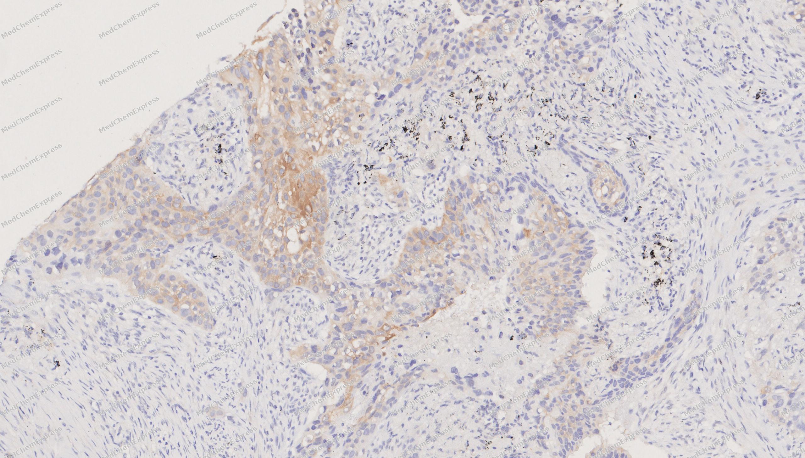 Immunohistochemical analysis of paraffin-embedded human lung cancer tissue using HSP27 antibody was performed. The section was pretreated using high-temperature mediated EDTA antigen retrieval buffer (pH 9.0), for 20 minutes. The tissues were incubated with primary antibody (HY-P80709, 1:100 dilution) at room temperature for 20 minutes. Detection was performed using an HRP conjugated compact polymer system. DAB was used as the chromogen. The tissues were counterstained with hematoxylin and mounted with neutral balsam mounting medium.
 -
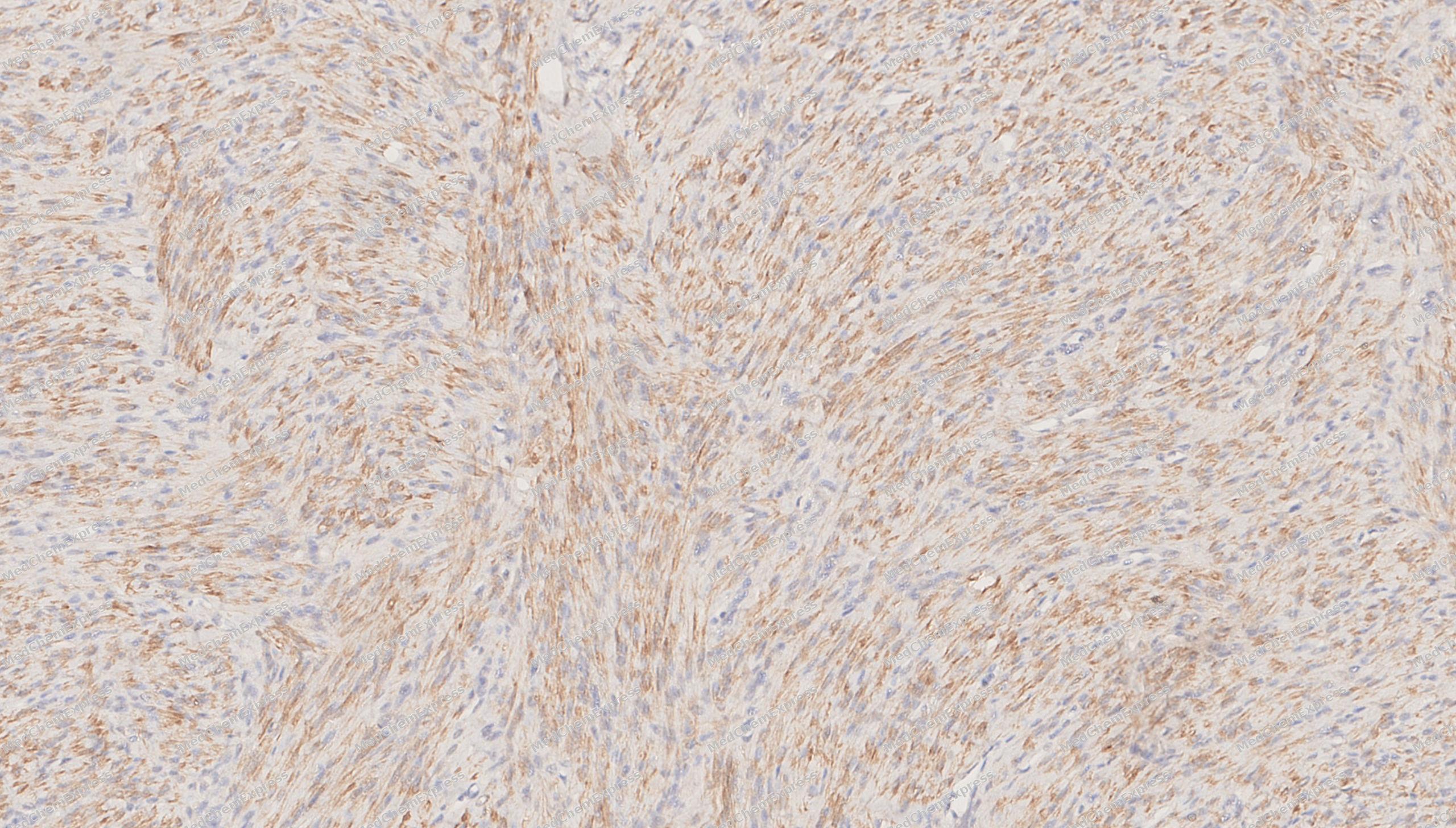 Immunohistochemical analysis of paraffin-embedded human Uterine leiomyoma tissue using HSP27 antibody was performed. The section was pretreated using high-temperature mediated EDTA antigen retrieval buffer (pH 9.0), for 20 minutes. The tissues were incubated with primary antibody (HY-P80709, 1:100 dilution) at room temperature for 20 minutes. Detection was performed using an HRP conjugated compact polymer system. DAB was used as the chromogen. The tissues were counterstained with hematoxylin and mounted with neutral balsam mounting medium.
 -
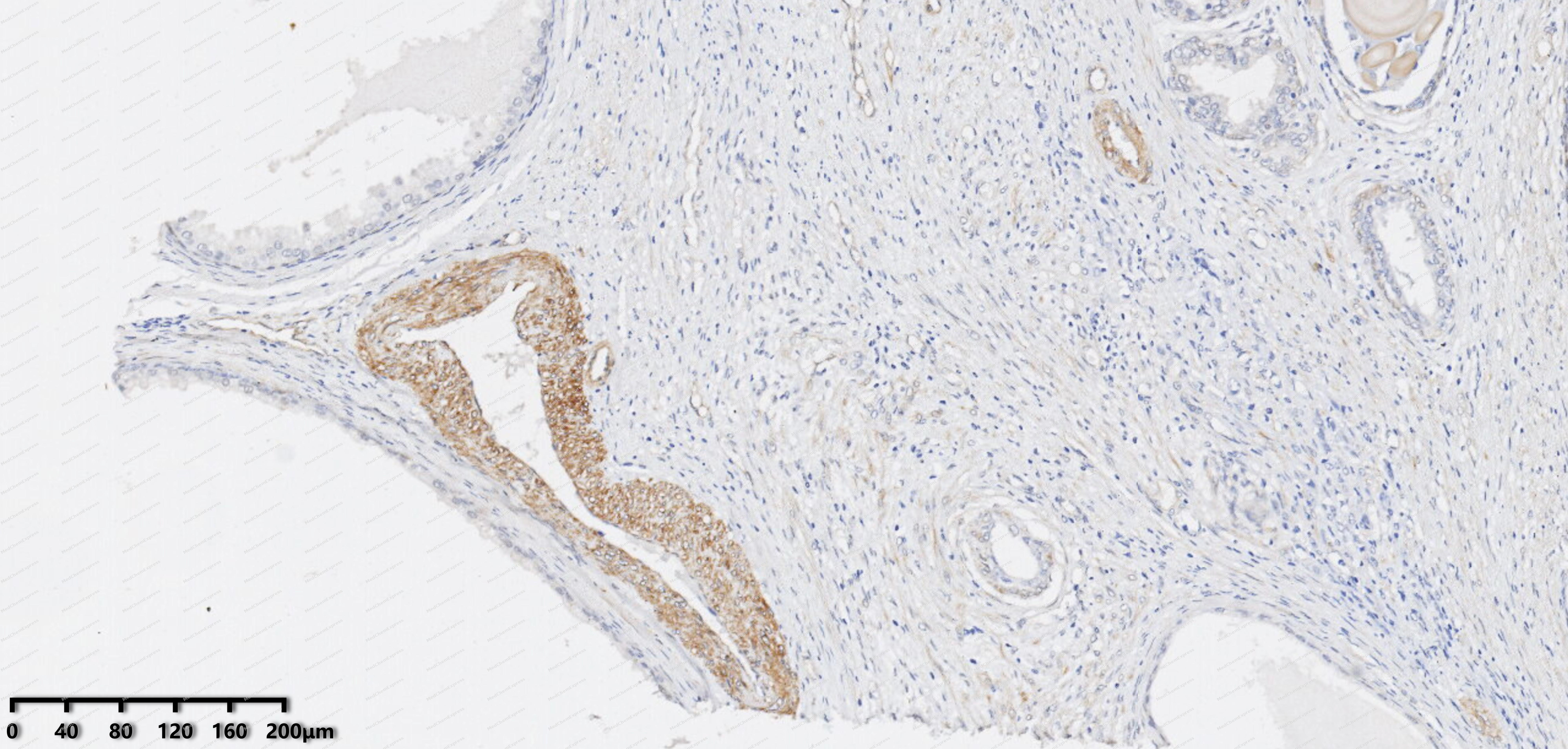 Immunohistochemical analysis of paraffin-embedded human Prostate cancer tissue using HSP27 antibody was performed. The section was pretreated using high-temperature mediated EDTA antigen retrieval buffer (pH 9.0), for 20 minutes. The tissues were incubated with primary antibody (HY-P80709, 1:100 dilution) at room temperature for 20 minutes. Detection was performed using an HRP conjugated compact polymer system. DAB was used as the chromogen. The tissues were counterstained with hematoxylin and mounted with neutral balsam mounting medium.
 -
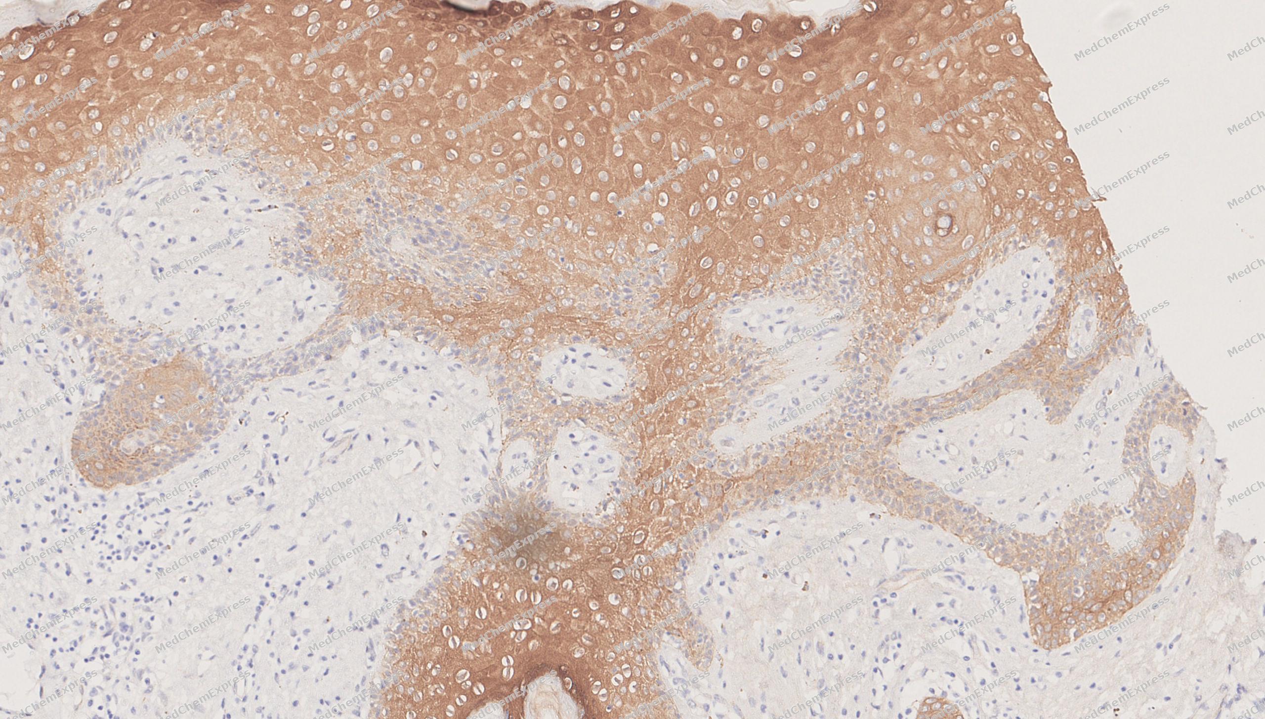 Immunohistochemical analysis of paraffin-embedded human breast cancer tissue using HSP27 antibody was performed. The section was pretreated using high-temperature mediated EDTA antigen retrieval buffer (pH 9.0), for 20 minutes. The tissues were incubated with primary antibody (HY-P80709, 1:100 dilution) at room temperature for 20 minutes. Detection was performed using an HRP conjugated compact polymer system. DAB was used as the chromogen. The tissues were counterstained with hematoxylin and mounted with neutral balsam mounting medium.
 -
 Immunocytochemistry analysis of Hela cells labeling Hsp27 with Hsp27 Antibody (HY-P80709) at 1/100 dilution. Cells were fixed in 4% paraformaldehyde for 15 minutes at room temperature, permeabilized with 0.1% Triton X-100 in PBS for 15 minutes at room temperature, then blocked with quick block buffer for 10 minutes at room temperature. Cells were then incubated with Hsp27 Antibody (HY-P80709) at 1/100 dilution in quick block buffer overnight at 4 ℃.AF488-conjugated Goat Anti-Mouse IgG H&L (HY-P8005, Green) was used as the secondary antibody at 1/1,000 dilution. PBS instead of the primary antibody was used as the secondary antibody only control. The Nuclear counterstain was DAPI (Blue).
 -
 Immunocytochemistry analysis of MCF-7 cells labeling Hsp27 with Hsp27 Antibody (HY-P80709) at 1/100 dilution. Cells were fixed in 4% paraformaldehyde for 15 minutes at room temperature, permeabilized with 0.1% Triton X-100 in PBS for 15 minutes at room temperature, then blocked with quick block buffer for 10 minutes at room temperature. Cells were then incubated with Hsp27 Antibody (HY-P80709) at 1/100 dilution in quick block buffer overnight at 4 ℃.AF488-conjugated Goat Anti-Mouse IgG H&L (HY-P8005, Green) was used as the secondary antibody at 1/1,000 dilution. PBS instead of the primary antibody was used as the secondary antibody only control. The Nuclear counterstain was DAPI (Blue).


Background
-
Function
Hsp27 is a Small heat shock protein which functions as a molecular chaperone probably maintaining denatured proteins in a folding-competent state. Plays a role in stress resistance and actin organization. Through its molecular chaperone activity may regulate numerous biological processes including the phosphorylation and the axonal transport of neurofilament proteins[1][2][3][4].
-
Subcellular Localization
Cytoplasm; Nucleus; Cytoplasm, cytoskeleton, spindle
-
Expression
Tissue_specificity:The substance was detected in all tissues tested: skeletal muscle, heart, aorta, large intestine, small intestine, stomach, esophagus, bladder, adrenal glands, thyroid gland, pancreas, testes, adipose tissue, kidneys, liver, spleen, cerebral cortex, serum, and cerebrospinal fluid. The highest concentrations were found in the heart and tissues composed of striated and smooth muscle.
Induction:Up-regulated in response to environmental stresses such as heat shock (PubMed:8325890) . Up-regulated by estrogen stimulation (PubMed:2743305) ; (Microbial infection) Up-regulated in response to enterovirus 71 (EV71) infection (at protein level) -
Subunit
Homooligomer (PubMed:10383393). Homodimer; becomes monomeric upon activation (PubMed:20178975). Heterooligomer; with HSPB6 (PubMed:23948568). Associates with alpha- and beta-tubulin (PubMed:10777697). Interacts with TGFB1I1 (By similarity). Interacts with CRYAB (PubMed:1560006). Interacts with HSPB8 (PubMed:11342557). Interacts with HSPBAP1 (PubMed:10751411)
-
SwissProt ID
-
Synonyms
HSPB1; HSP27; HSP28; Heat shock protein beta-1; HspB1; 28 kDa heat shock protein; Estrogen-regulated 24 kDa protein; Heat shock 27 kDa protein; HSP 27; Stress-responsive protein 27; SRP27
-
Research Field
Signal Transduction


Documentation
-
Data Sheet (261 KB)
-
SDS (252 KB)
- English - EN (252 KB)
- Français - FR (252 KB)
- Deutsch - DE (252 KB)
- Norwegian - NO (252 KB)
- Español - ES (252 KB)
- Swedish - SV (252 KB)
- Italian - IT (252 KB)
- Korean - KR (252 KB)
- Portuguese - PT (252 KB)
-
User Guide for Antibodies (1077 KB)
References
[1]. Rogalla T, et al. Regulation of Hsp27 oligomerization, chaperone function, and protective activity against oxidative stress/tumor necrosis factor alpha by phosphorylation. J Biol Chem. 1999 Jul 2;274(27):18947-56. [Content Brief]
[2]. Almeida-Souza L, et al. Increased monomerization of mutant HSPB1 leads to protein hyperactivity in Charcot-Marie-Tooth neuropathy. J Biol Chem. 2010 Apr 23;285(17):12778-86. [Content Brief]
[3]. Kostenko S, et al. PKA-induced F-actin rearrangement requires phosphorylation of Hsp27 by the MAPKAP kinase MK5. Cell Signal. 2009 May;21(5):712-8. [Content Brief]
[4]. Holmgren A, et al. Charcot-Marie-Tooth causing HSPB1 mutations increase Cdk5-mediated phosphorylation of neurofilaments. Acta Neuropathol. 2013 Jul;126(1):93-108. [Content Brief]

 Powered by Bioz
