



IL17A Antibody
(Synonyms: Interleukin-17A; CTLA-8; CTLA 8; CTLA8; Cytotoxic T lymphocyte associated antigen 8; Cytotoxic T lymphocyte associated serine esterase 8; IL 17A; IL-17A; IL-17; Interleukin 17; Interleukin-17; Interleukin 17A; Interleukin17; Interleukin17A; IL17_HUMAN; IL17_MOUSE.)Based on 2 publication(s) in Google Scholar
IL17A Antibody is a Rabbit-derived and non-conjugated IgG polyclonal antibody, targeting to IL17A.
-
Host:
Rabbit
-
Isotype:
IgG
-
Application:
WB, ELISA, IHC-P, IHC-F, ICC/IF
-
Reactivity :
Mouse, Rat,
-
Formulation:
Supplied in 0.01M TBS (pH7.4) with 1% BSA, 0.02% Proclin300 and 50% Glycerol.
-
Conjugation:
Non-conjugated


To place orders, for customer services and technical support, please contact: MedChemExpress USA
Tel: 609-228-6898 E-mail: [email protected] [email protected]



Publications Citing Use of MedChemExpress (MCE) IL17A Antibody
More

Applications
| Application |
WB

WB: Western Blot
|
ELISA
ELISA: Enzyme Linked Immunosorbent Assay
|
IHC-P
IHC-P: Immunohistochemistry-Paraffin
|
IHC-F
IHC-F: Immunohistochemistry-Frozen
|
ICC/IF
ICC/IF: Immunocytochemistry/
Immunofluorescence |
|---|---|---|---|---|---|
| Dilution Ratio | 1:500-2000 | 1:5000-10000 | 1:100-500 | 1:100-500 | 1:100-500 |


Product Details
IL17A Antibody is a Rabbit-derived and non-conjugated IgG polyclonal antibody, targeting to IL17A.
-
Host Rabbit

-
Clonality Polyclonal

-
Species ReactivityMouse, Rat,
-
Observed Molecular WeightObserved band size: 22 kDa
Note: Due to possible protein modifications or aggregation, the molecular weight should be confirmed by actual measurement, and the predicted value is for reference only.

-
Calculated Molecular Weight Predicted band size: 15 kDa

Entrez Gene: 16171 Human ; 16171 Mouse
SwissProt: Q62386 Mouse ; Q61453 Rat ;
KLH conjugated synthetic peptide derived from mouse IL-17: 85-150/150
Endogenous
affinity purified
Non-conjugated
Unmodified
IgG


Product Properties
-
Appearance
Solution
-
Formulation
Supplied in 0.01M TBS (pH7.4) with 1% BSA, 0.02% Proclin300 and 50% Glycerol.
-
Concentration
Batch-dependent, Please check the COA for the concentration of each lot. Check Lot Concentration
-
Storage & Stability
Stored at -20°C for 1 year. Avoid repeated freeze / thaw cycles.
-
Shipping
Shipping with blue ice.


Publications (2)
-
Journal Impact Factor
-
Most Recent
-
Sci Rep
Osthol ameliorates obesity-associated lipid metabolic disorders by inhibiting ADRA1D-dependent Th17 cell differentiation. [Abstract]2025 Oct 21;15(1):36691. PMID: 41120583 -
Sci Rep
Sodium propionate protects against bronchopulmonary dysplasia by inhibiting IL-17-mediated apoptosis of alveolar epithelial cells. [Abstract]2025 Apr 5;15(1):11722. PMID: 40188136


Verification Images
-
 Western blot analysis of extracts from Mouse liver tissue (lane2, 40μg), Rat pancreas tissue (lane3, 40μg) using IL17A Antibody (HY-P81114). Proteins were transferred to a PVDF membrane and blocked with 5% non-fat milk in TBST for 2 hour at room temperature. The primary antibody (1/500) and Loading control antibody (Beta Actin, HY-P80993, 1/10,000) was used in 5% non-fat milk in TBST at 4°C overnight. Goat Anti-Rabbit IgG-HRP Secondary Antibody (HY-P8001,1/10,000) was used for 1 hour at room temperature.
 -
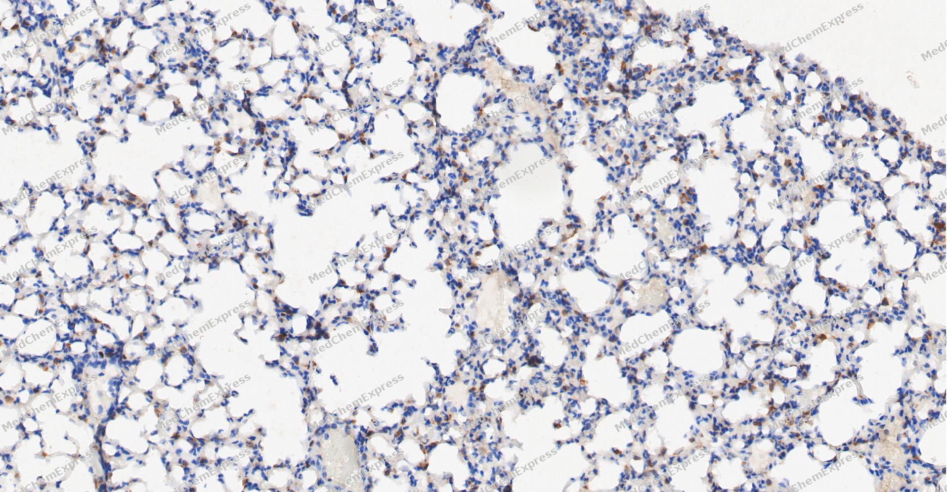 Immunohistochemical analysis of paraffin-embedded Mouse Lung tissue using IL17A antibody was performed. The section was pretreated using high-temperature and high-pressure mediated EDTA antigen retrieval buffer (pH 9.0), for 5 minutes. The tissues were incubated with primary antibody (HY-P81114, 1:500 dilution) at room temperature for 60 minutes. Detection was performed using an HRP conjugated compact polymer system. DAB was used as the chromogen. The tissues were counterstained with hematoxylin and mounted with neutral balsam mounting medium.
 -
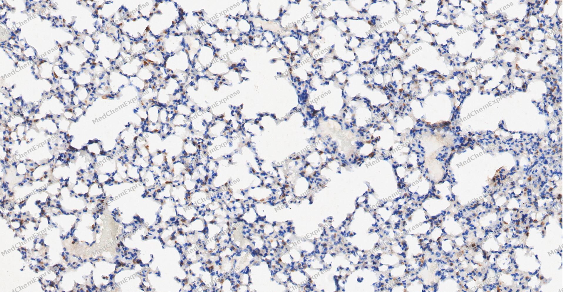 Immunohistochemical analysis of paraffin-embedded Mouse Lung tissue using IL17A antibody was performed. The section was pretreated using high-temperature and high-pressure mediated EDTA antigen retrieval buffer (pH 9.0), for 5 minutes. The tissues were incubated with primary antibody (HY-P81114, 1:500 dilution) at room temperature for 60 minutes. Detection was performed using an HRP conjugated compact polymer system. DAB was used as the chromogen. The tissues were counterstained with hematoxylin and mounted with neutral balsam mounting medium.
 -
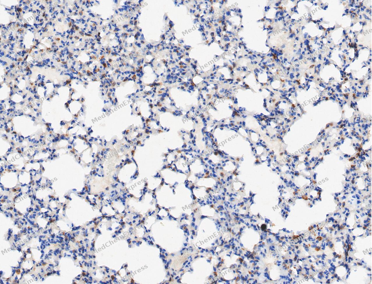 Immunohistochemical analysis of paraffin-embedded Mouse Lung tissue using IL17A antibody was performed. The section was pretreated using high-temperature and high-pressure mediated EDTA antigen retrieval buffer (pH 9.0), for 5 minutes. The tissues were incubated with primary antibody (HY-P81114, 1:500 dilution) at room temperature for 60 minutes. Detection was performed using an HRP conjugated compact polymer system. DAB was used as the chromogen. The tissues were counterstained with hematoxylin and mounted with neutral balsam mounting medium.
 -
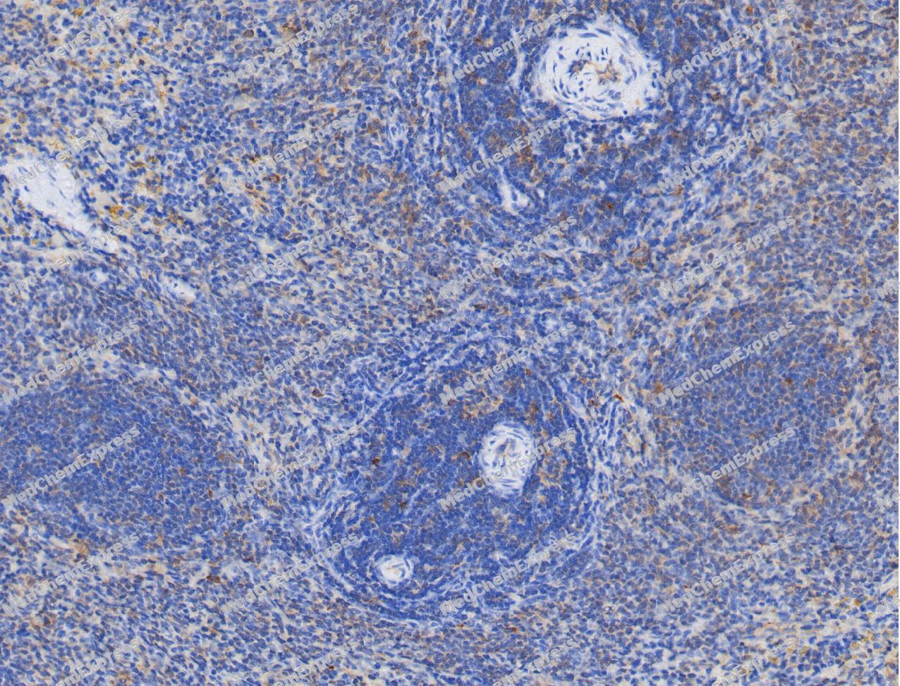 Immunohistochemical analysis of paraffin-embedded Rat spleen tissue using IL17A antibody was performed. The section was pretreated using high-temperature and high-pressure mediated EDTA antigen retrieval buffer (pH 9.0), for 5 minutes. The tissues were incubated with primary antibody (HY-P81114, 1:500 dilution) at room temperature for 60 minutes. Detection was performed using an HRP conjugated compact polymer system. DAB was used as the chromogen. The tissues were counterstained with hematoxylin and mounted with neutral balsam mounting medium.
 -
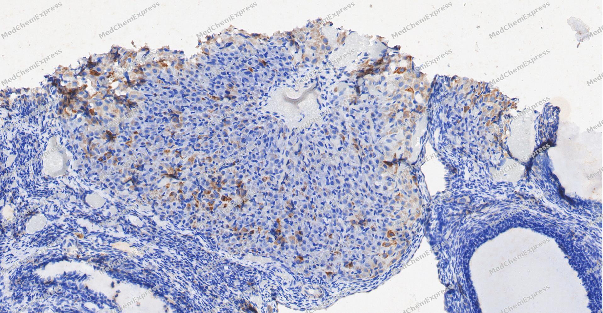 Immunohistochemical analysis of paraffin-embedded Rat Ovary tissue using IL17A antibody was performed. The section was pretreated using high-temperature and high-pressure mediated EDTA antigen retrieval buffer (pH 9.0), for 5 minutes. The tissues were incubated with primary antibody (HY-P81114, 1:500 dilution) at room temperature for 60 minutes. Detection was performed using an HRP conjugated compact polymer system. DAB was used as the chromogen. The tissues were counterstained with hematoxylin and mounted with neutral balsam mounting medium.
 -
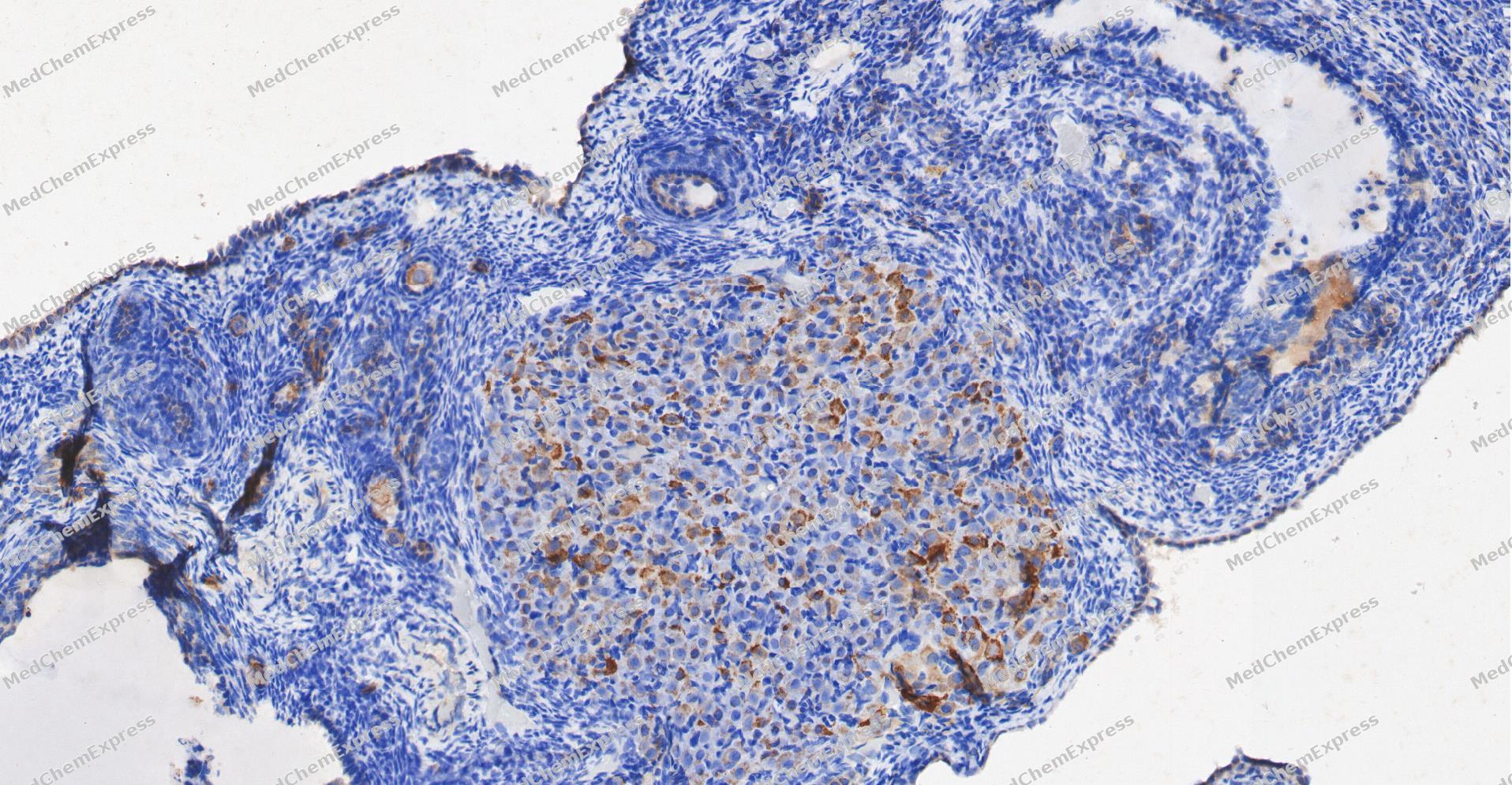 Immunohistochemical analysis of paraffin-embedded Rat Ovary tissue using IL17A antibody was performed. The section was pretreated using high-temperature and high-pressure mediated EDTA antigen retrieval buffer (pH 9.0), for 5 minutes. The tissues were incubated with primary antibody (HY-P81114, 1:500 dilution) at room temperature for 60 minutes. Detection was performed using an HRP conjugated compact polymer system. DAB was used as the chromogen. The tissues were counterstained with hematoxylin and mounted with neutral balsam mounting medium.
 -
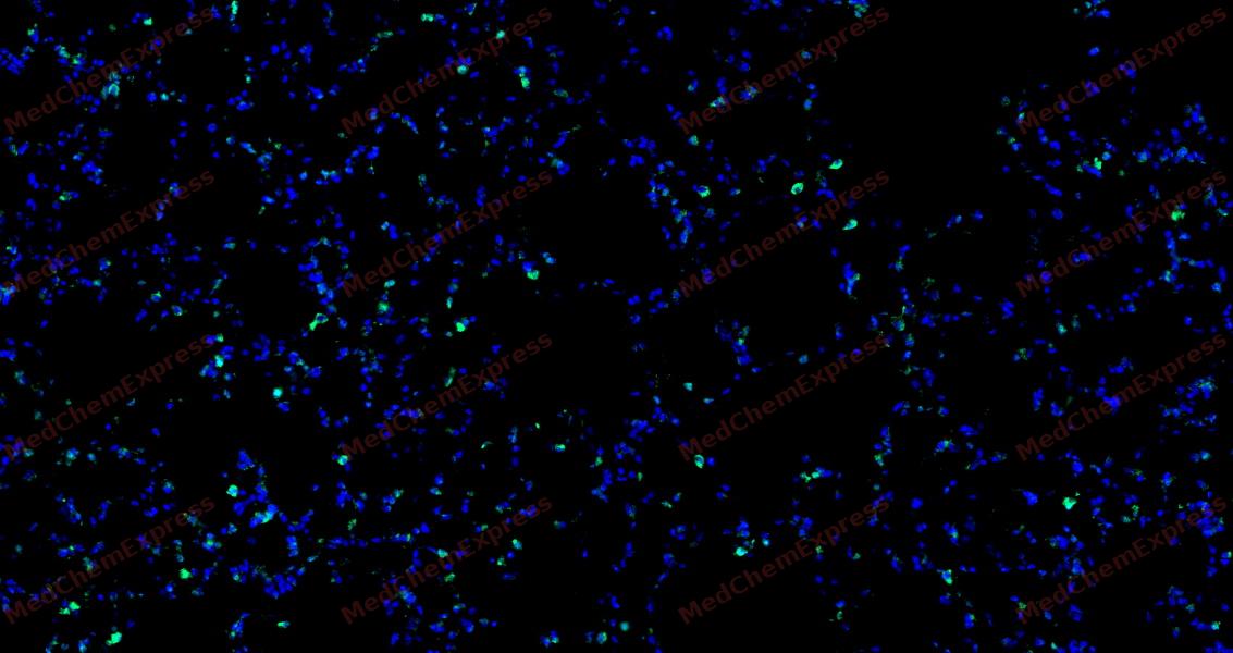 Tyramide signaling amplification based immunofluorescence was performed on paraffin-embedded Rat lung tissue using IL17A antibody. Antigen retrieval was performed in EDTA buffer pH 9.0 (95 °C, 20 min) followed by cooling to RT. Then incubated with primary antibody (HY-P81114, 1:500 dilution) at room temperature for 60 minutes and HRP conjugated secondary antibody for 10 minutes. Fluorescence was then developed with Vari Fluor 532 TSA (200×)(HY-D1832). The tissues were counterstained with DAPI and mounted with Anti-fade mounting medium.
 -
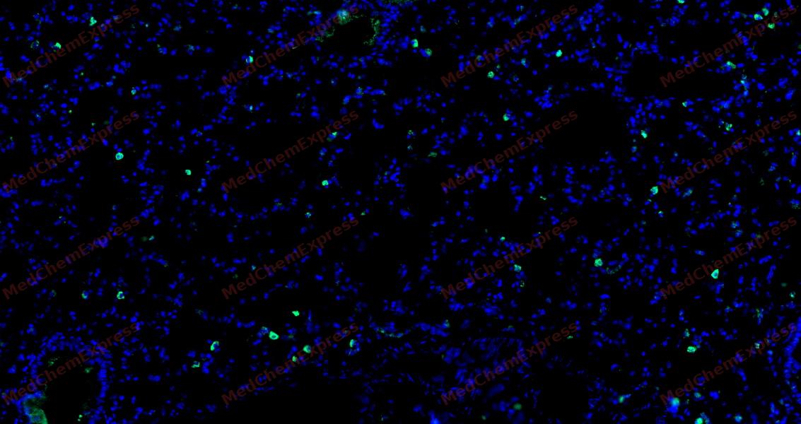 Tyramide signaling amplification based immunofluorescence was performed on paraffin-embedded Rat lung tissue using IL17A antibody. Antigen retrieval was performed in EDTA buffer pH 9.0 (95 °C, 20 min) followed by cooling to RT. Then incubated with primary antibody (HY-P81114, 1:500 dilution) at room temperature for 60 minutes and HRP conjugated secondary antibody for 10 minutes. Fluorescence was then developed with Vari Fluor 532 TSA (200×)(HY-D1832). The tissues were counterstained with DAPI and mounted with Anti-fade mounting medium.
 -
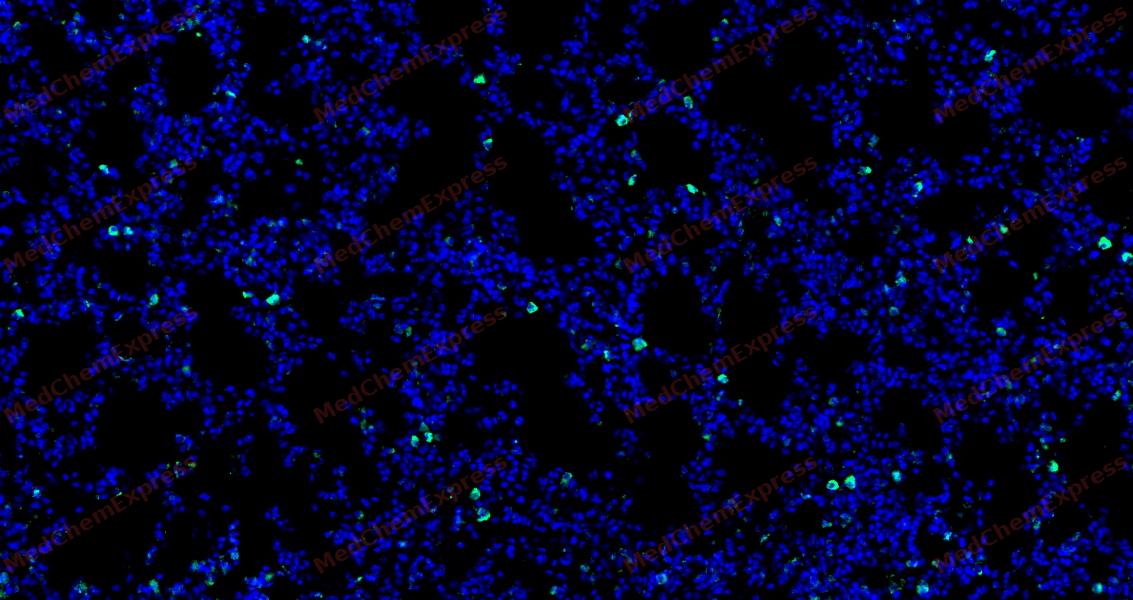 Tyramide signaling amplification based immunofluorescence was performed on paraffin-embedded Rat lung tissue using IL17A antibody. Antigen retrieval was performed in EDTA buffer pH 9.0 (95 °C, 20 min) followed by cooling to RT. Then incubated with primary antibody (HY-P81114, 1:500 dilution) at room temperature for 60 minutes and HRP conjugated secondary antibody for 10 minutes. Fluorescence was then developed with Vari Fluor 532 TSA (200×)(HY-D1832). The tissues were counterstained with DAPI and mounted with Anti-fade mounting medium.
 -
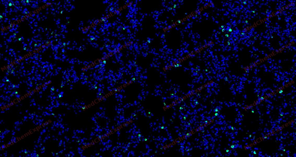 Tyramide signaling amplification based immunofluorescence was performed on paraffin-embedded Rat lung tissue using IL17A antibody. Antigen retrieval was performed in EDTA buffer pH 9.0 (95 °C, 20 min) followed by cooling to RT. Then incubated with primary antibody (HY-P81114, 1:500 dilution) at room temperature for 60 minutes and HRP conjugated secondary antibody for 10 minutes. Fluorescence was then developed with Vari Fluor 532 TSA (200×)(HY-D1832). The tissues were counterstained with DAPI and mounted with Anti-fade mounting medium.
 -
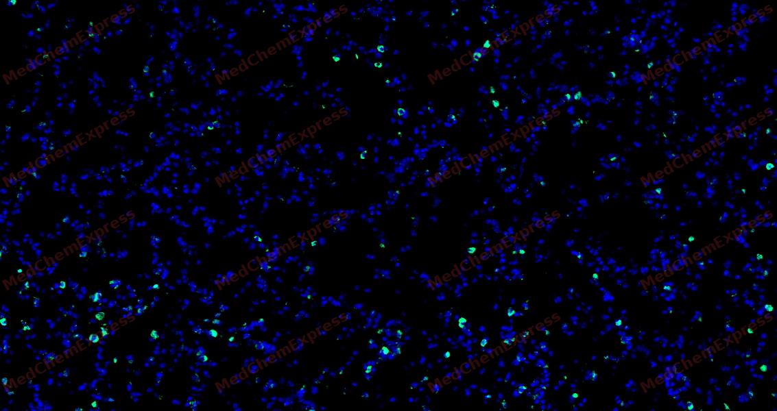 Tyramide signaling amplification based immunofluorescence was performed on paraffin-embedded Rat lung tissue using IL17A antibody. Antigen retrieval was performed in EDTA buffer pH 9.0 (95 °C, 20 min) followed by cooling to RT. Then incubated with primary antibody (HY-P81114, 1:500 dilution) at room temperature for 60 minutes and HRP conjugated secondary antibody for 10 minutes. Fluorescence was then developed with Vari Fluor 532 TSA (200×)(HY-D1832). The tissues were counterstained with DAPI and mounted with Anti-fade mounting medium.
 -
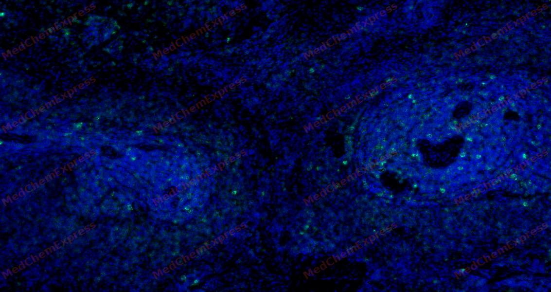 Tyramide signaling amplification based immunofluorescence was performed on paraffin-embedded Rat spleen tissue using IL17A antibody. Antigen retrieval was performed in EDTA buffer pH 9.0 (95 °C, 20 min) followed by cooling to RT. Then incubated with primary antibody (HY-P81114, 1:500 dilution) at room temperature for 60 minutes and HRP conjugated secondary antibody for 10 minutes. Fluorescence was then developed with Vari Fluor 532 TSA (200×)(HY-D1832). The tissues were counterstained with DAPI and mounted with Anti-fade mounting medium.


Background
-
Function
Interleukin-17A (IL-17A), also known as CTLA-8, belongs to the IL-17 cytokine family. IL-17A is expressed in memory Th17 cells and is a product of memory CD4+ T cells[1]. In addition to Th17 cells, IL-17A is also produced by a wide variety of immune cells, including CD8+ T cells, γδT cells, natural killer T (NKT) cells, monocytes, and neutrophils[2][3]. IL-17A plays a critical role in host defense mechanisms against many bacterial and fungal pathogens as well as allergic and autoimmune responses[1][2]. IL-17A induces the production of antimicrobial peptides (defensins and S100 proteins), cytokines (IL-6, G-CSF, and GM-CSF), chemokines (CXCL1, CXCL5, IL-8, CCL2, and CCL7), and matrix metalloproteinases (MMP1, MMP3, and MMP13) [2]. IL-17A is detrimental in viral infection through promoting neutrophilic inflammation[1]. IL-17A is a homodimeric cytokine and shares similar biological activities with IL-17F. IL-17A binds to IL-17RA with high affinity, and IL-17RA is required for the biological activity of IL-17A. IL-17A cannot bind to T cells, B cells, and myeloid cells that are deficient in IL-17RA. IL-17A has been implicated in a variety of autoimmune diseases, such as rheumatoid arthritis, multiple sclerosis, inflammatory bowel disease, asthma, and psoriasis[4]. IL-17A also plays a pathogenic role in cancer. In tumorigenesis, IL-17A recruits myeloid derived suppressor cells (MDSCs) to dampen anti-tumor immunity. IL-17A also enhances tumor growth in vivo through the induction of IL-6[1]. Murine IL-17A is a 21 kDa glycoprotein with 147 amino acids and 63% amino acid homology with human IL-17A (155 amino acids) [2].
-
Subcellular Localization
Secreted
-
Expression
Tissue_specificity:Expressed by Th17 cell lineage (at protein level) . The expression pattern reflects the differentiation state, with IL17A-IL17F heterodimers produced at higher levels than IL17A-IL17A and IL17F-IL17F dimers in fully differentiated Th17 cells (PubMed:16990136, PubMed:18025225) . Expressed in innate lymphoid cells (at protein level) (PubMed:23255360, PubMed:28709803) . Expressed in gamma-delta T cell subsets (at protein level) (PubMed:17372004, PubMed:20364087, PubMed:26431948, PubMed:28709803) . Expressed in iNKT cells (at protein level) (PubMed:17470641)
Induction:Induced upon differentiation of CD4-positive T cells toward Th17 effector cells upon antigen receptor binding in the presence of IL6 and TGFB1 (PubMed:16200068, PubMed:16990136, PubMed:18025225) . Up-regulated by IL23A-IL12B, IL1B and TNF and inhibited by IFNG and IL4 (PubMed:16200068, PubMed:18025225) . Up-regulated by pro-inflammatory cytokines in response to microbes in various immune cells: induced in innate lymphoid cells upon fungal infection, in Vdelta4-positive gamma-delta T cells upon C.mastitidis infection, in Vdelta5-positive gamma-delta T cells upon S.aureus infection and in Vdelta1-positive gamma-delta T cells upon E.coli infection (PubMed:17372004, PubMed:20364087, PubMed:23255360, PubMed:28709803) . Induced in gamma-delta T cells in intestinal lamina propria upon acute injury (PubMed:26431948) . Induced in KLRB1/NK1.1-negative iNKT cell subset upon CD1D stimulation (PubMed:17470641) -
Subunit
Homodimer (PubMed:18025225).
-
SwissProt ID
-
Synonyms
Interleukin-17A; CTLA-8; CTLA 8; CTLA8; Cytotoxic T lymphocyte associated antigen 8; Cytotoxic T lymphocyte associated serine esterase 8; IL 17A; IL-17A; IL-17; Interleukin 17; Interleukin-17; Interleukin 17A; Interleukin17; Interleukin17A; IL17_HUMAN; IL17_MOUSE.
Documentation
-
Data Sheet (262 KB)
-
SDS (252 KB)
- English - EN (252 KB)
- Français - FR (252 KB)
- Deutsch - DE (252 KB)
- Norwegian - NO (252 KB)
- Español - ES (252 KB)
- Swedish - SV (252 KB)
- Italian - IT (252 KB)
- Korean - KR (252 KB)
- Portuguese - PT (252 KB)
-
User Guide for Antibodies (1077 KB)
References
[1]. Chen K, et al. Interluekin-17A (IL17A). Gene. 2017 May 30;614:8-14. [Content Brief]
[2]. Iwakura Y, et al. The roles of IL-17A in inflammatory immune responses and host defense against pathogens. Immunol Rev. 2008 Dec;226:57-79. [Content Brief]
[3]. Cua DJ, et al. Innate IL-17-producing cells: the sentinels of the immune system. Nat Rev Immunol. 2010 Jul;10(7):479-89. [Content Brief]
[4]. Wright JF, et al. The human IL-17F/IL-17A heterodimeric cytokine signals through the IL-17RA/IL-17RC receptor complex. J Immunol. 2008 Aug 15;181(4):2799-805. [Content Brief]

 Powered by Bioz
