



IRF5 Antibody (YA1884)
(Synonyms: Interferon regulatory factor 5; Interferon regulatory factor 5 bone marrow variant; IRF 5; SLEB10)
Based on 1 Customer Validation
IRF5 Antibody (YA1884) is a Rabbit-derived and non-conjugated IgG monoclonal antibody, targeting to IRF5.
-
Host:
Rabbit
-
Isotype:
IgG
-
Application:
WB, IHC-P, ICC/IF, IP, FC
-
Reactivity :
Human, Mouse, Rat
-
Formulation:
Supplied in 50mM Tris-Glycine(pH 7.4), 0.15M NaCl, 40%Glycerol, 0.01% sodium azide and 0.05% BSA.
-
Conjugation:
Non-conjugated


To place orders, for customer services and technical support, please contact: MedChemExpress USA
Tel: 609-228-6898 E-mail: [email protected] [email protected]





Applications
| Application |
WB

WB: Western Blot
|
IHC-P
IHC-P: Immunohistochemistry-Paraffin
|
ICC/IF
ICC/IF: Immunocytochemistry/
Immunofluorescence |
IP
IP: Immunoprecipitation
|
FC
FC: Flow Cytometry
|
|---|---|---|---|---|---|
| Dilution Ratio | 1:500-1:1000 | 1:50-1:100 | 1:50-1:200 | 1:100 | 1:50-1:100 |


Product Details
IRF5 Antibody (YA1884) is a Rabbit-derived and non-conjugated IgG monoclonal antibody, targeting to IRF5.
-
Host Rabbit

-
Clonality Recombinant,Monoclonal

-
Species ReactivityHuman, Mouse, Rat
-
Observed Molecular WeightObserved band size: 56 kDa
Note: Due to possible protein modifications or aggregation, the molecular weight should be confirmed by actual measurement, and the predicted value is for reference only.

-
Calculated Molecular Weight Predicted band size: 56 kDa

Entrez Gene: 3663 Human ; 27056 Mouse ; 296953 Rat
SwissProt: Q13568 Human ; P56477 Mouse ;
OMIM: 612245 Human
A synthesized peptide derived from human IRF5
Endogenous
Affinity Chromatography
Non-conjugated
Unmodified
IgG


Product Properties
-
Appearance
Liquid
-
Formulation
Supplied in 50mM Tris-Glycine(pH 7.4), 0.15M NaCl, 40%Glycerol, 0.01% sodium azide and 0.05% BSA.
-
Concentration
Batch-dependent, Please check the COA for the concentration of each lot. Check Lot Concentration
-
Storage & Stability
Stored at -20°C for 1 year. Avoid repeated freeze / thaw cycles.
-
Shipping
Shipping with blue ice.


Verification Images
-
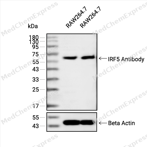 Western blot analysis of extracts from Raw264.7(lane 2(20ug) and Raw264.7(lane 3(40ug) using IRF5 Antibody (HY-P82139) Rabbit mAb. Proteins were transferred to a PVDF membrane and blocked with 5% non-fat milk in TBST for 2 hour at room temperature. The primary antibody (1/1000) and Loading control antibody (Beta Actin, HY-P80438, 1/10000) was used in 5% non-fat milk in TBST at 4°C overnight. Goat Anti-Mouse/Rabbit IgG-HRP Secondary Antibody (1/10000) was used for 1 hour at room temperature.
 -
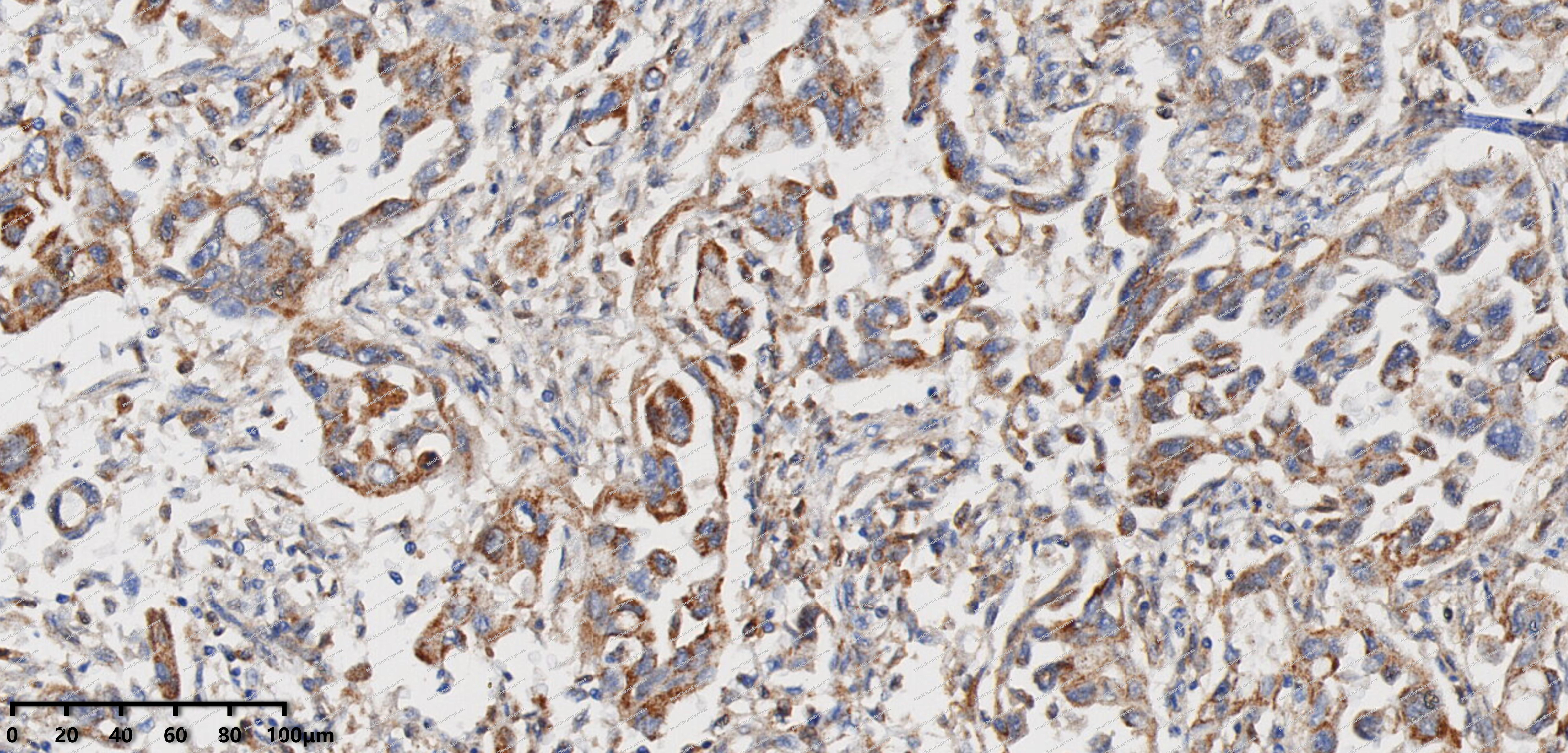 Immunohistochemical analysis of paraffin-embedded human Pancreatic cancer tissue using IRF5 antibody was performed. The section was pretreated using high-temperature mediated EDTA antigen retrieval buffer (pH 9.0), for 20 minutes. The tissues were incubated with primary antibody (HY-P82139, 1:100 dilution) at room temperature for 20 minutes. Detection was performed using an HRP conjugated compact polymer system. DAB was used as the chromogen. The tissues were counterstained with hematoxylin and mounted with neutral balsam mounting medium.
 -
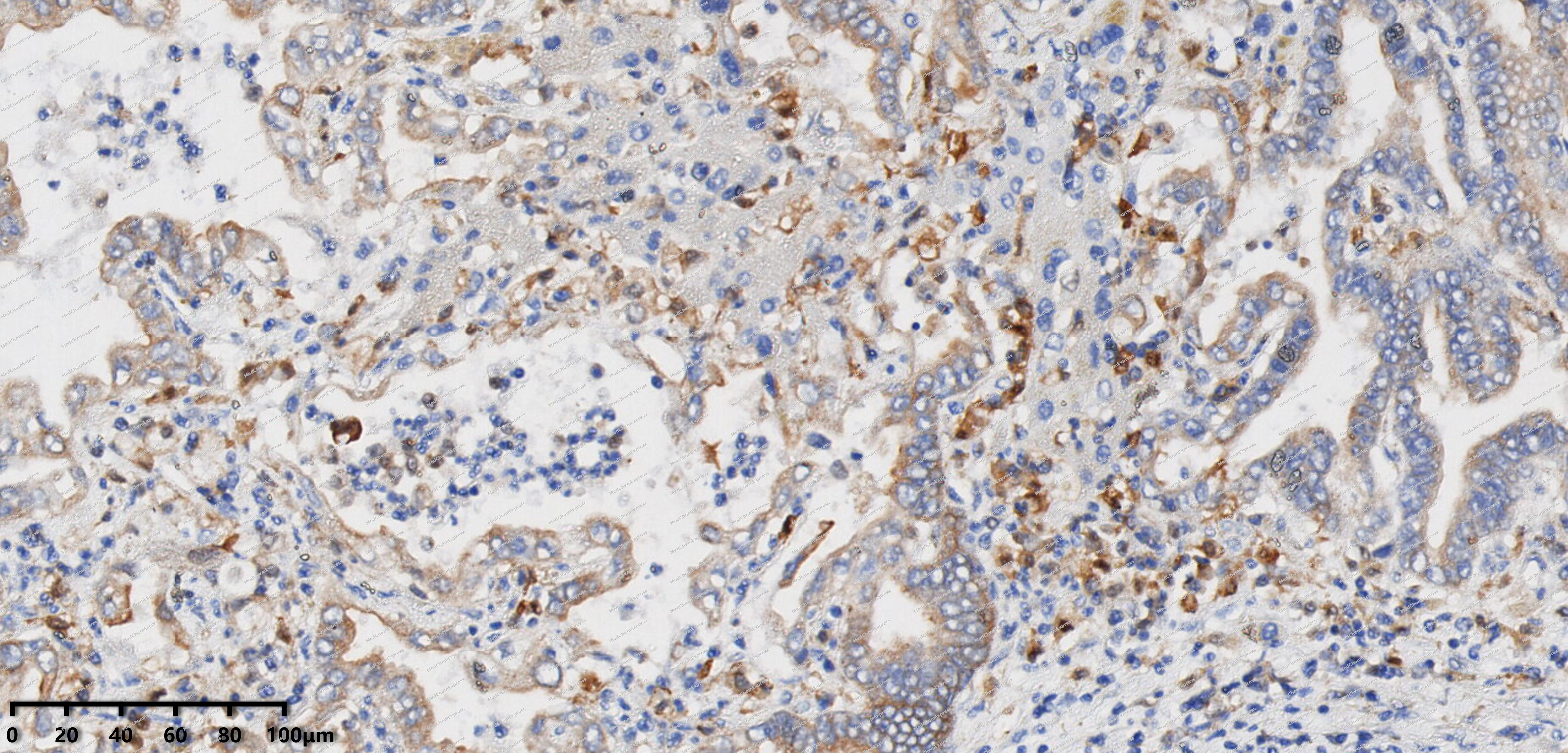 Immunohistochemical analysis of paraffin-embedded human Intrahepatic cholangiocarcinoma tissue using IRF5 antibody was performed. The section was pretreated using high-temperature mediated EDTA antigen retrieval buffer (pH 9.0), for 20 minutes. The tissues were incubated with primary antibody (HY-P82139, 1:100 dilution) at room temperature for 20 minutes. Detection was performed using an HRP conjugated compact polymer system. DAB was used as the chromogen. The tissues were counterstained with hematoxylin and mounted with neutral balsam mounting medium.
 -
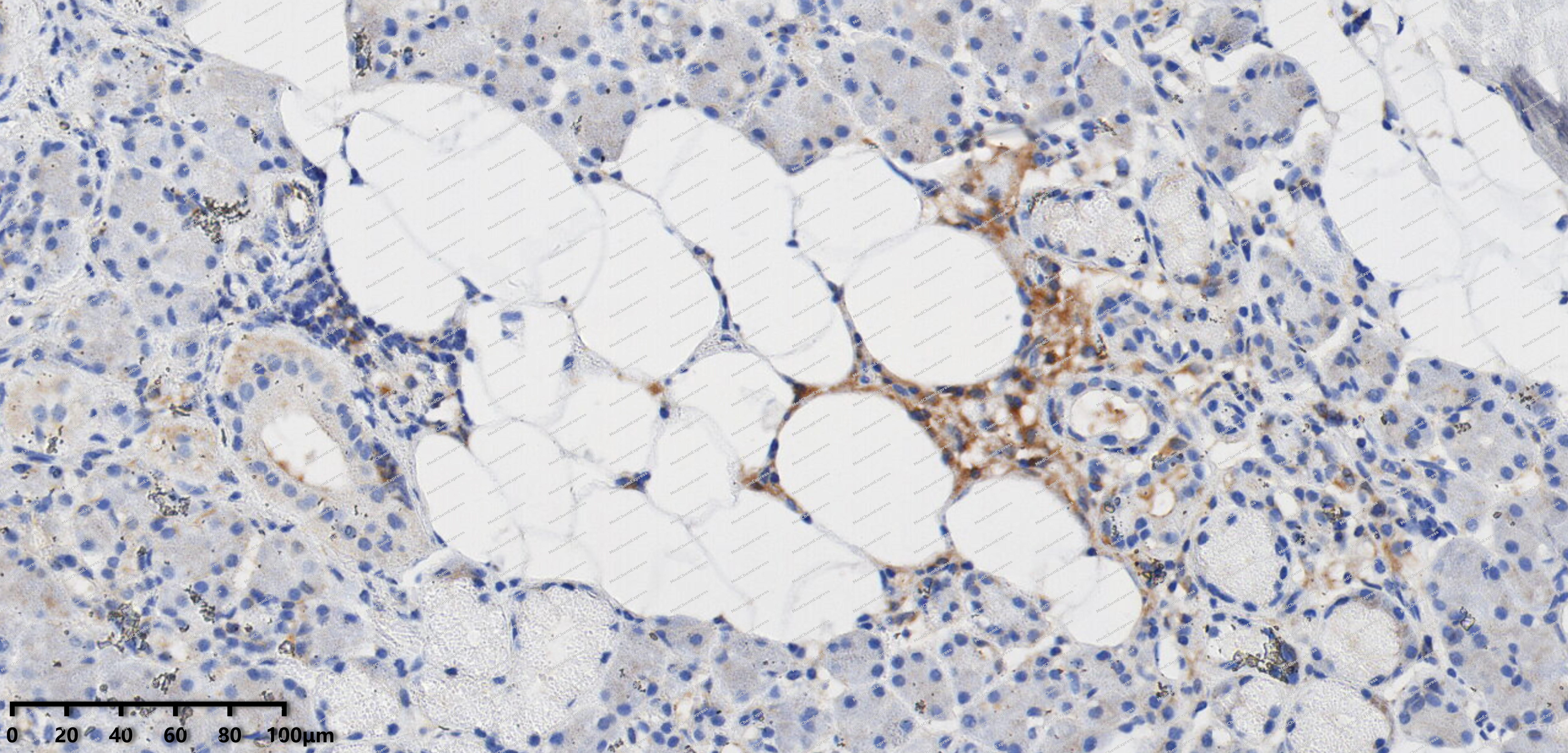 Immunohistochemical analysis of paraffin-embedded human pleomorphic adenoma tissue using IRF5 antibody was performed. The section was pretreated using high-temperature mediated EDTA antigen retrieval buffer (pH 9.0), for 20 minutes. The tissues were incubated with primary antibody (HY-P82139, 1:100 dilution) at room temperature for 20 minutes. Detection was performed using an HRP conjugated compact polymer system. DAB was used as the chromogen. The tissues were counterstained with hematoxylin and mounted with neutral balsam mounting medium.
 -
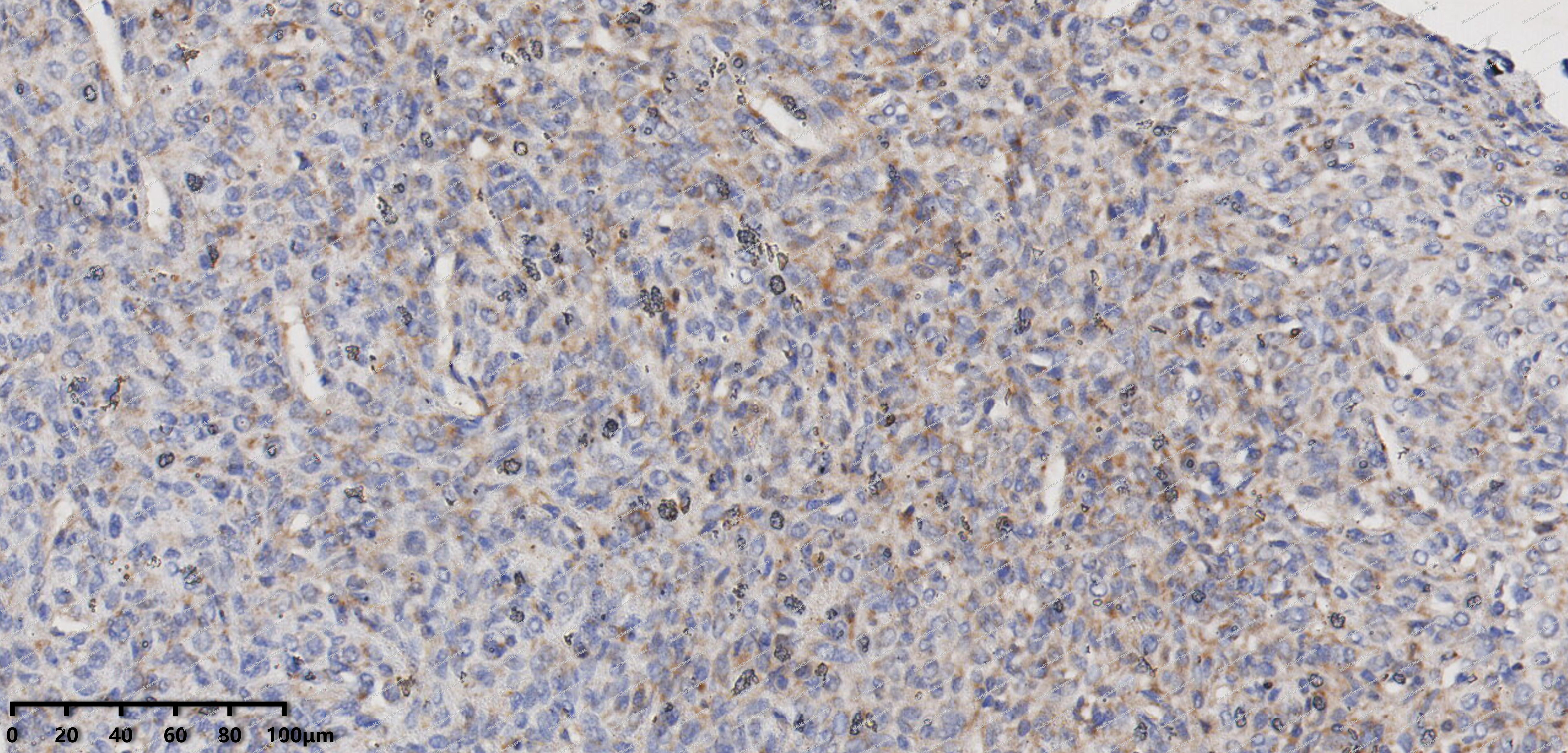 Immunohistochemical analysis of paraffin-embedded human solitary fibrous tumor tissue using IRF5 antibody was performed. The section was pretreated using high-temperature mediated EDTA antigen retrieval buffer (pH 9.0), for 20 minutes. The tissues were incubated with primary antibody (HY-P82139, 1:100 dilution) at room temperature for 20 minutes. Detection was performed using an HRP conjugated compact polymer system. DAB was used as the chromogen. The tissues were counterstained with hematoxylin and mounted with neutral balsam mounting medium.
 -
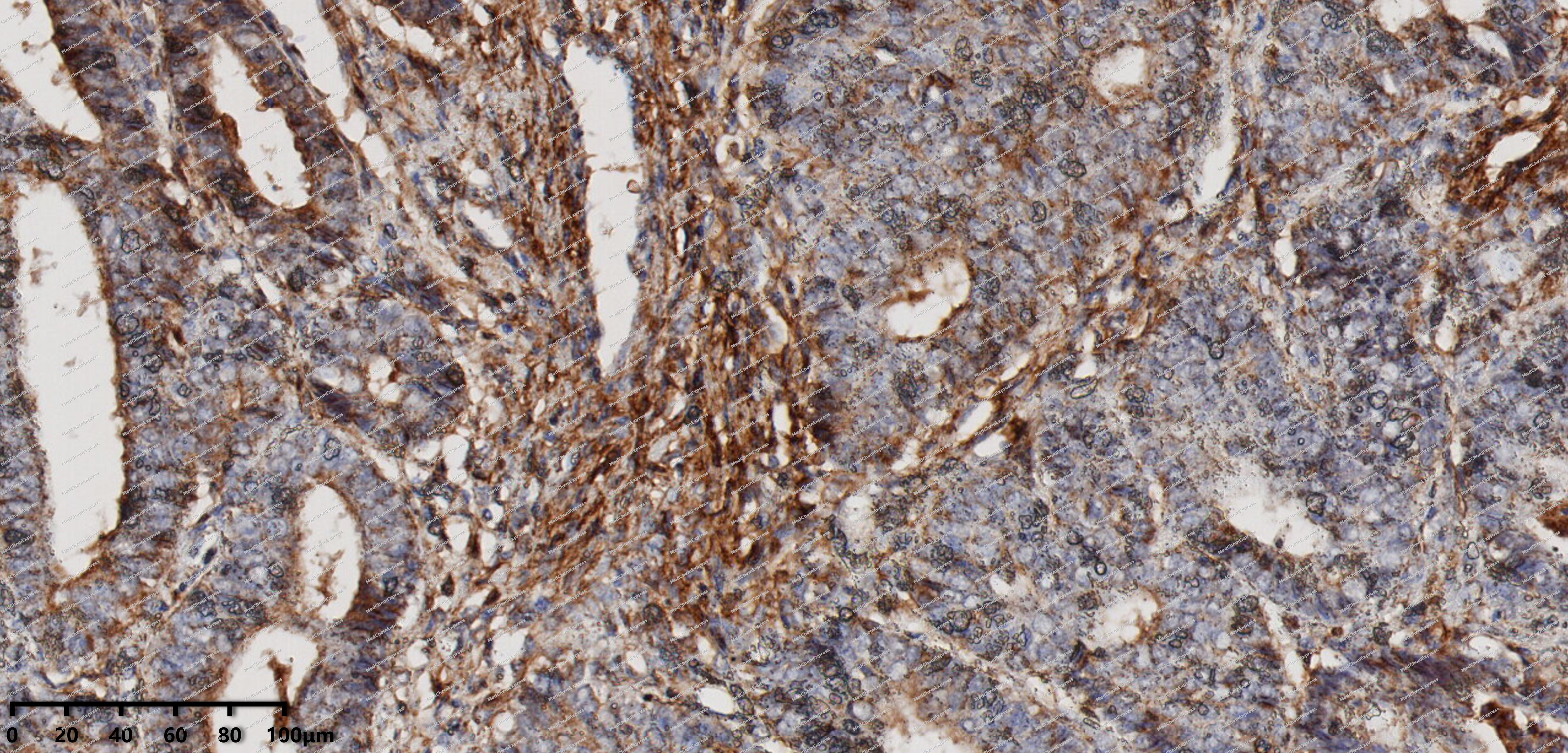 Immunohistochemical analysis of paraffin-embedded human Endometrial carcinoma tissue using IRF5 antibody was performed. The section was pretreated using high-temperature mediated EDTA antigen retrieval buffer (pH 9.0), for 20 minutes. The tissues were incubated with primary antibody (HY-P82139, 1:100 dilution) at room temperature for 20 minutes. Detection was performed using an HRP conjugated compact polymer system. DAB was used as the chromogen. The tissues were counterstained with hematoxylin and mounted with neutral balsam mounting medium.
 -
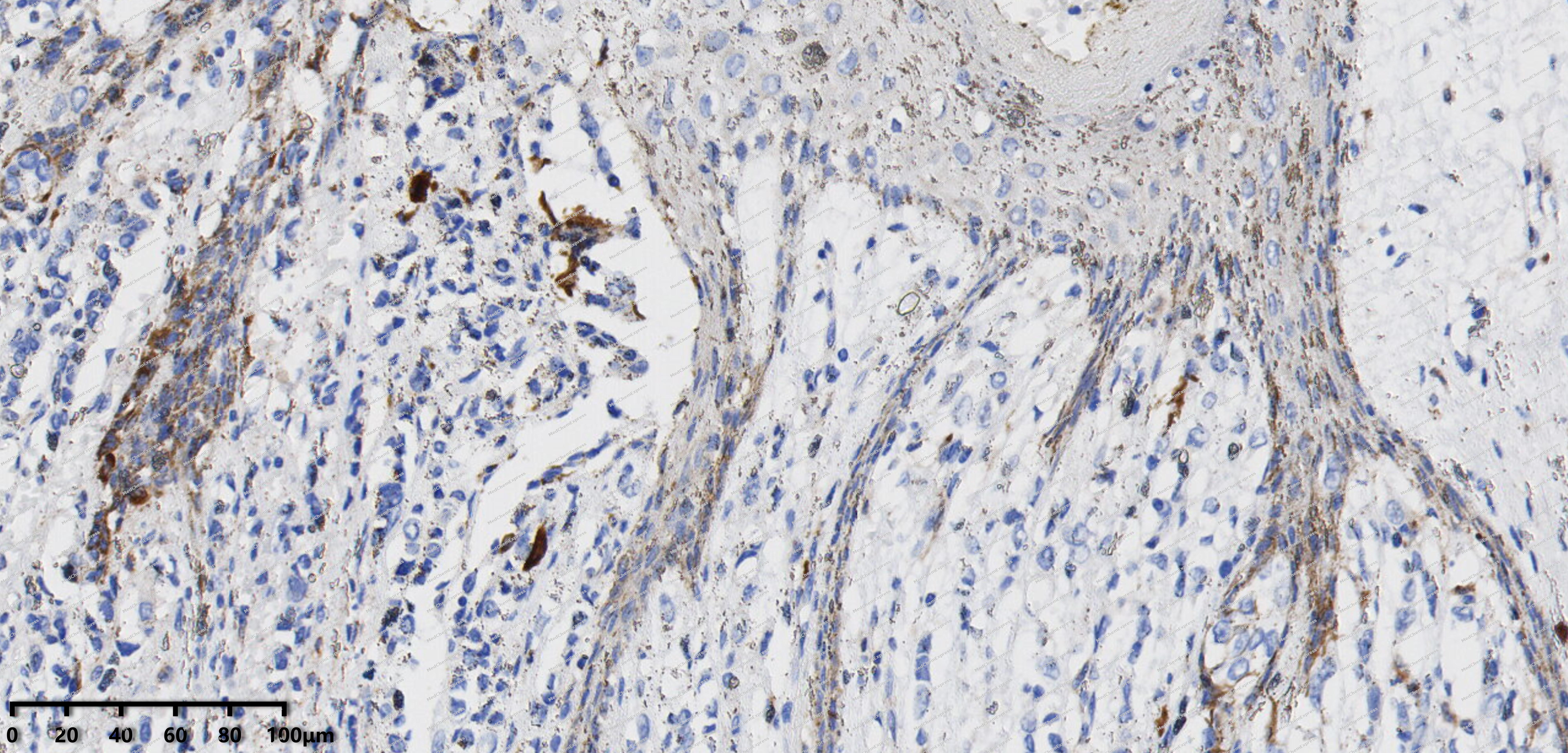 Immunohistochemical analysis of paraffin-embedded human breast cancer tissue using IRF5 antibody was performed. The section was pretreated using high-temperature mediated EDTA antigen retrieval buffer (pH 9.0), for 20 minutes. The tissues were incubated with primary antibody (HY-P82139, 1:100 dilution) at room temperature for 20 minutes. Detection was performed using an HRP conjugated compact polymer system. DAB was used as the chromogen. The tissues were counterstained with hematoxylin and mounted with neutral balsam mounting medium.
 -
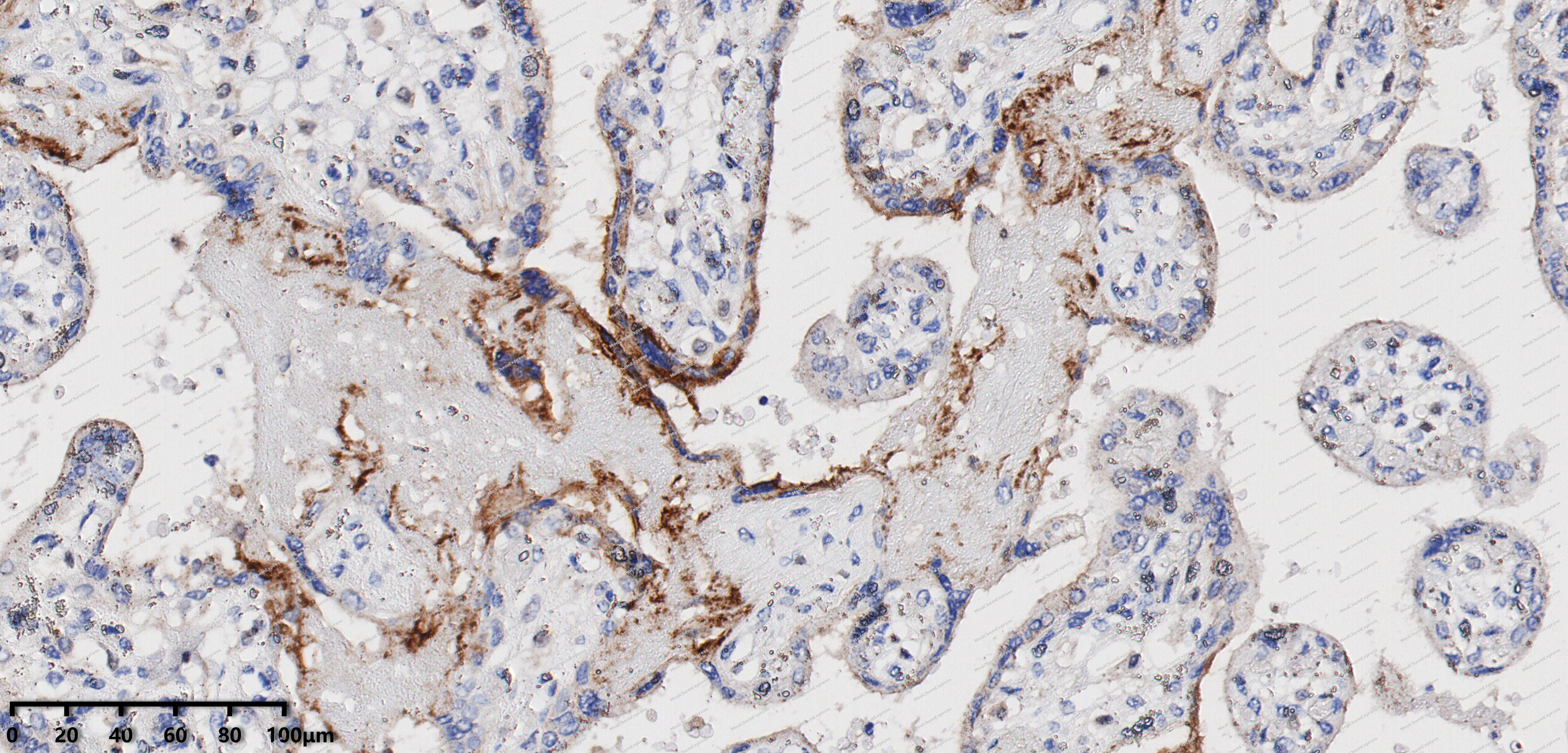 Immunohistochemical analysis of paraffin-embedded human placenta tissue using IRF5 antibody was performed. The section was pretreated using high-temperature mediated EDTA antigen retrieval buffer (pH 9.0), for 20 minutes. The tissues were incubated with primary antibody (HY-P82139, 1:100 dilution) at room temperature for 20 minutes. Detection was performed using an HRP conjugated compact polymer system. DAB was used as the chromogen. The tissues were counterstained with hematoxylin and mounted with neutral balsam mounting medium.
 -
 Flow cytometric analysis of 1X10^6 Hela cells labeling IRF5 Antibody (YA1884) (HY-P82139, red). Cells were fixed with 4% paraformaldehyde and permeabilised with 90% methanol. Then stained with the primary antibody at 1/50 dilution for an hour at 4℃. Alexa Fluor® 488-conjugated AffiniPure Goat Anti-Rabbit IgG H&L (HY-P8002) was used as the secondary antibody at 1/1,000 dilution for 30 minutes at 4℃. Rabbit IgG Isotype Control (HY-P80879, blue) was used as the isotype control, cells without incubation with primary antibody were used as the unlabeled control (black).


Background
-
Function
IRF5 transcription factor that plays a critical role in innate immunity by activating expression of type I interferon (IFN) IFNA and INFB and inflammatory cytokines downstream of endolysosomal toll-like receptors TLR7, TLR8 and TLR9. Regulates the transcription of type I IFN genes (IFN-alpha and IFN-beta) and IFN-stimulated genes (ISG) by binding to an interferon-stimulated response element (ISRE) in their promoters. Can efficiently activate both the IFN-beta (IFNB) and the IFN-alpha (IFNA) genes and mediate their induction downstream of the TLR-activated, MyD88-dependent pathway. Key transcription factor regulating the IFN response during SARS-CoV-2 infection[1][2][3][4][5][6].
-
Subcellular Localization
Cytoplasm; Nucleus
-
Isoforms & Post-Translational Modification
Q13568 has 6 isomers: Q13568-1: 56044 Da (predicted); Q13568-2: 57770 Da (predicted); Q13568-3: 56669 Da (predicted); Q13568-4: 54943 Da (predicted); Q13568-5: 47035 Da (predicted); Q13568-6: 17020 Da (predicted).
Phosphorylation of serine and threonine residues by IKBKB in a C-terminal autoinhibitory region, stimulates dimerization, transport into the nucleus, assembly with the coactivator CBP/EP300 and initiation of transcription;'Lys-63'-linked polyubiquitination by TRAF6 is required for activation -
Subunit
Homodimer, when phosphorylated (PubMed:18836453, PubMed:25326418). Interacts with TASL (via pLxIS motif); interaction takes place downstream of TLR7, TLR8 or TLR9, leading to its activation (PubMed:32433612). Interacts with MYD88 and TRAF6 (By similarity)
-
SwissProt ID
-
Synonyms
Interferon regulatory factor 5; Interferon regulatory factor 5 bone marrow variant; IRF 5; SLEB10
-
Research Field
Immunology


Documentation
-
Data Sheet (234 KB)
-
SDS (252 KB)
- English - EN (252 KB)
- Français - FR (252 KB)
- Deutsch - DE (252 KB)
- Norwegian - NO (252 KB)
- Español - ES (252 KB)
- Swedish - SV (252 KB)
- Italian - IT (252 KB)
- Korean - KR (252 KB)
- Portuguese - PT (252 KB)
-
User Guide for Antibodies (1077 KB)

 Powered by Bioz
