



Lamin A/C Antibody (YA6050)
(Synonyms: Prelamin-A/C [Cleaved into: Lamin-A/C (70 kDa lamin) (Renal carcinoma antigen NY-REN-32)])
Based on 1 Customer Validation
Lamin A/C Antibody (YA6050) is a Rabbit-derived and non-conjugated IgG monoclonal antibody, targeting to Lamin A/C.
-
Host:
Rabbit
-
Isotype:
IgG
-
Application:
WB, IHC-P, ICC/IF, IP, ELISA
-
Reactivity :
Human, Mouse, Rat
-
Formulation:
Supplied in PBS, 50% glycerol, 0.05% Proclin 300, 0.05%BSA
-
Conjugation:
Non-conjugated


To place orders, for customer services and technical support, please contact: MedChemExpress USA
Tel: 609-228-6898 E-mail: [email protected] [email protected]





Applications
| Application |
IHC-P

IHC-P: Immunohistochemistry-Paraffin
|
WB
WB: Western Blot
|
ICC/IF
ICC/IF: Immunocytochemistry/
Immunofluorescence |
ELISA
ELISA: Enzyme Linked Immunosorbent Assay
|
IP
IP: Immunoprecipitation
|
|---|---|---|---|---|---|
| Dilution Ratio | 1:1000-1:4000 | 1:2000-1:10000 | 1:200-1:1000 | 1:5000-1:20000 | 1:50-1:200 |


Product Details
Lamin A/C Antibody (YA6050) is a Rabbit-derived and non-conjugated IgG monoclonal antibody, targeting to Lamin A/C.
-
Host Rabbit

-
Clonality Monoclonal

-
Species ReactivityHuman, Mouse, Rat
-
Observed Molecular WeightObserved band size: 74 kDa,63 kDa
Note: Due to possible protein modifications or aggregation, the molecular weight should be confirmed by actual measurement, and the predicted value is for reference only.

-
Calculated Molecular Weight Predicted band size: 74 kDa,63 kDa

Protein A
Non-conjugated
Unmodified
IgG


Product Properties
-
Appearance
Liquid
-
Formulation
Supplied in PBS, 50% glycerol, 0.05% Proclin 300, 0.05%BSA
-
Concentration
Batch-dependent, Please check the COA for the concentration of each lot. Check Lot Concentration
-
Storage & Stability
Stored at -20°C for 1 year. Avoid repeated freeze / thaw cycles.
-
Shipping
Shipping with blue ice.


Verification Images
-
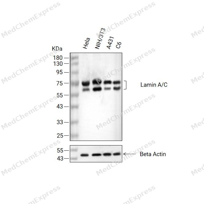 Western blot analysis of extracts from Hela(lane 2(20μg) , NIH/3T3(lane 3(20μg) ,A431(lane 4(20μg)and C6( lane 5(20μg) using Lamin A/C Antibody (HY-P86358). Proteins were transferred to a PVDF membrane and blocked with 5% non-fat milk in TBST for 2 hour at room temperature. The primary antibody (1/5000) and Loading control antibody (Beta Actin, HY-P80993,1/10000) was used in 5% non-fat milk in TBST at 4°C overnight. Goat Anti-Rabbit IgG-HRP Secondary Antibody (HY-P8001,1/10,000) was used for 1 hour at room temperature.
 -
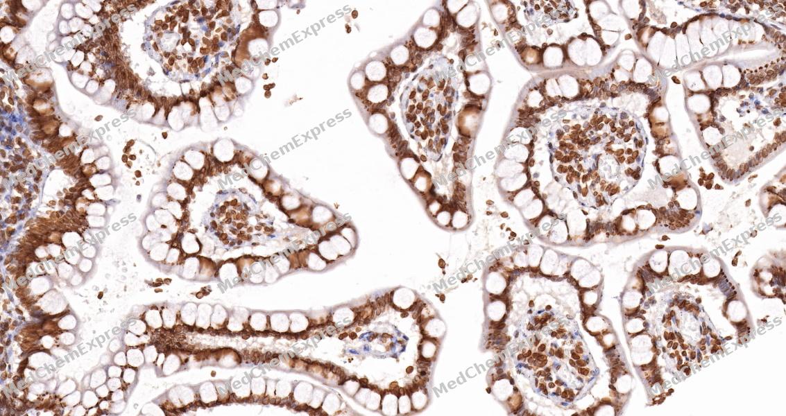 Immunohistochemical analysis of paraffin-embedded human small intestine tissue using Lamin A/C Antibody (HY-P86358, 1/2000). The section was pretreated using heat mediated antigen retrieval with sodium citrate buffer (pH 6.0) for 8 minutes. The tissues were blocked with quick block buffer for 0.5 hours at room temperature, washed with PBS and PBST, and then incubated with the primary antibody overnight at 4℃. The detection was performed using an HRP conjugated compact polymer system. DAB was used as the chromogen. Tissues were counterstained with hematoxylin and mounted with DPX.
 -
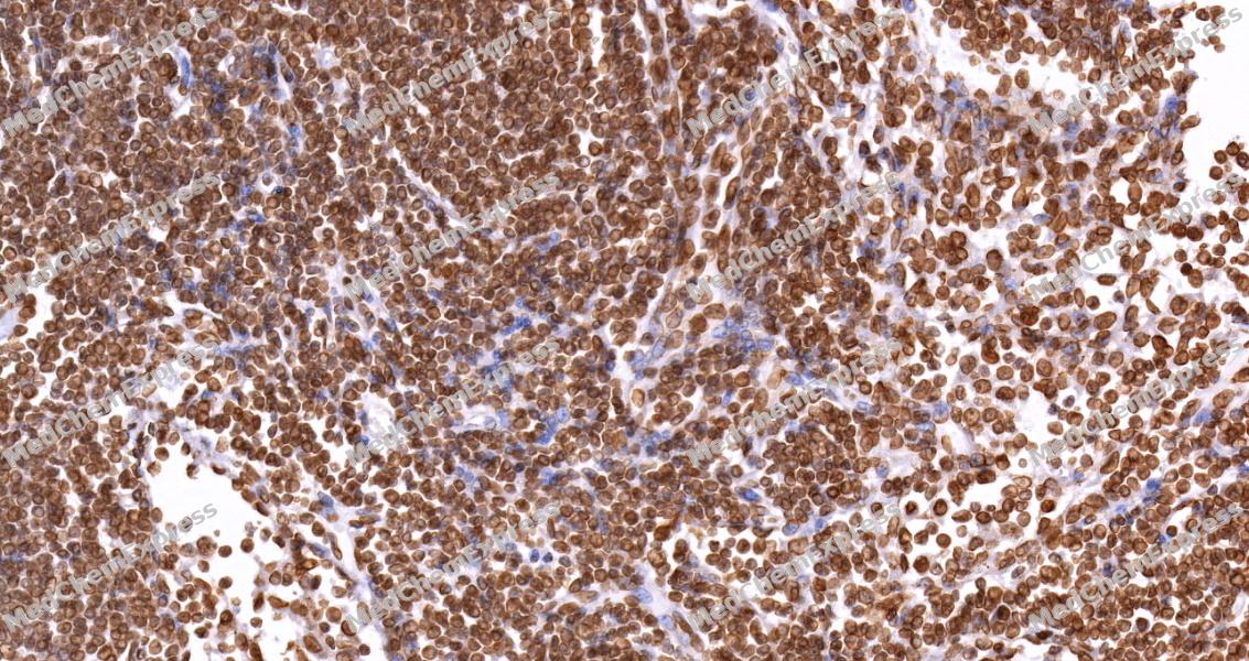 Immunohistochemical analysis of paraffin-embedded human lymph node tissue using Lamin A/C Antibody (HY-P86358, 1/2000). The section was pretreated using heat mediated antigen retrieval with sodium citrate buffer (pH 6.0) for 8 minutes. The tissues were blocked with quick block buffer for 0.5 hours at room temperature, washed with PBS and PBST, and then incubated with the primary antibody overnight at 4℃. The detection was performed using an HRP conjugated compact polymer system. DAB was used as the chromogen. Tissues were counterstained with hematoxylin and mounted with DPX.
 -
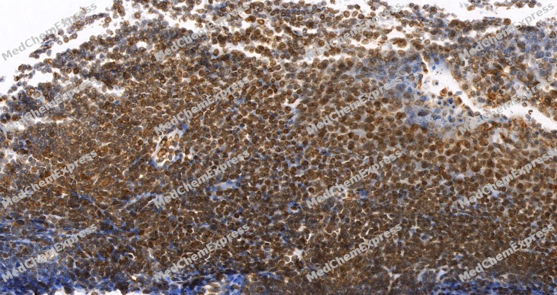 Immunohistochemical analysis of paraffin-embedded human tonsil tissue using Lamin A/C Antibody (HY-P86358, 1/2000). The section was pretreated using heat mediated antigen retrieval with sodium citrate buffer (pH 6.0) for 8 minutes. The tissues were blocked with quick block buffer for 0.5 hours at room temperature, washed with PBS and PBST, and then incubated with the primary antibody overnight at 4℃. The detection was performed using an HRP conjugated compact polymer system. DAB was used as the chromogen. Tissues were counterstained with hematoxylin and mounted with DPX.
 -
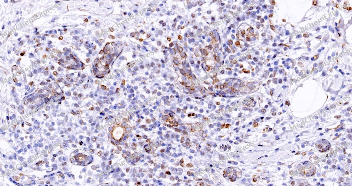 Immunohistochemical analysis of paraffin-embedded human breast tissue using Lamin A/C Antibody (HY-P86358, 1/2000). The section was pretreated using heat mediated antigen retrieval with sodium citrate buffer (pH 6.0) for 8 minutes. The tissues were blocked with quick block buffer for 0.5 hours at room temperature, washed with PBS and PBST, and then incubated with the primary antibody overnight at 4℃. The detection was performed using an HRP conjugated compact polymer system. DAB was used as the chromogen. Tissues were counterstained with hematoxylin and mounted with DPX.
 -
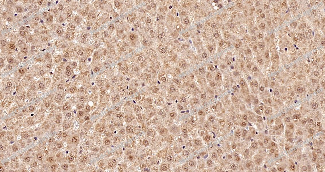 Immunohistochemical analysis of paraffin-embedded rat liver tissue using Lamin A/C Antibody (HY-P86358, 1/2000). The section was pretreated using heat mediated antigen retrieval with sodium citrate buffer (pH 6.0) for 8 minutes. The tissues were blocked with quick block buffer for 0.5 hours at room temperature, washed with PBS and PBST, and then incubated with the primary antibody overnight at 4℃. The detection was performed using an HRP conjugated compact polymer system. DAB was used as the chromogen. Tissues were counterstained with hematoxylin and mounted with DPX.
 -
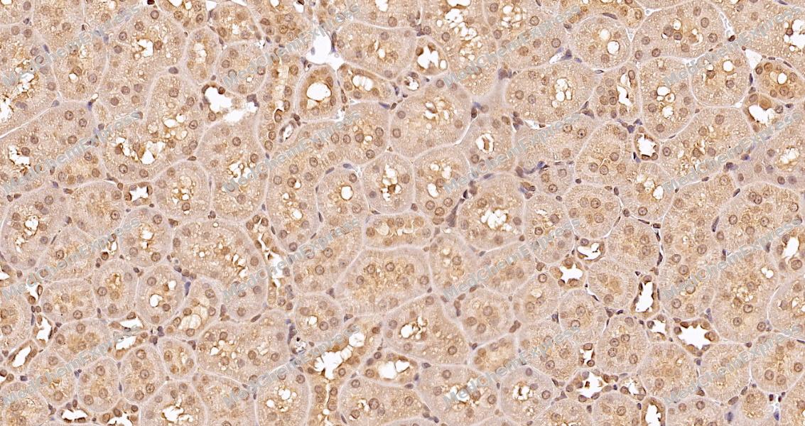 Immunohistochemical analysis of paraffin-embedded rat kidney tissue using Lamin A/C Antibody (HY-P86358, 1/2000). The section was pretreated using heat mediated antigen retrieval with sodium citrate buffer (pH 6.0) for 8 minutes. The tissues were blocked with quick block buffer for 0.5 hours at room temperature, washed with PBS and PBST, and then incubated with the primary antibody overnight at 4℃. The detection was performed using an HRP conjugated compact polymer system. DAB was used as the chromogen. Tissues were counterstained with hematoxylin and mounted with DPX.
 -
 Immunocytochemistry analysis of Hela cells labeling Lamin A/C with Lamin A/C Antibody (HY-P86358) at 1/200 dilution. Cells were fixed in 4% paraformaldehyde for 15 minutes at room temperature, permeabilized with 0.1% Triton X-100 in PBS for 15 minutes at room temperature, then blocked with quick block buffer for 10 minutes at room temperature. Cells were then incubated with Lamin A/C Antibody (HY-P86358) at 1/200 dilution in quick block buffer overnight at 4 ℃.AF488-conjugated Goat Anti-Rabbit IgG H&L (HY-P8002, Green) was used as the secondary antibody at 1/1,000 dilution. PBS instead of the primary antibody was used as the secondary antibody only control. The Nuclear counterstain was DAPI (Blue).
 -
 Immunocytochemistry analysis of Hela cells labeling Lamin A/C with Lamin A/C Antibody (HY-P86358) at 1/500 dilution. Cells were fixed in 4% paraformaldehyde for 15 minutes at room temperature, permeabilized with 0.1% Triton X-100 in PBS for 15 minutes at room temperature, then blocked with quick block buffer for 10 minutes at room temperature. Cells were then incubated with Lamin A/C Antibody (HY-P86358) at 1/500 dilution in quick block buffer overnight at 4 ℃.AF488-conjugated Goat Anti-Rabbit IgG H&L (HY-P8002, Green) was used as the secondary antibody at 1/1,000 dilution. PBS instead of the primary antibody was used as the secondary antibody only control. The Nuclear counterstain was DAPI (Blue).


Background
-
Function
Lamin A/C is a Lamins are intermediate filament proteins that assemble into a filamentous meshwork, and which constitute the major components of the nuclear lamina, a fibrous layer on the nucleoplasmic side of the inner nuclear membrane. Lamins provide a framework for the nuclear envelope, bridging the nuclear envelope and chromatin, thereby playing an important role in nuclear assembly, chromatin organization, nuclear membrane and telomere dynamics. Lamin A and C also regulate matrix stiffness by conferring nuclear mechanical properties. The structural integrity of the lamina is strictly controlled by the cell cycle, as seen by the disintegration and formation of the nuclear envelope in prophase and telophase, respectively. Lamin A and C are present in equal amounts in the lamina of mammals. Also invoved in DNA repair: recruited by DNA repair proteins XRCC4 and IFFO1 to the DNA double-strand breaks (DSBs) to prevent chromosome translocation by immobilizing broken DNA ends. Required for normal development of peripheral nervous system and skeletal muscle and for muscle satellite cell proliferation. Required for osteoblastogenesis and bone formation. Also prevents fat infiltration of muscle and bone marrow, helping to maintain the volume and strength of skeletal muscle and bone. Required for cardiac homeostasis; Prelamin-A/C can accelerate smooth muscle cell senescence. It acts to disrupt mitosis and induce DNA damage in vascular smooth muscle cells (VSMCs), leading to mitotic failure, genomic instability, and premature senescence[1][2][3][4][5][6][7][8][9][10][11][12][13][14][15][16][17][18][19][20][21][22].
-
Subcellular Localization
Nucleus lamina; Nucleus envelope; Nucleus, nucleoplasm; Nucleus matrix; Nucleus speckle
-
Expression
Tissue_specificity:In arteries, the accumulation of prelamin A/C is not observed in young, healthy vessels, but it is prevalent in vascular smooth muscle cells (VSMCs) and atherosclerotic lesions in older adults, and often co-localizes with aging and degenerated VSMCs. The expression of prelamin A/C increases with age and disease progression. During normal aging, the accumulation of prelamin A/C is partly due to the downregulation of ZMPSTE24/FACE1 expression caused by oxidative stress. -
Isoforms & Post-Translational Modification
P02545 has 6 isomers: P02545-1: 74139 Da (predicted); P02545-2: 65135 Da (predicted); P02545-3: 70661 Da (predicted); P02545-4: 63893 Da (predicted); P02545-5: 62853 Da (predicted); P02545-6: 69249 Da (predicted).
Proteolytic cleavage of the C-terminal of 18 residues of prelamin-A/C results in the production of lamin-A/C (PubMed:20458013, PubMed:8175923, PubMed:9030603). The prelamin-A/C maturation pathway includes farnesylation of CAAX motif by protein farnesyltransferase (FNTA and FNTB), removal of the last three amino acids (-AAX) by RCE1/FACE2 and/or ZMPSTE24, methylation of the C-terminal cysteine by ICMT and endoproteolytic removal of the last 15 C-terminal amino acids by ZMPSTE24 (PubMed:20458013, PubMed:8175923, PubMed:9030603). Proteolytic cleavage requires prior farnesylation and methylation, and absence of these blocks cleavage (PubMed:20458013, PubMed:8175923, PubMed:9030603);Farnesylation of prelamin-A/C facilitates nuclear envelope targeting;Phosphorylation plays a key role in lamin organization, subcellular localization and nuclear envelope disintegration (PubMed:2188730, PubMed:2344612, PubMed:24741066, PubMed:37788673, PubMed:37832547). Phosphorylation by CDK1 at Ser-22 and Ser-392 at the onset of mitosis drives lamin disassembly and nuclear envelope breakdown (PubMed:2188730, PubMed:2344612). Phosphorylation at Ser-22 and Ser-392 during interphase promotes localization to the nucleoplasm and regulates lamina assembly (PubMed:24741066). Phosphorylation at Ser-22, Ser-392 and Ser-628 during interphase causes redistribution between the nucleus and the cytoplasm (PubMed:24741066). Phosphorylation at Ser-22 by CDK1 regulates matrix stiffness (PubMed:25127216). Phosphorylation status of Ser-22 determines its localization between double-strand break (DSB) sites and the nuclear matrix (PubMed:31548606). Phosphorylated by ATR at Ser-282 in response to DNA damage, leading to lamin disassembly and nuclear envelope rupture (PubMed:37832547). Phosphorylation also regulates stability in micronuclei arising from genome instability: phosphorylation at Ser-395 by ATR in response to genome instability and double-stranded DNA breaks primes LMNA for subsequent phosphorylation at Ser-392 by CDK1 and micronuclei envelope rupture (PubMed:37788673). The rupture of micronuclear envelope triggers the cGAS-STING pathway thereby activating the type I interferon response and innate immunity (PubMed:37788673);Acetylation by KAT8 is required for nuclear architecture;Sumoylation is necessary for the localization to the nuclear envelope -
Subunit
Homodimer of lamin A and lamin C (PubMed:15476822, PubMed:31434876, PubMed:33706103). Lamin dimers then assemble into dimeric head-to-tail polymers (PubMed:31434876).
-
SwissProt ID
-
Synonyms
Prelamin-A/C [Cleaved into: Lamin-A/C (70 kDa lamin) (Renal carcinoma antigen NY-REN-32)]


Documentation
-
Data Sheet (235 KB)
-
SDS (252 KB)
- English - EN (252 KB)
- Français - FR (252 KB)
- Deutsch - DE (252 KB)
- Norwegian - NO (252 KB)
- Español - ES (252 KB)
- Swedish - SV (252 KB)
- Italian - IT (252 KB)
- Korean - KR (252 KB)
- Portuguese - PT (252 KB)
-
User Guide for Antibodies (1077 KB)

 Powered by Bioz
