



LAMP1 Antibody (YA311)
(Synonyms: CD107 antigen like family member A antibody; CD107 antigen-like family member A antibody; CD107a antibody; CD107a antigen antibody; LAMP 1 antibody; LAMP-1 antibody; LAMP1 antibody; LAMP1_HUMAN antibody; LAMPA antibody; LGP120 antibody; lgpA antibody; Lysosomal membrane glycoprotein 120KD antibody; Lysosomal Associated Membrane Protein 1 antibody; Lysosome associated membrane glycoprotein 1 antibody; Lysosome-associated membrane glycoprotein 1 antibody; Lysosome-associated membrane protein 1 antibody; OTTHUMP00000040663 antibody; )Based on 1 publication(s) in Google Scholar
LAMP1 Antibody (YA311) is a Rabbit-derived and non-conjugated IgG monoclonal antibody, targeting to LAMP1.
-
Host:
Rabbit
-
Isotype:
IgG
-
Application:
WB, IHC-P
-
Reactivity :
Human
-
Formulation:
Supplied in 1*TBS (pH7.4), 0.05% BSA and 40% Glycerol. Preservative: 0.05% Sodium Azide.
-
Conjugation:
Non-conjugated


To place orders, for customer services and technical support, please contact: MedChemExpress USA
Tel: 609-228-6898 E-mail: [email protected] [email protected]



Publications Citing Use of MedChemExpress (MCE) LAMP1 Antibody (YA311)
More

Applications
| Application |
WB

WB: Western Blot
|
IHC-P
IHC-P: Immunohistochemistry-Paraffin
|
|---|---|---|
| Dilution Ratio | 1:1000-1:2000 | 1:50-1:200 |


Product Details
LAMP1 Antibody (YA311) is a Rabbit-derived and non-conjugated IgG monoclonal antibody, targeting to LAMP1.
-
Host Rabbit

-
Clonality Recombinant,Monoclonal

-
Species ReactivityHuman
-
Observed Molecular WeightObserved band size: 100-120 kDa
Note: Due to possible protein modifications or aggregation, the molecular weight should be confirmed by actual measurement, and the predicted value is for reference only.

-
Calculated Molecular Weight Predicted band size: 45 kDa

Synthetic peptide corresponding to Human LAMP1.AA range:84-206.
Endogenous
Protein A affinity purified.
Non-conjugated
Unmodified
IgG


Product Properties
-
Appearance
Solution
-
Formulation
Supplied in 1*TBS (pH7.4), 0.05% BSA and 40% Glycerol. Preservative: 0.05% Sodium Azide.
-
Concentration
Batch-dependent, Please check the COA for the concentration of each lot. Check Lot Concentration
-
Storage & Stability
Stored at -20°C for 1 year. Avoid repeated freeze / thaw cycles.
-
Shipping
Shipping with blue ice.


Publications (1)
-
Journal Impact Factor
-
Most Recent


Verification Images
-
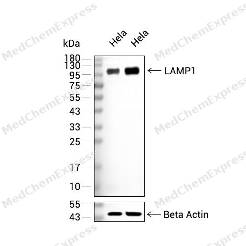 Western blot analysis of extracts from Hela (lane 2(20μg), Hela (lane 3(40μg), using LAMP1 Antibody. Proteins were transferred to a PVDF membrane and blocked with 5% BSA in TBST for 2 hour at room temperature. The primary antibody and Loading control antibody (Beta Actin, HY-P80438, 1/3000) was used in 5% BSA in TBST at 4°C overnight. Goat Anti-Mouse/Rabbit IgG-HRP Secondary Antibody (HY-P8004/HY-P8001, 1/10,000) was used for 1 hour at room temperature.
 -
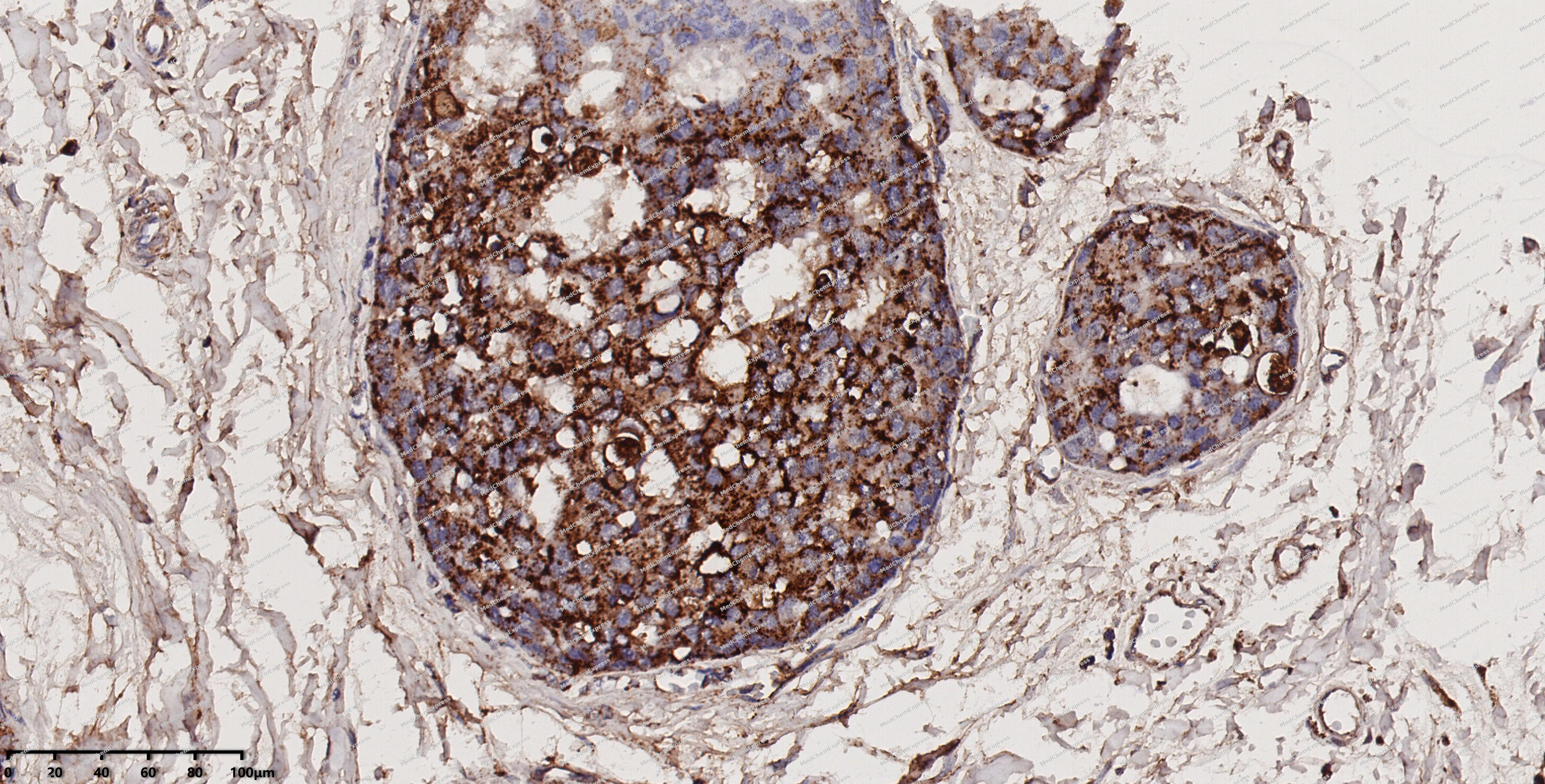 Immunohistochemical analysis of paraffin-embedded human breast cancer using LAMP1 antibody. The section was pre-treated using heat mediated antigen retrieval with EDTA (pH 9.0) for 14 minutes. The tissues were blocked in 5% BSA for 30 minutes at room temperature, washed with TBST, and then probed with the primary antibody (HY-P80206, 1/200) for 30 minutes at room temperature. The detection was performed using Polymer HRP-conjugated Goat Anti-Mouse/Rabbit lgG(H&L) secondary antibody (HY-P83652). DAB was used as the chromogen. Tissues were counterstained with hematoxylin and mounted with DPX.
 -
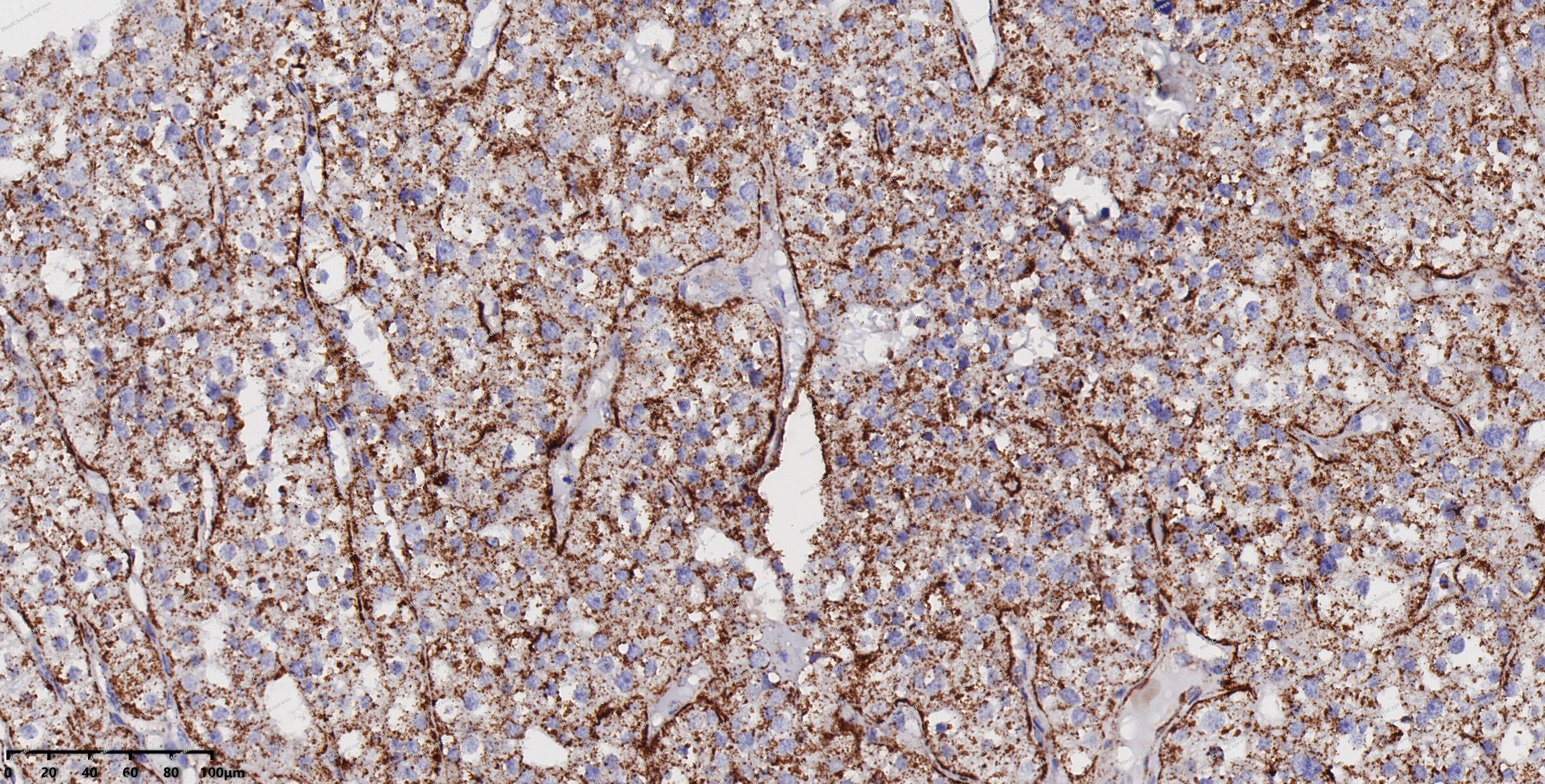 Immunohistochemical analysis of paraffin-embedded human thyroid cancer using LAMP1 antibody. The section was pre-treated using heat mediated antigen retrieval with EDTA (pH 9.0) for 14 minutes. The tissues were blocked in 5% BSA for 30 minutes at room temperature, washed with TBST, and then probed with the primary antibody (HY-P80206, 1/200) for 30 minutes at room temperature. The detection was performed using Polymer HRP-conjugated Goat Anti-Mouse/Rabbit lgG(H&L) secondary antibody (HY-P83652). DAB was used as the chromogen. Tissues were counterstained with hematoxylin and mounted with DPX.
 -
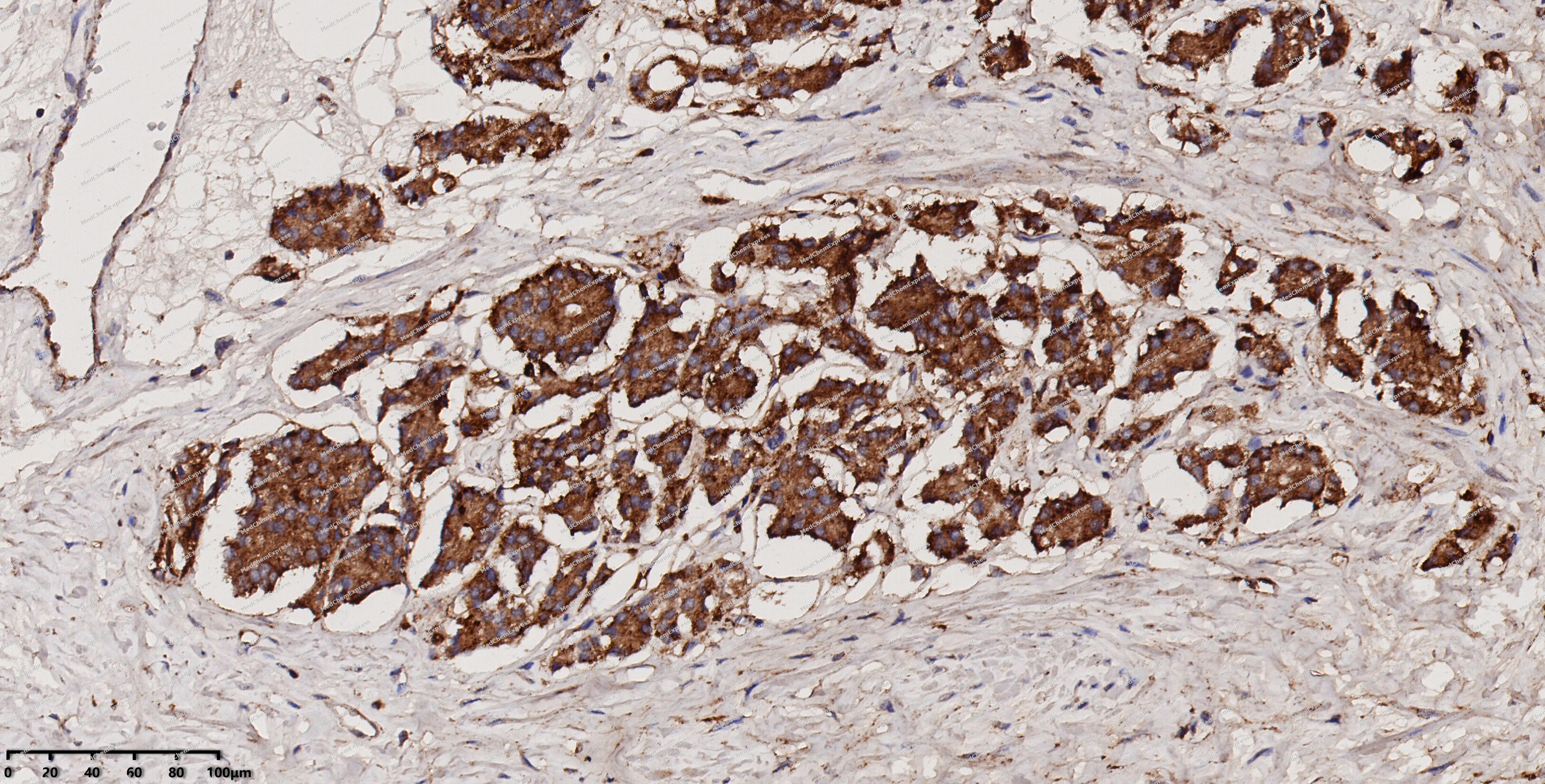 Immunohistochemical analysis of paraffin-embedded human prostate cancer using LAMP1 antibody. The section was pre-treated using heat mediated antigen retrieval with EDTA (pH 9.0) for 14 minutes. The tissues were blocked in 5% BSA for 30 minutes at room temperature, washed with TBST, and then probed with the primary antibody (HY-P80206, 1/200) for 30 minutes at room temperature. The detection was performed using Polymer HRP-conjugated Goat Anti-Mouse/Rabbit lgG(H&L) secondary antibody (HY-P83652). DAB was used as the chromogen. Tissues were counterstained with hematoxylin and mounted with DPX.
 -
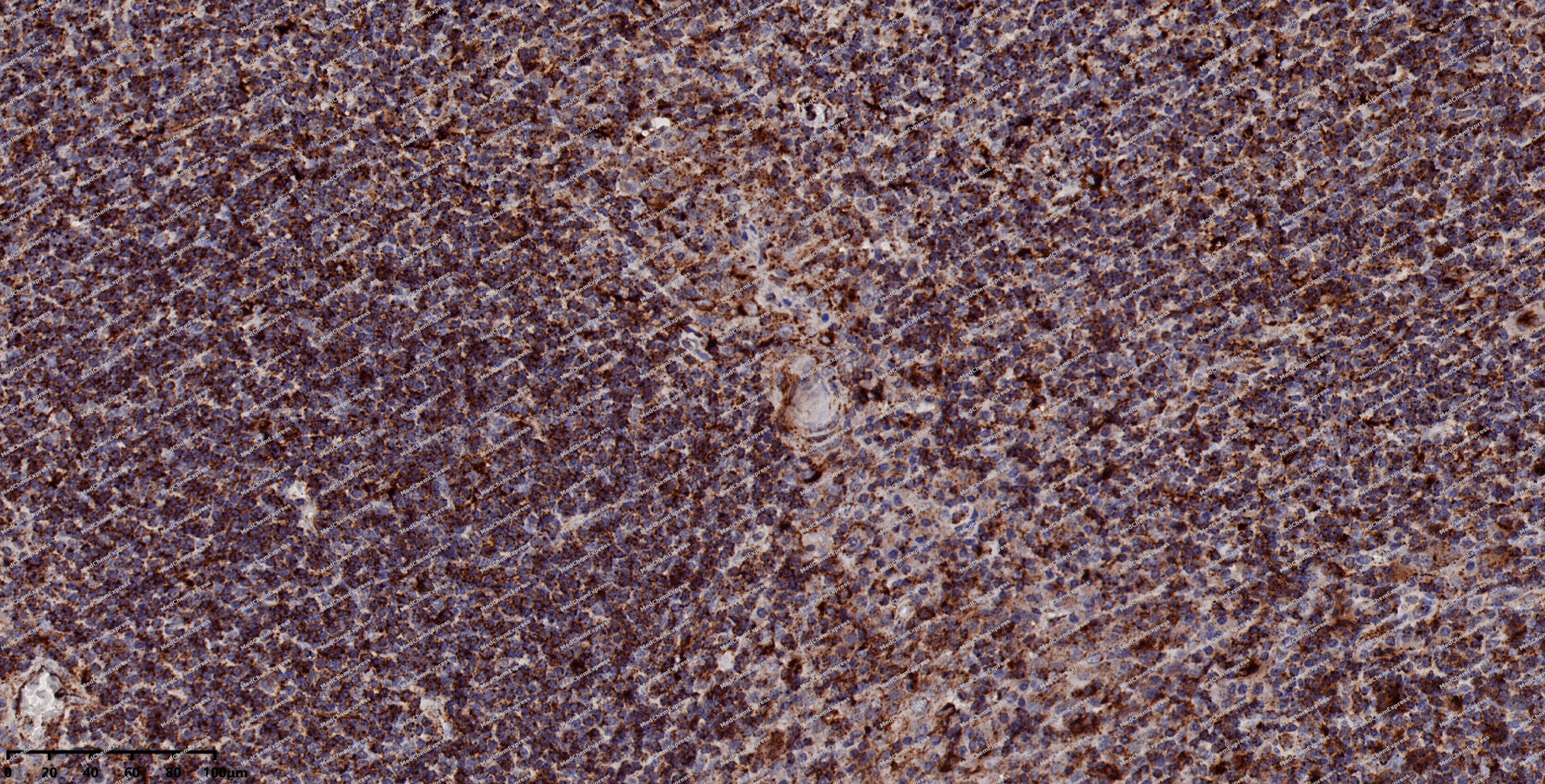 Immunohistochemical analysis of paraffin-embedded human tonsil using LAMP1 antibody. The section was pre-treated using heat mediated antigen retrieval with EDTA (pH 9.0) for 14 minutes. The tissues were blocked in 5% BSA for 30 minutes at room temperature, washed with TBST, and then probed with the primary antibody (HY-P80206, 1/200) for 30 minutes at room temperature. The detection was performed using Polymer HRP-conjugated Goat Anti-Mouse/Rabbit lgG(H&L) secondary antibody (HY-P83652). DAB was used as the chromogen. Tissues were counterstained with hematoxylin and mounted with DPX.
 -
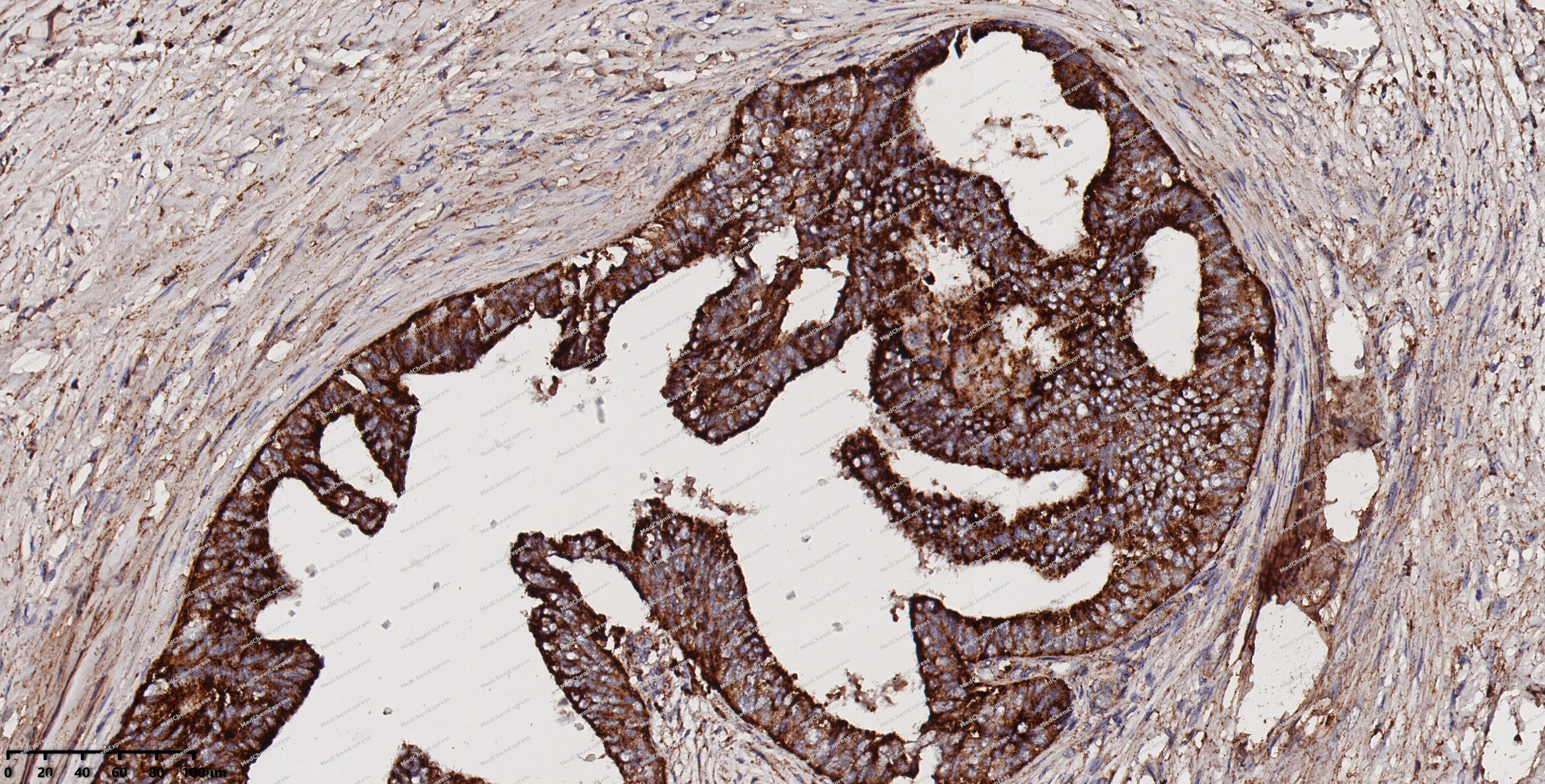 Immunohistochemical analysis of paraffin-embedded human cervical cancer using LAMP1 antibody. The section was pre-treated using heat mediated antigen retrieval with EDTA (pH 9.0) for 14 minutes. The tissues were blocked in 5% BSA for 30 minutes at room temperature, washed with TBST, and then probed with the primary antibody (HY-P80206, 1/200) for 30 minutes at room temperature. The detection was performed using Polymer HRP-conjugated Goat Anti-Mouse/Rabbit lgG(H&L) secondary antibody (HY-P83652). DAB was used as the chromogen. Tissues were counterstained with hematoxylin and mounted with DPX.
 -
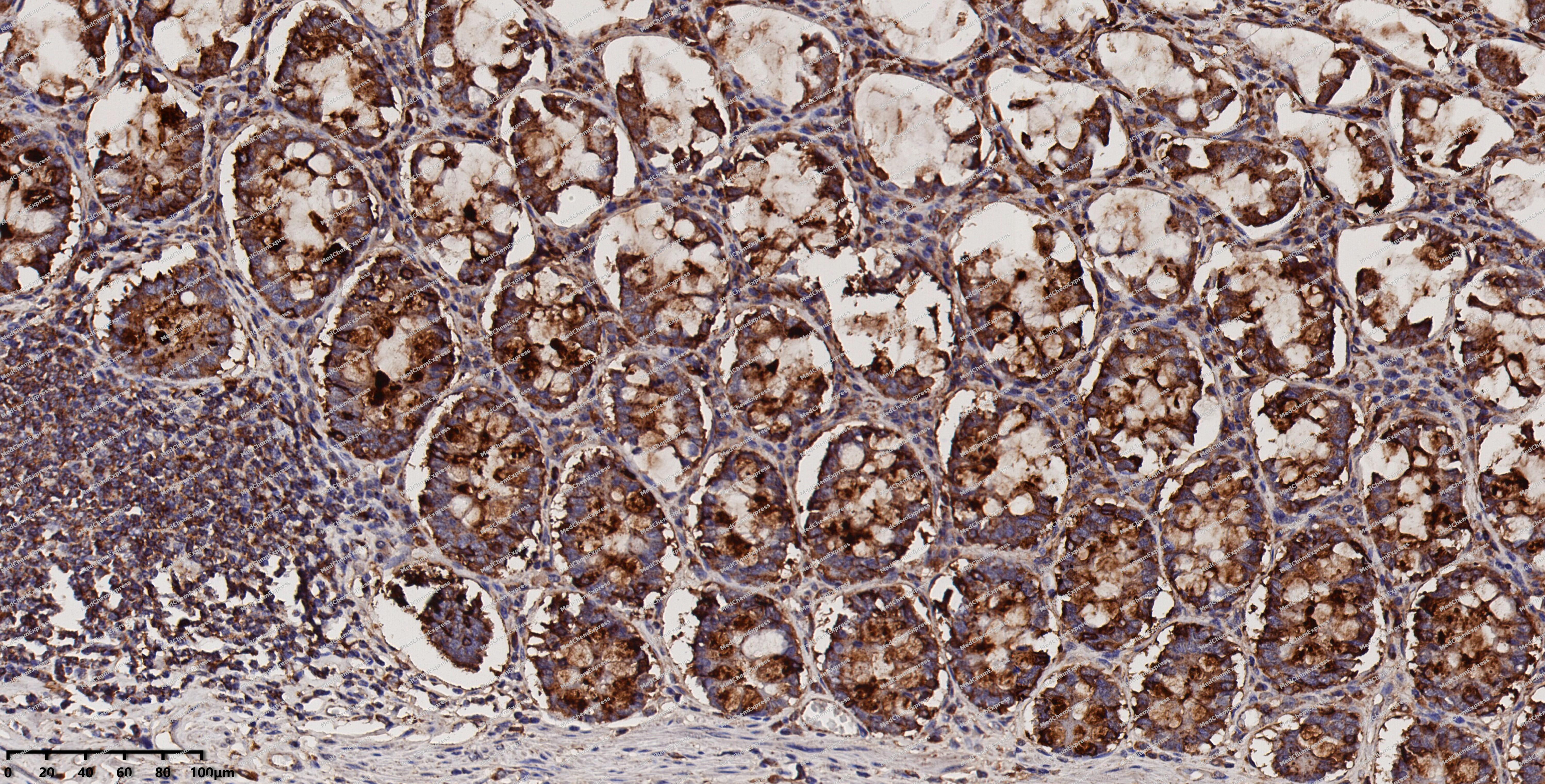 Immunohistochemical analysis of paraffin-embedded human colon using LAMP1 antibody. The section was pre-treated using heat mediated antigen retrieval with EDTA (pH 9.0) for 14 minutes. The tissues were blocked in 5% BSA for 30 minutes at room temperature, washed with TBST, and then probed with the primary antibody (HY-P80206, 1/200) for 30 minutes at room temperature. The detection was performed using Polymer HRP-conjugated Goat Anti-Mouse/Rabbit lgG(H&L) secondary antibody (HY-P83652). DAB was used as the chromogen. Tissues were counterstained with hematoxylin and mounted with DPX.
 -
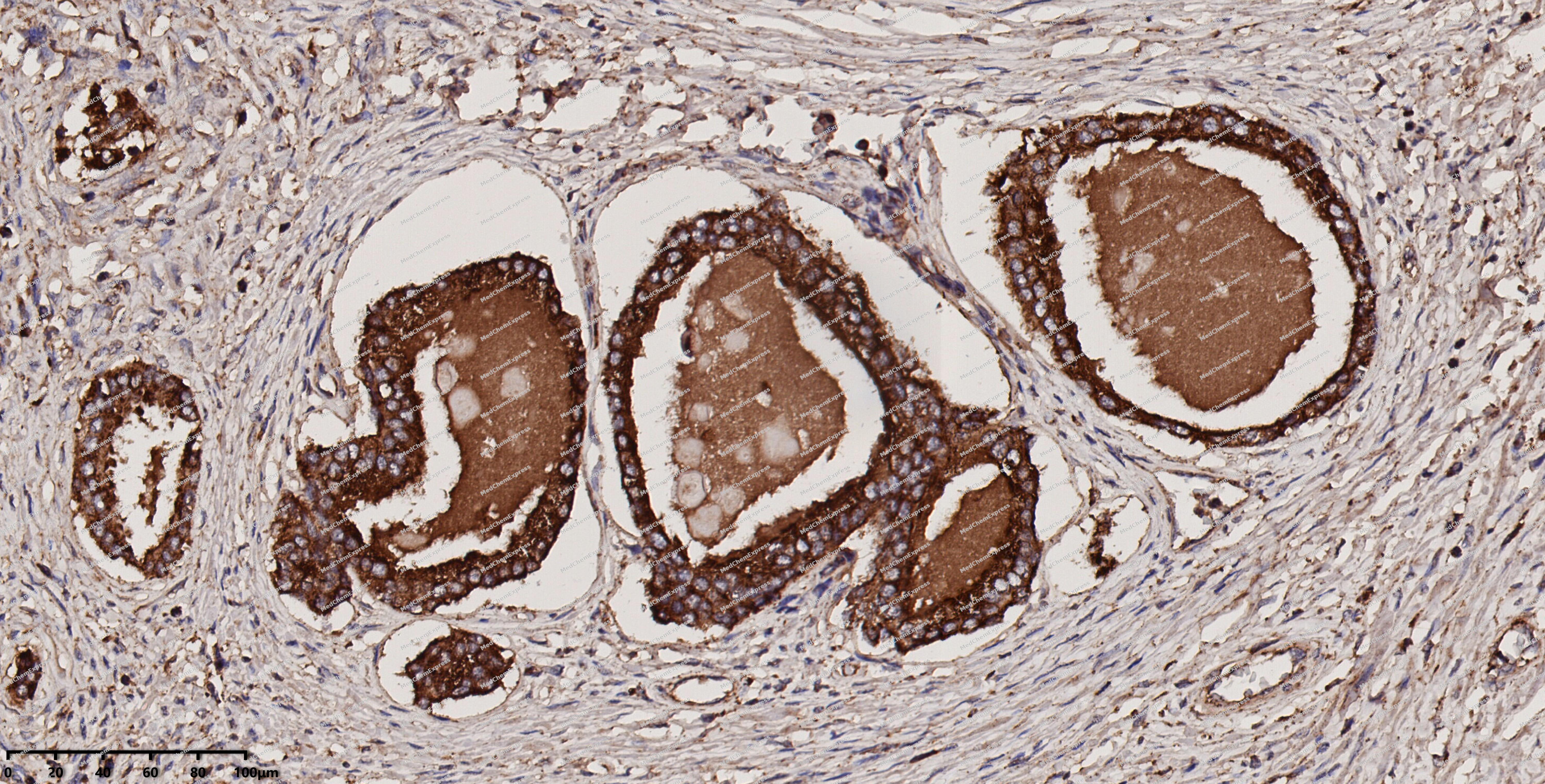 Immunohistochemical analysis of paraffin-embedded human prostate using LAMP1 antibody. The section was pre-treated using heat mediated antigen retrieval with EDTA (pH 9.0) for 14 minutes. The tissues were blocked in 5% BSA for 30 minutes at room temperature, washed with TBST, and then probed with the primary antibody (HY-P80206, 1/200) for 30 minutes at room temperature. The detection was performed using Polymer HRP-conjugated Goat Anti-Mouse/Rabbit lgG(H&L) secondary antibody (HY-P83652). DAB was used as the chromogen. Tissues were counterstained with hematoxylin and mounted with DPX.
 -
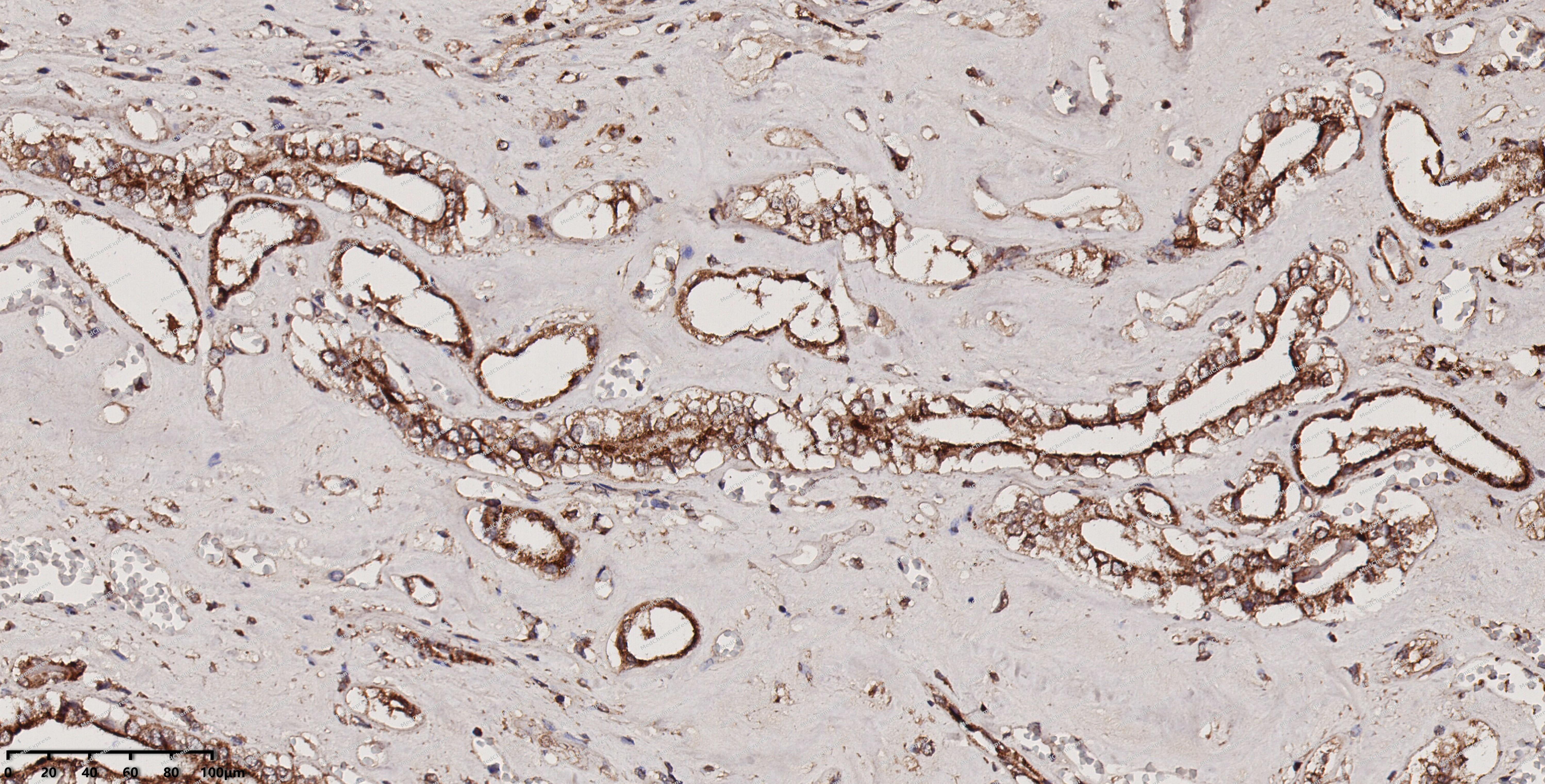 Immunohistochemical analysis of paraffin-embedded human kidney using LAMP1 antibody. The section was pre-treated using heat mediated antigen retrieval with EDTA (pH 9.0) for 14 minutes. The tissues were blocked in 5% BSA for 30 minutes at room temperature, washed with TBST, and then probed with the primary antibody (HY-P80206, 1/200) for 30 minutes at room temperature. The detection was performed using Polymer HRP-conjugated Goat Anti-Mouse/Rabbit lgG(H&L) secondary antibody (HY-P83652). DAB was used as the chromogen. Tissues were counterstained with hematoxylin and mounted with DPX.
 -
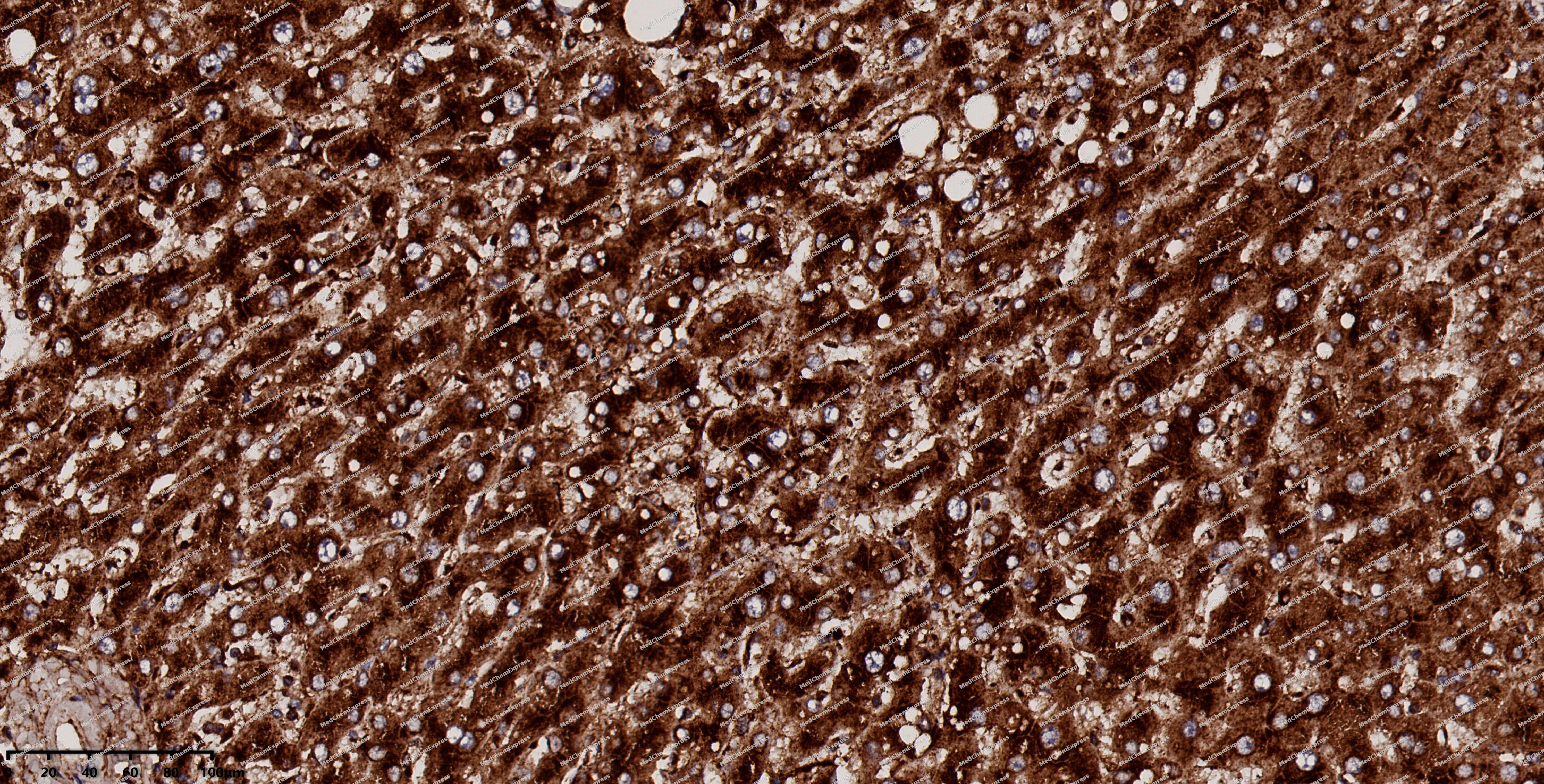 Immunohistochemical analysis of paraffin-embedded human liver using LAMP1 antibody. The section was pre-treated using heat mediated antigen retrieval with EDTA (pH 9.0) for 14 minutes. The tissues were blocked in 5% BSA for 30 minutes at room temperature, washed with TBST, and then probed with the primary antibody (HY-P80206, 1/200) for 30 minutes at room temperature. The detection was performed using Polymer HRP-conjugated Goat Anti-Mouse/Rabbit lgG(H&L) secondary antibody (HY-P83652). DAB was used as the chromogen. Tissues were counterstained with hematoxylin and mounted with DPX.


Background
-
Function
LAMP1 is a Lysosomal membrane glycoprotein which plays an important role in lysosome biogenesis, lysosomal pH regulation, autophagy and cholesterol homeostasis. Acts as an important regulator of lysosomal lumen pH regulation by acting as a direct inhibitor of the proton channel TMEM175, facilitating lysosomal acidification for optimal hydrolase activity. Also plays an important role in NK-cells cytotoxicity. Mechanistically, participates in cytotoxic granule movement to the cell surface and perforin trafficking to the lytic granule. In addition, protects NK-cells from degranulation-associated damage induced by their own cytotoxic granule content. Presents carbohydrate ligands to selectins; (Microbial infection) Acts as a receptor for Lassa virus glycoprotein. Also promotes fusion of the virus with host membrane in less acidic endosomes; (Microbial infection) Supports the FURIN-mediated cleavage of mumps virus fusion protein F by interacting with both FURIN and the unprocessed form but not the processed form of the viral protein F[1][2][3][4][5][6][7][8][9][10].
-
Subcellular Localization
Lysosome membrane; Single-pass type I membrane protein; Endosome membrane; Single-pass type I membrane protein; Late endosome membrane; Single-pass type I membrane protein; Cell membrane; Single-pass type I membrane protein; Cytolytic granule membrane; Single-pass type I membrane protein
-
Isoforms & Post-Translational Modification
P11279 has 2 isomers: P11279-1: 44882 Da (predicted); P11279-2: 38986 Da (predicted).
O- and N-glycosylated; some of the 18 N-linked glycans are polylactosaminoglycans;(Microbial infection) The glycosylation of Asn-76 is essential for Lassa virus entry into cells -
Subunit
Interacts with ABCB9; this interaction strongly stabilizes ABCB9 and protects ABCB9 against lysosomal degradation (PubMed:22641697). Interacts with FURIN (PubMed:32295904). Interacts with TMEM175; inhibiting the proton channel activity of TMEM175 (PubMed:37390818)
-
SwissProt ID
-
Synonyms
CD107 antigen like family member A antibody; CD107 antigen-like family member A antibody; CD107a antibody; CD107a antigen antibody; LAMP 1 antibody; LAMP-1 antibody; LAMP1 antibody; LAMP1_HUMAN antibody; LAMPA antibody; LGP120 antibody; lgpA antibody; Lysosomal membrane glycoprotein 120KD antibody; Lysosomal Associated Membrane Protein 1 antibody; Lysosome associated membrane glycoprotein 1 antibody; Lysosome-associated membrane glycoprotein 1 antibody; Lysosome-associated membrane protein 1 antibody; OTTHUMP00000040663 antibody;
-
Research Field
Neuroscience
Documentation
-
Data Sheet (262 KB)
-
SDS (254 KB)
- English - EN (254 KB)
- Français - FR (254 KB)
- Deutsch - DE (254 KB)
- Norwegian - NO (254 KB)
- Español - ES (254 KB)
- Swedish - SV (254 KB)
- Italian - IT (254 KB)
- Korean - KR (254 KB)
- Portuguese - PT (254 KB)
-
User Guide for Antibodies (1077 KB)
References
[1]. Zhang J, et al. Lysosomal LAMP proteins regulate lysosomal pH by direct inhibition of the TMEM175 channel. Mol Cell. 2023 Jul 20;83(14):2524-2539.e7. [Content Brief]
[2]. Peters PJ, et al. Cytotoxic T lymphocyte granules are secretory lysosomes, containing both perforin and granzymes. J Exp Med. 1991 May 1;173(5):1099-109. [Content Brief]
[3]. Krzewski K, et al. LAMP1/CD107a is required for efficient perforin delivery to lytic granules and NK-cell cytotoxicity. Blood. 2013 Jun 6;121(23):4672-83. [Content Brief]
[4]. Cohnen A, et al. Surface CD107a/LAMP-1 protects natural killer cells from degranulation-associated damage. Blood. 2013 Aug 22;122(8):1411-8. [Content Brief]
[5]. Sawada R, et al. E-selectin-dependent adhesion efficiency of colonic carcinoma cells is increased by genetic manipulation of their cell surface lysosomal membrane glycoprotein-1 expression levels. J Biol Chem. 1993 Jun 15;268(17):12675-81. [Content Brief]
[6]. Jae LT, et al. Virus entry. Lassa virus entry requires a trigger-induced receptor switch. Science. 2014 Jun 27;344(6191):1506-10. [Content Brief]
[7]. Cohen-Dvashi H, et al. Molecular Mechanism for LAMP1 Recognition by Lassa Virus. J Virol. 2015 Aug;89(15):7584-92. [Content Brief]
[8]. Cohen-Dvashi H, et al. Role of LAMP1 Binding and pH Sensing by the Spike Complex of Lassa Virus. J Virol. 2016 Nov 15;90(22):10329-10338. [Content Brief]
[9]. Israeli H, et al. Mapping of the Lassa virus LAMP1 binding site reveals unique determinants not shared by other old world arenaviruses. PLoS Pathog. 2017 Apr;13(4):e1006337. [Content Brief]
[10]. Hulseberg CE, et al. Lamp1 Increases the Efficiency of Lassa Virus Infection by Promoting Fusion in Less Acidic Endosomal Compartments. mBio. 2018 Jan 2;9(1):. [Content Brief]

 Powered by Bioz
