



SQSTM1/p62 Antibody (YA062)
(Synonyms: p60; p62; A170; DMRV; OSIL; PDB3; ZIP3; p62B; NADGP; FTDALS3)Based on 2 publication(s) in Google Scholar
SQSTM1/p62 Antibody (YA062) is a Rabbit-derived and non-conjugated IgG monoclonal antibody, targeting to SQSTM1/p62.
-
Host:
Rabbit
-
Isotype:
IgG
-
Application:
WB, IHC-F, IHC-P, ICC/IF, IP
-
Reactivity :
Human
-
Formulation:
1.Supplied in 50 mM Tris-Glycine (pH 7.4), 0.15 M NaCl, 40% Glycerol and 0.05% BSA. Preservative: 0.01% Sodium azide.
2.Supplied in 1xPBS(pH 7.4), 150mM NaCl, 50% Glycerol, 0.02% Sodium azide and 0.05% BSA.
-
Conjugation:
Non-conjugated



To place orders, for customer services and technical support, please contact: MedChemExpress USA
Tel: 609-228-6898 E-mail: [email protected] [email protected]



-

HY-P80741
LC3A/B Antibody -
HY-P80209
LC3B Antibody (YA308) -

HY-100558
Bafilomycin A1 -
HY-100941
CCCP -
HY-P80137
GAPDH Antibody (YA418) -
HY-P81051
Nrf2 Antibody(YA895) -
HY-P80742
LC3B Antibody -
HY-P8001
HRP-conjugated Goat Anti-Rabbit IgG H&L -
HY-P8004
HRP-conjugated Goat Anti-Mouse IgG H&L -
HY-P80344
STAT3 Antibody (YA056) -
HY-P80779
Parkin Antibody -
HY-P80568
Beclin 1 Antibody (YA587)
Publications Citing Use of MedChemExpress (MCE) SQSTM1/p62 Antibody (YA062)
More
 2 Publications Citing Use of MCE SQSTM1/p62 Antibody (YA062) (1)
-
WB


Applications
| Application |
WB

WB: Western Blot
|
IHC-P
IHC-P: Immunohistochemistry-Paraffin
|
IHC-F
IHC-F: Immunohistochemistry-Frozen
|
ICC/IF
ICC/IF: Immunocytochemistry/
Immunofluorescence |
IP
IP: Immunoprecipitation
|
|---|---|---|---|---|---|
| Dilution Ratio | 1:500-1:1000 | 1:50-1:100 | 1:50-1:100 | 1:50-1:200 | 1:20 |


Product Details
SQSTM1/p62 Antibody (YA062) is a Rabbit-derived and non-conjugated IgG monoclonal antibody, targeting to SQSTM1/p62.
-
Host Rabbit

-
Clonality Recombinant,Monoclonal

-
Species ReactivityHuman
-
Observed Molecular WeightObserved band size: 62 kDa
Note: Due to possible protein modifications or aggregation, the molecular weight should be confirmed by actual measurement, and the predicted value is for reference only.

-
Calculated Molecular Weight Predicted band size: 48 kDa

Entrez Gene: 8878 Human ; 18412 Mouse ; 113894 Rat
SwissProt: Q13501 Human ; Q64337 Mouse ; O08623 Rat
OMIM: 167250 Human
Synthetic peptide corresponding to Human SQSTM1.The exact sequence is proprietary to MCE.
Endogenous
affinity purified
Non-conjugated
Unmodified
IgG


Product Properties
-
Appearance
Solution
-
Formulation
1.Supplied in 50 mM Tris-Glycine (pH 7.4), 0.15 M NaCl, 40% Glycerol and 0.05% BSA. Preservative: 0.01% Sodium azide.
2.Supplied in 1xPBS(pH 7.4), 150mM NaCl, 50% Glycerol, 0.02% Sodium azide and 0.05% BSA. -
Concentration
Batch-dependent, Please check the COA for the concentration of each lot. Check Lot Concentration
-
Storage & Stability
Stored at -20°C for 1 year. Avoid repeated freeze / thaw cycles.
-
Shipping
Shipping with blue ice.


Publications (2)
-
Journal Impact Factor
-
Most Recent
-
Naunyn Schmiedebergs Arch Pharmacol
Dehydrocostus lactone induces apoptosis and mitophagy in gastric cancer cells through the ROS-mediated mitochondrial pathway. [Abstract]2025 Oct 3. PMID: 41039061 -
Viruses
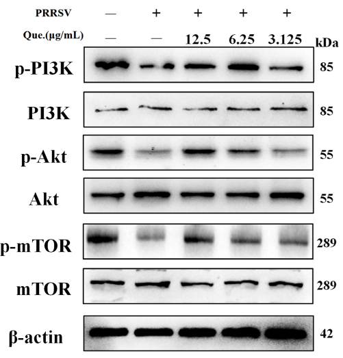
Quercetin Regulates Autophagy to Inhibit PRRSV Replication Through the PI3K/Akt/mTOR Signaling Pathway. [Abstract]2025 Dec 17;17(12):1637. PMID: 41472306
SQSTM1/p62 Antibody (YA062) purchased from MedChemExpress. Usage Cited in: Viruses. 2025 Dec 17;17(12):1637. [Abstract]
Quercetin suppresses the PI3K/Akt/mTOR signaling pathway. MARC-145 cells were infected with PRRSV (MOI of 0.8) and treated with quercetin for 48 h.


Verification Images
-
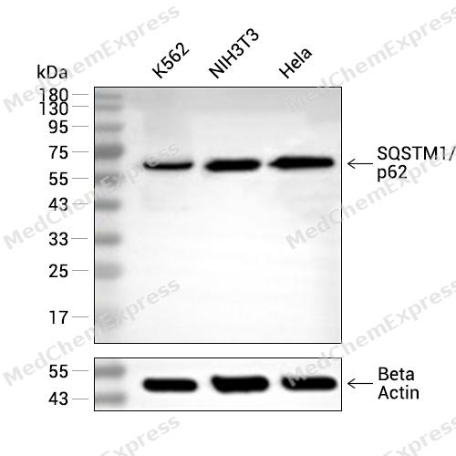 Western blot analysis of extracts from K562 (lane 2(20μg), NIH3T3 (lane 3(20μg) and Hela (lane 4(20μg) using SQSTM1/p62 (HY-P80899) Rabbit mAb. Proteins were transferred to a PVDF membrane and blocked with 5% non-fat milk in TBST for 2 hour at room temperature. The primary antibody (1/1000) and Loading control antibody (Beta Actin, HY-P80438, 1/10000) was used in 5% non-fat milk in TBST at 4°C overnight. Goat Anti-Mouse/Rabbit IgG-HRP Secondary Antibody (1/10000) was used for 1 hour at room temperature.
 -
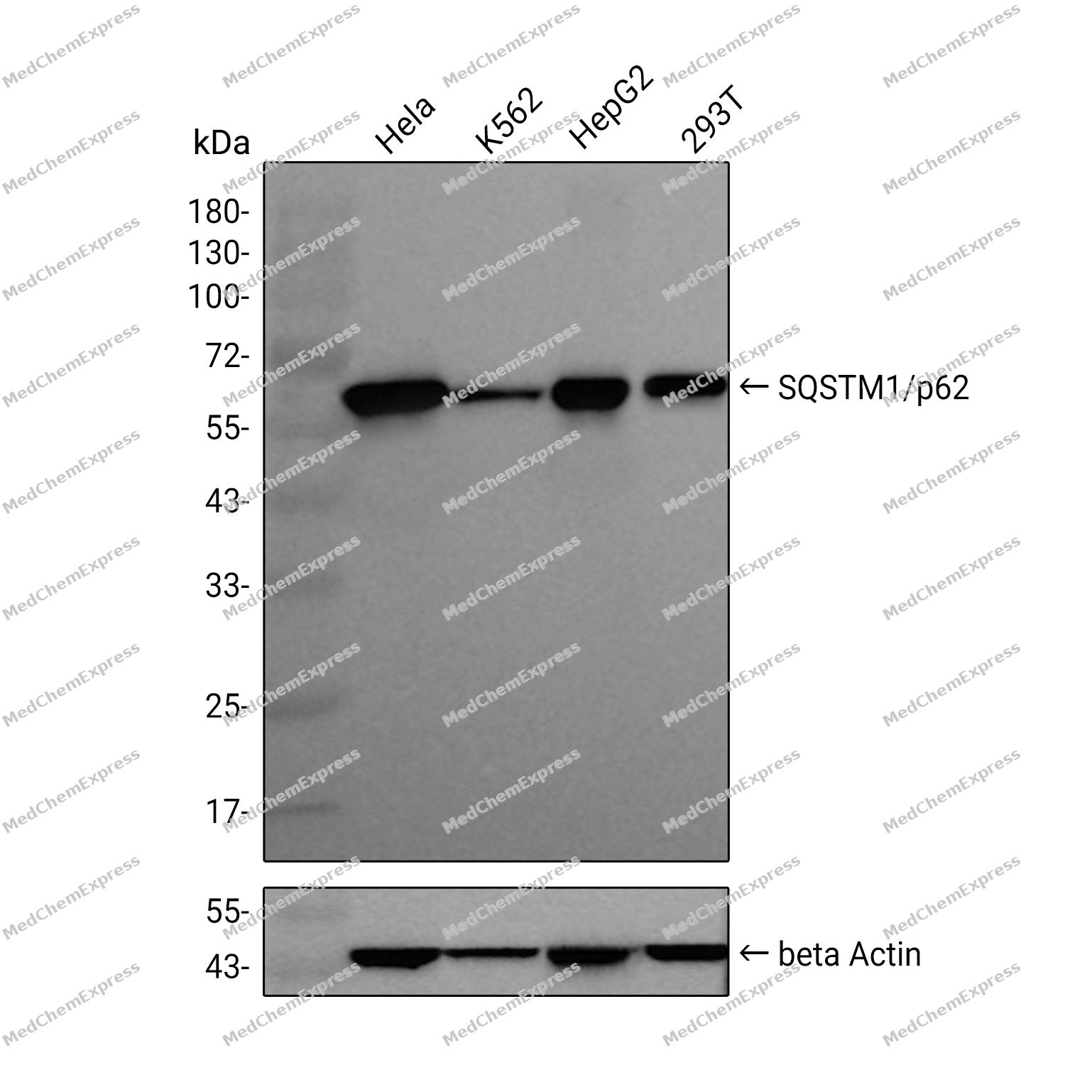
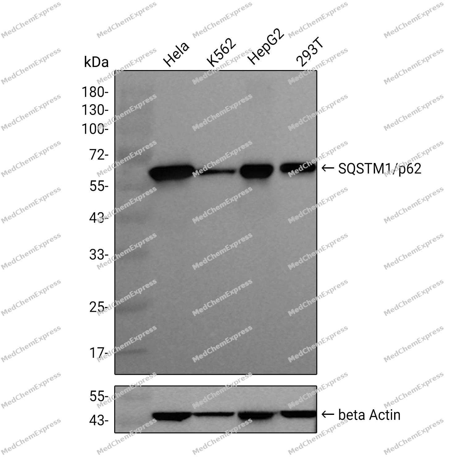 Western blot analysis was performed on extracts from Hela (lane 1, 15 μg), K562 (lane 2, 15 μg), HepG2 (lane 3, 15 μg), and 293T (lane 4, 15 μg) using SQSTM1/p62 Rabbit mAb. Proteins were transferred to a PVDF membrane and blocked with 5% non - fat milk in TBST at 4°C overnight. The primary antibody (1:1000 dilution) and the loading control antibody (beta-Actin(HRP), HY-P80993, 1:5000 dilution) was incubated in 5% non-fat milk in TBST for 1 hour at 37°C. Goat Anti - Rabbit IgG - HRP Secondary Antibody (1:20000 dilution) was then applied for 40 minutes at 37°C.
 -
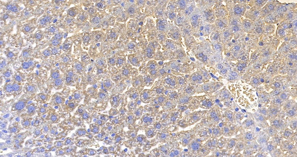 Immunohistochemical analysis of paraffin-embedded mouse liver tissue using SQSTM1p62 Antibody. The section was pre-treated using heat mediated antigen retrieval with sodium citrate buffer (pH 6.0) for 8 minutes. The tissues were blocked in QuickBlock for 20 minutes at room temperature, washed with ddH2O and PBS, and then probed with the primary antibody at 1/100 dilution in 4℃ overnight. The detection was performed using an HRP conjugated compact polymer system. DAB was used as the chromogen. Tissues were counterstained with hematoxylin and mounted with DPX.
 -
Immunohistochemical analysis of paraffin-embedded mouse liver tissue using SQSTM1p62 Antibody. The section was pre-treated using heat mediated antigen retrieval with sodium citrate buffer (pH 6.0) for 8 minutes. The tissues were blocked in QuickBlock for 20 minutes at room temperature, washed with ddH2O and PBS, and then probed with the primary antibody at 1/100 dilution in 4℃ overnight. The detection was performed using an HRP conjugated compact polymer system. DAB was used as the chromogen. Tissues were counterstained with hematoxylin and mounted with DPX.
-
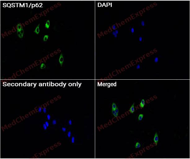 Immunocytochemistry analysis of A549 cells labeling SQSTM1/p62 with SQSTM1/p62 Antibody (HY-P80899) at 1/50 dilution. Cells were fixed in 4% paraformaldehyde for 15 minutes at room temperature, permeabilized with 0.1% Triton X-100 for 10 minutes at room temperature, then blocked with QuickBlock™ Blocking Buffer for Immunol Staining for 10 min at room temperature. Cells were then incubated with SQSTM1/p62 antibody (HY-P80899) at 1/50 dilution in QuickBlock™ Blocking Buffer for Immunol Staining at 4 ℃. Alexa Fluor® 488-conjugated AffiniPure Goat Anti-Rabbit IgG H&L(HY-P8002, Green) was used as the secondary antibody at 1/1,000 dilution. PBS instead of the primary antibody was used as the secondary antibody only control. The Nuclear counterstain was DAPI (Blue).
 -
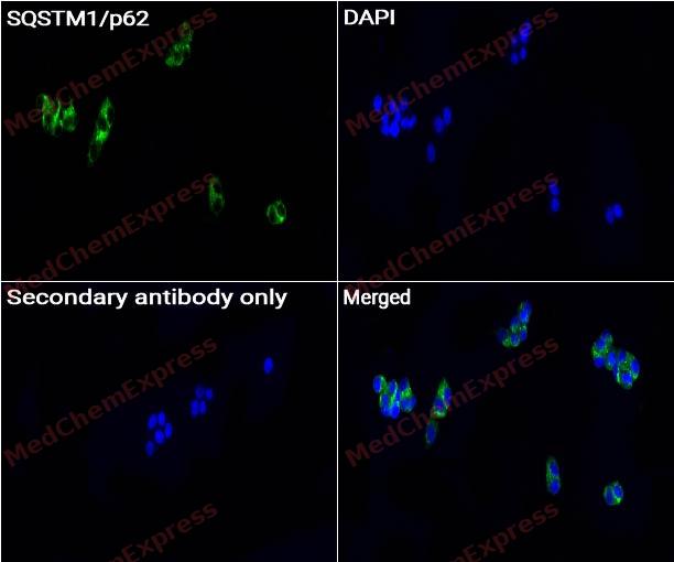 Immunocytochemistry analysis of HepG2 cells labeling SQSTM1/p62 with SQSTM1/p62 Antibody (HY-P80899) at 1/50 dilution. Cells were fixed in 4% paraformaldehyde for 15 minutes at room temperature, permeabilized with 0.1% Triton X-100 for 10 minutes at room temperature, then blocked with QuickBlock™ Blocking Buffer for Immunol Staining for 10 min at room temperature. Cells were then incubated with SQSTM1/p62 antibody (HY-P80899) at 1/200 dilution in QuickBlock™ Blocking Buffer for Immunol Staining at 4 ℃. Alexa Fluor® 488-conjugated AffiniPure Goat Anti-Rabbit IgG H&L(HY-P8002, Green) was used as the secondary antibody at 1/1,000 dilution. PBS instead of the primary antibody was used as the secondary antibody only control. The Nuclear counterstain was DAPI (Blue).


Background
-
Function
SQSTM1/p62 is a Molecular adapter required for selective macroautophagy (aggrephagy) by acting as a bridge between polyubiquitinated proteins and autophagosomes. Promotes the recruitment of ubiquitinated cargo proteins to autophagosomes via multiple domains that bridge proteins and organelles in different steps. SQSTM1 first mediates the assembly and removal of ubiquitinated proteins by undergoing liquid-liquid phase separation upon binding to ubiquitinated proteins via its UBA domain, leading to the formation of insoluble cytoplasmic inclusions, known as p62 bodies. SQSTM1 then interacts with ATG8 family proteins on autophagosomes via its LIR motif, leading to p62 body recruitment to autophagosomes, followed by autophagic clearance of ubiquitinated proteins. SQSTM1 is itself degraded along with its ubiquitinated cargos. Also required to recruit ubiquitinated proteins to PML bodies in the nucleus. Also involved in autophagy of peroxisomes (pexophagy) in response to reactive oxygen species (ROS) by acting as a bridge between ubiquitinated PEX5 receptor and autophagosomes. Acts as an activator of the NFE2L2/NRF2 pathway via interaction with KEAP1: interaction inactivates the BCR(KEAP1) complex by sequestering the complex in inclusion bodies, promoting nuclear accumulation of NFE2L2/NRF2 and subsequent expression of cytoprotective genes. Promotes relocalization of 'Lys-63'-linked ubiquitinated STING1 to autophagosomes. Involved in endosome organization by retaining vesicles in the perinuclear cloud: following ubiquitination by RNF26, attracts specific vesicle-associated adapters, forming a molecular bridge that restrains cognate vesicles in the perinuclear region and organizes the endosomal pathway for efficient cargo transport. Sequesters tensin TNS2 into cytoplasmic puncta, promoting TNS2 ubiquitination and proteasomal degradation. May regulate the activation of NFKB1 by TNF-alpha, nerve growth factor (NGF) and interleukin-1. May play a role in titin/TTN downstream signaling in muscle cells. Adapter that mediates the interaction between TRAF6 and CYLD[1][2][3][4][5][6][7][8][9][10][11][12][13][14][15][16][17][18][19][20][21][22][23][24][25][26][27][28][29][30][31][32][33][34].
-
Subcellular Localization
Cytoplasmic vesicle, autophagosome; Preautophagosomal structure; Cytoplasm, cytosol; Nucleus, PML body; Late endosome; Lysosome; Nucleus; Endoplasmic reticulum; Cytoplasm, myofibril, sarcomere
-
Expression
Tissue_specificity:Ubiquitously expressed.
Induction:By proteasomal inhibitor PSI and prostaglandin J2 (PGJ2) (at protein level) . By phorbol 12-myristate 13-acetate (PMA) . Expression is directly activated by NFE2L2/NRF2; creating a positive feedback loop (PubMed:20452972) -
Isoforms & Post-Translational Modification
Q13501 has 2 isomers: Q13501-1: 47687 Da (predicted); Q13501-2: 38629 Da (predicted).
Phosphorylation at Ser-407 by ULK1 destabilizes the UBA dimer interface and increases binding affinity to ubiquitinated proteins (By similarity). Phosphorylation at Ser-407 also primes for subsequent phosphorylation at Ser-403 (By similarity). Phosphorylation at Ser-403 by CK2 or ULK1 promotes binding to ubiquitinated proteins by increasing the affinity between the UBA domain and polyubiquitin chains (PubMed:22017874, PubMed:25040165). Phosphorylation at Ser-403 by ULK1 is stimulated by SESN2 (PubMed:25040165). Phosphorylated at Ser-403 by TBK1, leading to promote relocalization of 'Lys-63'-linked ubiquitinated STING1 to autophagosomes (PubMed:29496741). Phosphorylation at Ser-349 by ULK1 promotes interaction with KEAP1 and inactivation of the BCR(KEAP1) complex, promoting NFE2L2/NRF2 nuclear accumulation and expression of phase II detoxifying enzymes (PubMed:37306101). Phosphorylated in vitro by TTN (PubMed:15802564);Ubiquitinated by UBE2J1 and RNF26 at Lys-435: ubiquitinated SQSTM1 attracts specific vesicle-associated adapters, forming a molecular bridge that restrains cognate vesicles in the perinuclear region and organizes the endosomal pathway for efficient cargo transport (PubMed:27368102, PubMed:33472082). Ubiquitination by UBE2D2 and UBE2D3 increases its ability to bind polyubiquitin chains by destabilizing the UBA dimer interface (PubMed:28322253). Deubiquitination by USP15 releases target vesicles for fast transport into the cell periphery (PubMed:27368102). Ubiquitinated by the BCR(KEAP1) complex at Lys-420, increasing SQSTM1 sequestering activity and promoting its degradation (PubMed:28380357). Ubiquitinated via 'Lys-29' and 'Lys-33'-linked polyubiquitination leading to xenophagic targeting of bacteria and inhibition of their replication (PubMed:27880896);Acetylated at Lys-420 and Lys-435 by KAT5/TIP60, promotes activity by destabilizing the UBA dimer interface and increases binding affinity to ubiquitinated proteins (PubMed:31857589). Deacetylated by HDAC6 (PubMed:31857589);Palmitoylation at Cys-289 and Cys-290 by ZDHHC19 is required for efficient autophagic degradation of SQSTM1-cargo complexes by promoting affinity for ATG8 proteins and recruitment of p62 bodies to autophagosomes (PubMed:37802024). Dealmitoylated at Cys-289 and Cys-290 by LYPLA1 (PubMed:37802024);(Microbial infection) Cleaved by S.pyogenes SpeB protease; leading to its degradation (PubMed:24331465). Degradation by SpeB prevents autophagy, promoting to S.pyogenes intracellular replication (PubMed:24331465);(Microbial infection) Deubiquitinated by Epstein-Barr virus BPLF1; leading to inhibition of the recruitment of MAP1LC3A/LC3 to SQSTM1-positive structures -
Subunit
Homooligomer or heterooligomer; may form homotypic arrays (PubMed:12887891, PubMed:19931284). Dimerization interferes with ubiquitin binding (PubMed:19931284). Component of a ternary complex with PAWR and PRKCZ (PubMed:11755531). Forms a complex with JUB/Ajuba, PRKCZ and TRAF6 (PubMed:15870274). Identified in a complex with TRAF6 and CYLD (By similarity). Identified in a heterotrimeric complex with ubiquitin and ZFAND5, where ZFAND5 and SQSTM1 both interact with the same ubiquitin molecule (PubMed:21923101). Interacts (via LIR motif) with MAP1LC3A and MAP1LC3B, as well as with other ATG8 family members, including GABARAP, GABARAPL1 and GABARAPL2; these interactions are necessary for the recruitment MAP1 LC3 family members to inclusion bodies containing polyubiquitinated protein aggregates and for their degradation by autophagy (PubMed:16286508, PubMed:17580304, PubMed:22421968, PubMed:24089205, PubMed:24668264). Interacts directly with PRKCI and PRKCZ (PubMed:10356400, PubMed:12813044, PubMed:12887891, PubMed:9566925). Interacts with EBI3, LCK, RASA1, NR2F2, NTRK1, NTRK2, NTRK3, NBR1, MAP2K5 and MAPKAPK5 (PubMed:10708586, PubMed:11244088, PubMed:12471037, PubMed:8551575, PubMed:8618896, PubMed:8650207, PubMed:8910285). Upon TNF-alpha stimulation, interacts with RIPK1 probably bridging IKBKB to the TNF-R1 complex composed of TNF-R1/TNFRSF1A, TRADD and RIPK1 (PubMed:10747026). Interacts with the proteasome subunits PSMD4 and PSMC2 (PubMed:15340068). Interacts with TRAF6 (PubMed:10747026). Interacts with 'Lys-63'-linked polyubiquitinated MAPT/TAU (PubMed:15953362). Interacts with FHOD3 (PubMed:21149568). Interacts with CYLD (PubMed:32185393). Interacts with SESN1 (PubMed:23274085). Interacts with SESN2 (PubMed:23274085, PubMed:25040165). Interacts with ULK1 (PubMed:25040165). Interacts with UBD (PubMed:25422469). Interacts with WDR81; the interaction is direct and regulates the interaction of SQSTM1 with ubiquitinated proteins (PubMed:28404643). Interacts with WDFY3; this interaction is required to recruit WDFY3 to cytoplasmic bodies and to PML bodies (PubMed:20168092). Interacts with LRRC25 (PubMed:29288164). Interacts with STING1; leading to relocalization of STING1 to autophagosomes (PubMed:29496741). Interacts (when phosphorylated at Ser-349) with KEAP1; the interaction is direct and inactivates the BCR(KEAP1) complex by sequestering KEAP1 in inclusion bodies, promoting its degradation (PubMed:20452972, PubMed:20495340, PubMed:37306101). Interacts with MOAP1; promoting dissociation of SQSTM1 inclusion bodies that sequester KEAP1 (PubMed:33393215). Interacts with GBP1 (By similarity). Interacts with TAX1BP1 (PubMed:34471133). Interacts with (ubiquitinated) PEX5; specifically binds PEX5 ubiquitinated at 'Lys-209' in response to reactive oxygen species (ROS) (PubMed:26344566). Interacts (via PB1 domain) with TNS2; the interaction leads to sequestration of TNS2 in cytoplasmic aggregates with SQSTM1 and promotes TNS2 ubiquitination and proteasomal degradation (PubMed:25101860). Interacts with IRS1; the interaction is disrupted by the presence of tensin TNS2 (PubMed:25101860). Interacts with TRIM5 (PubMed:20357094, PubMed:25127057). Interacts with TRIM11 (when ubiquitinated); promoting AIM2 recruitment to autophagosomes and autophagy-dependent degradation of AIM2 (PubMed:27498865). Interacts with TRIM13 (PubMed:22178386). Interacts with TRIM16 (PubMed:30143514). Interacts with TRIM23 (PubMed:28871090). Interacts with TRIM50 (PubMed:22792322). Interacts with TRIM55 (PubMed:15802564). Interacts with ECSIT; this interaction inhibits TLR4 signaling via functional regulation of the TRAF6-ECSIT complex (PubMed:31281713). Interacts with GABRR1, GABRR2 and GABRR3 (By similarity). Interacts with WDR83 (PubMed:38103557). Interacts with GRB2 (PubMed:35831301). Interacts with USP12; the interaction is independent of USP12 deubiquitinase activity and may be involved in regulation of autophagic flux (PubMed:30266909). Interacts with ASB6 (PubMed:34164402)
-
SwissProt ID
-
Synonyms
p60; p62; A170; DMRV; OSIL; PDB3; ZIP3; p62B; NADGP; FTDALS3
-
Research Field
Signal Transduction
Documentation
-
Data Sheet (268 KB)
-
SDS (252 KB)
- English - EN (252 KB)
- Français - FR (252 KB)
- Deutsch - DE (252 KB)
- Norwegian - NO (252 KB)
- Español - ES (252 KB)
- Swedish - SV (252 KB)
- Italian - IT (252 KB)
- Korean - KR (252 KB)
- Portuguese - PT (252 KB)
-
User Guide for Antibodies (1077 KB)
References
[1]. Seibenhener ML, et al. Sequestosome 1/p62 is a polyubiquitin chain binding protein involved in ubiquitin proteasome degradation. Mol Cell Biol. 2004 Sep;24(18):8055-68. [Content Brief]
[2]. Babu JR, et al. Sequestosome 1/p62 shuttles polyubiquitinated tau for proteasomal degradation. J Neurochem. 2005 Jul;94(1):192-203. [Content Brief]
[3]. Bjørkøy G, et al. p62/SQSTM1 forms protein aggregates degraded by autophagy and has a protective effect on huntingtin-induced cell death. J Cell Biol. 2005 Nov 21;171(4):603-14. [Content Brief]
[4]. Pankiv S, et al. p62/SQSTM1 binds directly to Atg8/LC3 to facilitate degradation of ubiquitinated protein aggregates by autophagy. J Biol Chem. 2007 Aug 17;282(33):24131-45. [Content Brief]
[5]. Clausen TH, et al. p62/SQSTM1 and ALFY interact to facilitate the formation of p62 bodies/ALIS and their degradation by autophagy. Autophagy. 2010 Apr;6(3):330-44. [Content Brief]
[6]. Matsumoto G, et al. Serine 403 phosphorylation of p62/SQSTM1 regulates selective autophagic clearance of ubiquitinated proteins. Mol Cell. 2011 Oct 21;44(2):279-89. [Content Brief]
[7]. Taillebourg E, et al. The deubiquitinating enzyme USP36 controls selective autophagy activation by ubiquitinated proteins. Autophagy. 2012 May 1;8(5):767-79. [Content Brief]
[8]. Isakson P, et al. TRAF6 mediates ubiquitination of KIF23/MKLP1 and is required for midbody ring degradation by selective autophagy. Autophagy. 2013 Dec;9(12):1955-64. [Content Brief]
[9]. Liu X, et al. The BEACH-containing protein WDR81 coordinates p62 and LC3C to promote aggrephagy. J Cell Biol. 2017 May 1;216(5):1301-1320. [Content Brief]
[10]. Zaffagnini G, et al. p62 filaments capture and present ubiquitinated cargos for autophagy. EMBO J. 2018 Mar 1;37(5):. [Content Brief]
[11]. Sun D, et al. Polyubiquitin chain-induced p62 phase separation drives autophagic cargo segregation. Cell Res. 2018 Apr;28(4):405-415. [Content Brief]
[12]. You Z, et al. Requirement for p62 acetylation in the aggregation of ubiquitylated proteins under nutrient stress. Nat Commun. 2019 Dec 19;10(1):5792. [Content Brief]
[13]. Ylä-Anttila P, et al. The Epstein-Barr virus deubiquitinase BPLF1 targets SQSTM1/p62 to inhibit selective autophagy. Autophagy. 2021 Nov;17(11):3461-3474. [Content Brief]
[14]. Turco E, et al. Reconstitution defines the roles of p62, NBR1 and TAX1BP1 in ubiquitin condensate formation and autophagy initiation. Nat Commun. 2021 Sep 1;12(1):5212. [Content Brief]
[15]. Heo AJ, et al. The N-terminal cysteine is a dual sensor of oxygen and oxidative stress. Proc Natl Acad Sci U S A. 2021 Dec 14;118(50):. [Content Brief]
[16]. Hou B, et al. Grb2 interacts with necrosome components and is involved in rasfonin-induced necroptosis. Cell Death Discov. 2022 Jul 13;8(1):319. [Content Brief]
[17]. Ikeda R, et al. Phosphorylation of phase-separated p62 bodies by ULK1 activates a redox-independent stress response. EMBO J. 2023 Jul 17;42(14):e113349. [Content Brief]
[18]. Huang X, et al. S-acylation of p62 promotes p62 droplet recruitment into autophagosomes in mammalian autophagy. Mol Cell. 2023 Oct 5;83(19):3485-3501.e11. [Content Brief]
[19]. Wang Z, et al. Inhibition of sequestosome 1/p62 up-regulation prevents aggregation of ubiquitinated proteins induced by prostaglandin J2 without reducing its neurotoxicity. Mol Cell Neurosci. 2005 Jun;29(2):222-31. [Content Brief]
[20]. Zhang J, et al. ATM functions at the peroxisome to induce pexophagy in response to ROS. Nat Cell Biol. 2015 Oct;17(10):1259-1269. [Content Brief]
[21]. Jain A, et al. p62/SQSTM1 is a target gene for transcription factor NRF2 and creates a positive feedback loop by inducing antioxidant response element-driven gene transcription. J Biol Chem. 2010 Jul 16;285(29):22576-91. [Content Brief]
[22]. Lee Y, et al. Keap1/Cullin3 Modulates p62/SQSTM1 Activity via UBA Domain Ubiquitination. Cell Rep. 2017 Apr 4;19(1):188-202. [Content Brief]
[23]. Tan CT, et al. MOAP-1-mediated dissociation of p62/SQSTM1 bodies releases Keap1 and suppresses Nrf2 signaling. EMBO Rep. 2021 Jan 7;22(1):e50854. [Content Brief]
[24]. Prabakaran T, et al. Attenuation of cGAS-STING signaling is mediated by a p62/SQSTM1-dependent autophagy pathway activated by TBK1. EMBO J. 2018 Apr 13;37(8):. [Content Brief]
[25]. Jongsma ML, et al. An ER-Associated Pathway Defines Endosomal Architecture for Controlled Cargo Transport. Cell. 2016 Jun 30;166(1):152-66. [Content Brief]
[26]. Cremer T, et al. The ER-embedded UBE2J1/RNF26 ubiquitylation complex exerts spatiotemporal control over the endolysosomal pathway. Cell Rep. 2021 Jan 19;34(3):108659. [Content Brief]
[27]. Koh A, et al. Regulation of C1-Ten protein tyrosine phosphatase by p62/SQSTM1-mediated sequestration and degradation. Cell Signal. 2014 Nov;26(11):2470-80. [Content Brief]
[28]. Sanz L, et al. The interaction of p62 with RIP links the atypical PKCs to NF-kappaB activation. EMBO J. 1999 Jun 1;18(11):3044-53. [Content Brief]
[29]. Sanz L, et al. The atypical PKC-interacting protein p62 channels NF-kappaB activation by the IL-1-TRAF6 pathway. EMBO J. 2000 Apr 3;19(7):1576-86. [Content Brief]
[30]. Wooten MW, et al. The atypical protein kinase C-interacting protein p62 is a scaffold for NF-kappaB activation by nerve growth factor. J Biol Chem. 2001 Mar 16;276(11):7709-12. [Content Brief]
[31]. Geetha T, et al. Association of the atypical protein kinase C-interacting protein p62/ZIP with nerve growth factor receptor TrkA regulates receptor trafficking and Erk5 signaling. J Biol Chem. 2003 Feb 14;278(7):4730-9. [Content Brief]
[32]. Wooten MW, et al. The p62 scaffold regulates nerve growth factor-induced NF-kappaB activation by influencing TRAF6 polyubiquitination. J Biol Chem. 2005 Oct 21;280(42):35625-9. [Content Brief]
[33]. Long J, et al. Dimerisation of the UBA domain of p62 inhibits ubiquitin binding and regulates NF-kappaB signalling. J Mol Biol. 2010 Feb 12;396(1):178-94. [Content Brief]
[34]. Lange S, et al. The kinase domain of titin controls muscle gene expression and protein turnover. Science. 2005 Jun 10;308(5728):1599-603. [Content Brief]

 Powered by Bioz
