



Myelin Basic Protein Antibody (YA5613)
(Synonyms: GDB; Golli MBP; Golli MBP; myelin basic protein; Hemopoietic MBP; HMBPR; HUGO; MBP; MBP_CAVPO; MBP_HUMAN; MGC99675; MLD; Myelin A1 protein; Myelin A1 Protein, basic; Myelin basic protein; Myelin Deficient; Myelin membrane encephalitogenic protein; OTTHUMP00000163776; OTTHUMP00000174387; OTTHUMP00000174388; SHI; Shiverer; SP)
Based on 1 Customer Validation
Myelin Basic Protein Antibody (YA5613) is a Mouse-derived and non-conjugated IgG1 monoclonal antibody, targeting to Myelin Basic Protein.
-
Host:
Mouse
-
Isotype:
IgG1
-
Application:
IHC-P, WB, ICC/IF, ELISA
-
Reactivity :
Human, Mouse, Rat
-
Formulation:
Supplied in PBS, 50% glycerol, 0.05% Proclin 300, 0.05%BSA
-
Conjugation:
Non-conjugated


To place orders, for customer services and technical support, please contact: MedChemExpress USA
Tel: 609-228-6898 E-mail: [email protected] [email protected]





Applications
| Application |
IHC-P

IHC-P: Immunohistochemistry-Paraffin
|
WB
WB: Western Blot
|
ICC/IF
ICC/IF: Immunocytochemistry/
Immunofluorescence |
ELISA
ELISA: Enzyme Linked Immunosorbent Assay
|
|---|---|---|---|---|
| Dilution Ratio | 1:200-1000 | 1:500-2000 | 1:100-500 | 1:1000-5000 |


Product Details
Myelin Basic Protein Antibody (YA5613) is a Mouse-derived and non-conjugated IgG1 monoclonal antibody, targeting to Myelin Basic Protein.
-
Host Mouse

-
Clonality Monoclonal

-
Species ReactivityHuman, Mouse, Rat
-
Observed Molecular WeightObserved band size: 33 kDa
Note: Due to possible protein modifications or aggregation, the molecular weight should be confirmed by actual measurement, and the predicted value is for reference only.

-
Calculated Molecular Weight Predicted band size: 33 kDa

Synthesized peptide derived from human Myelin Basic Protein(MBP) AA range: 150-250
Protein G
Non-conjugated
Unmodified
IgG1


Product Properties
-
Appearance
Solution
-
Formulation
Supplied in PBS, 50% glycerol, 0.05% Proclin 300, 0.05%BSA
-
Concentration
Batch-dependent, Please check the COA for the concentration of each lot. Check Lot Concentration
-
Storage & Stability
Stored at -20°C for 1 year. Avoid repeated freeze / thaw cycles.
-
Shipping
Shipping with blue ice.


Verification Images
-
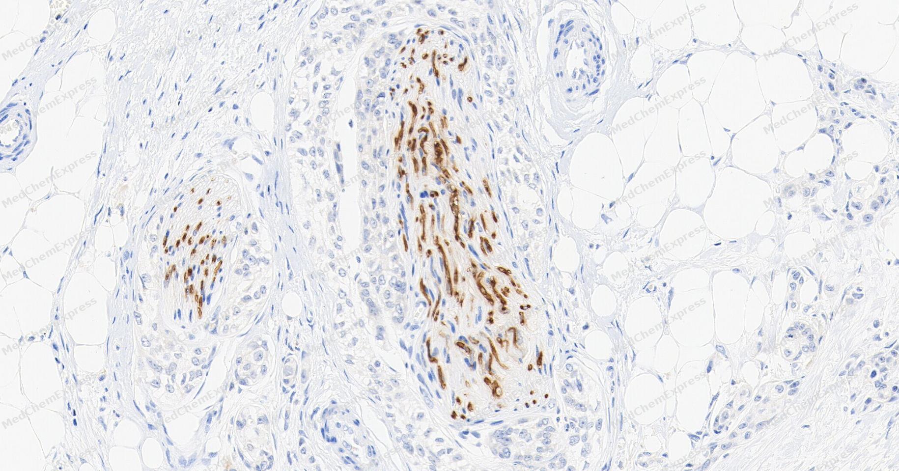 Immunohistochemical analysis of paraffin-embedded human bladder tissue using Myelin Basic Protein Antibody (YA5613). The section was pre-treated using heat mediated antigen retrieval with Tris/EDTA buffer (pH 9.0) for 20 minutes. The tissues were probed with the primary antibody (HY-P85921, 1/200) overnight at 4℃. The detection was performed using an HRP conjugated compact polymer system. DAB was used as the chromogen. Tissues were counterstained with hematoxylin and mounted with neutral balsam.
 -
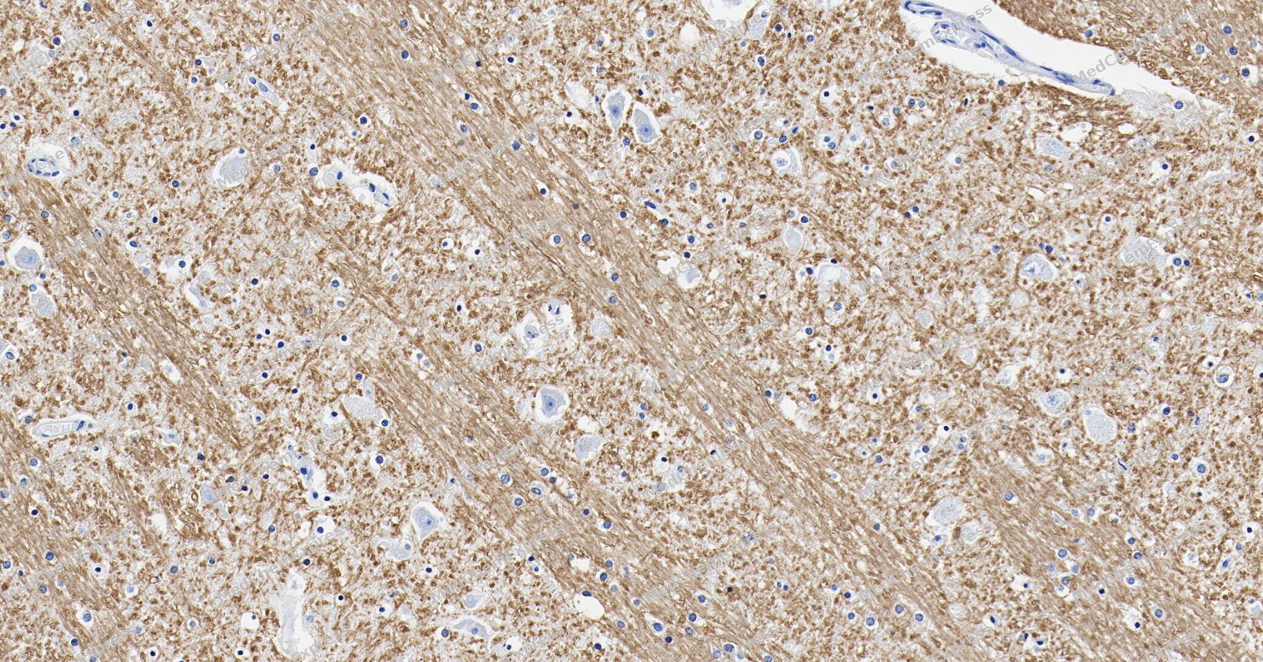 Immunohistochemical analysis of paraffin-embedded human brain tissue using Myelin Basic Protein Antibody (YA5613). The section was pre-treated using heat mediated antigen retrieval with Tris/EDTA buffer (pH 9.0) for 20 minutes. The tissues were probed with the primary antibody (HY-P85921, 1/200) overnight at 4℃. The detection was performed using an HRP conjugated compact polymer system. DAB was used as the chromogen. Tissues were counterstained with hematoxylin and mounted with neutral balsam.
 -
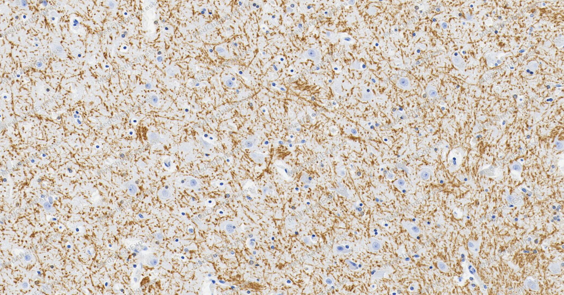 Immunohistochemical analysis of paraffin-embedded human thalamus tissue using Myelin Basic Protein Antibody (YA5613). The section was pre-treated using heat mediated antigen retrieval with Tris/EDTA buffer (pH 9.0) for 20 minutes. The tissues were probed with the primary antibody (HY-P85921, 1/200) overnight at 4℃. The detection was performed using an HRP conjugated compact polymer system. DAB was used as the chromogen. Tissues were counterstained with hematoxylin and mounted with neutral balsam.
 -
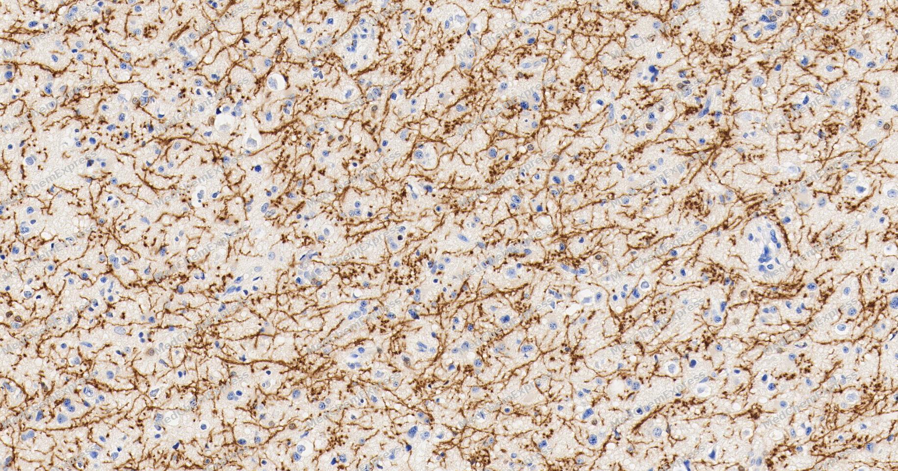 Immunohistochemical analysis of paraffin-embedded human astrocytoma tissue using Myelin Basic Protein Antibody (YA5613). The section was pre-treated using heat mediated antigen retrieval with Tris/EDTA buffer (pH 9.0) for 20 minutes. The tissues were probed with the primary antibody (HY-P85921, 1/200) overnight at 4℃. The detection was performed using an HRP conjugated compact polymer system. DAB was used as the chromogen. Tissues were counterstained with hematoxylin and mounted with neutral balsam.
 -
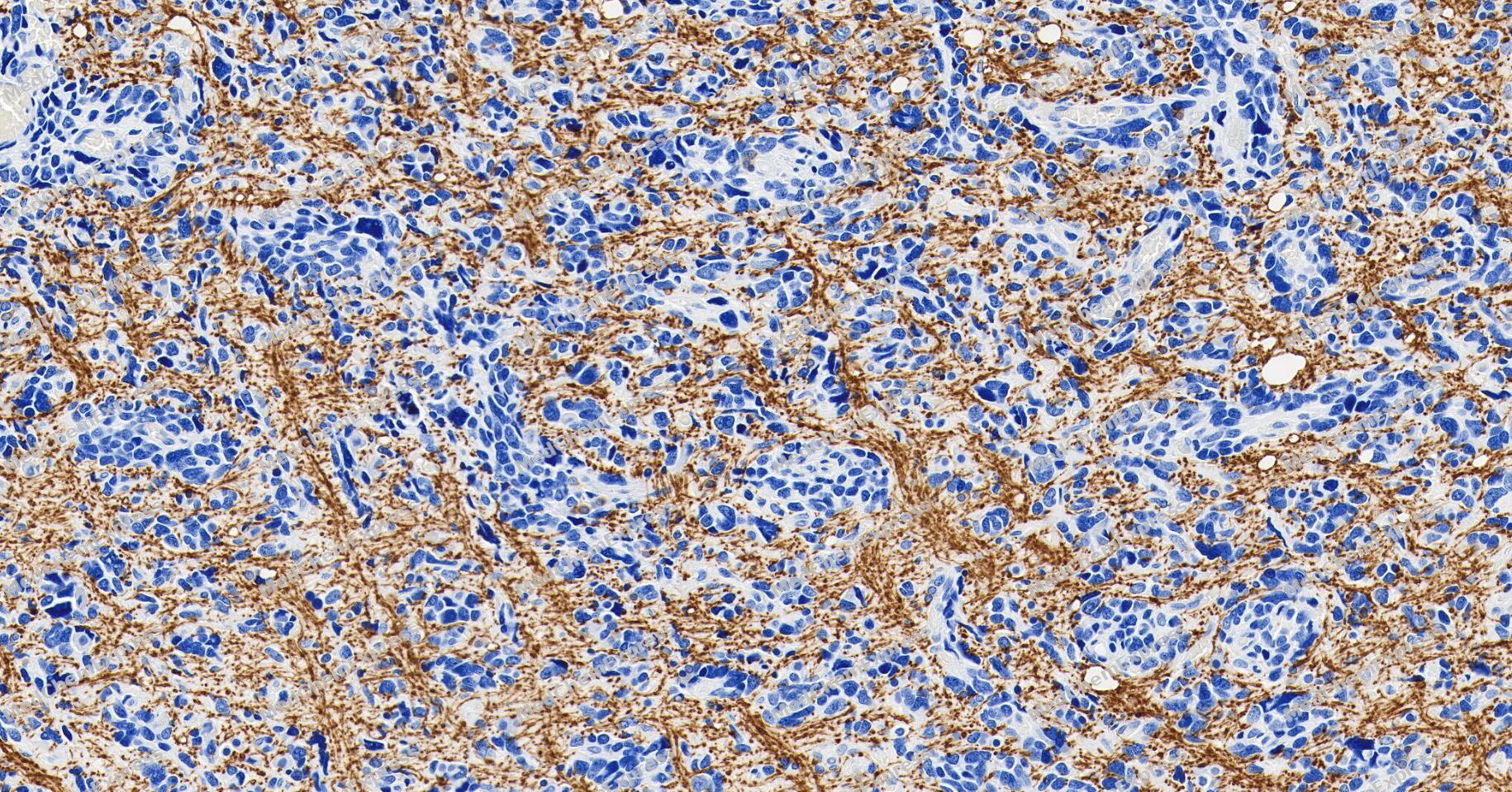 Immunohistochemical analysis of paraffin-embedded human brain cancer tissue using Myelin Basic Protein Antibody (YA5613). The section was pre-treated using heat mediated antigen retrieval with Tris/EDTA buffer (pH 9.0) for 20 minutes. The tissues were probed with the primary antibody (HY-P85921, 1/200) overnight at 4℃. The detection was performed using an HRP conjugated compact polymer system. DAB was used as the chromogen. Tissues were counterstained with hematoxylin and mounted with neutral balsam.
 -
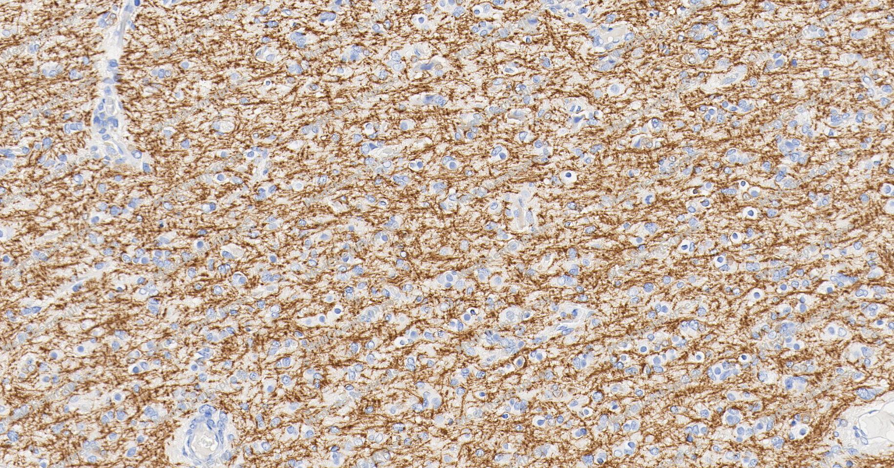 Immunohistochemical analysis of paraffin-embedded human glioma tissue using Myelin Basic Protein Antibody (YA5613). The section was pre-treated using heat mediated antigen retrieval with Tris/EDTA buffer (pH 9.0) for 20 minutes. The tissues were probed with the primary antibody (HY-P85921, 1/200) overnight at 4℃. The detection was performed using an HRP conjugated compact polymer system. DAB was used as the chromogen. Tissues were counterstained with hematoxylin and mounted with neutral balsam.
 -
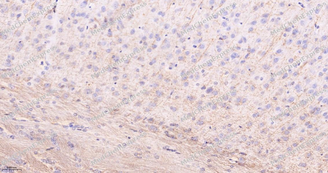 Immunohistochemical analysis of paraffin-embedded mouse hippocampus tissue using Myelin Basic Protein Antibody (HY-P85921, 1/1000). The section was pretreated using heat mediated antigen retrieval with sodium citrate buffer (pH 6.0) for 8 minutes. The tissues were blocked with quick block buffer for 0.5 hours at room temperature, washed with PBS and PBST, and then incubated with the primary antibody overnight at 4℃. The detection was performed using an HRP conjugated compact polymer system. DAB was used as the chromogen. Tissues were counterstained with hematoxylin and mounted with DPX.
 -
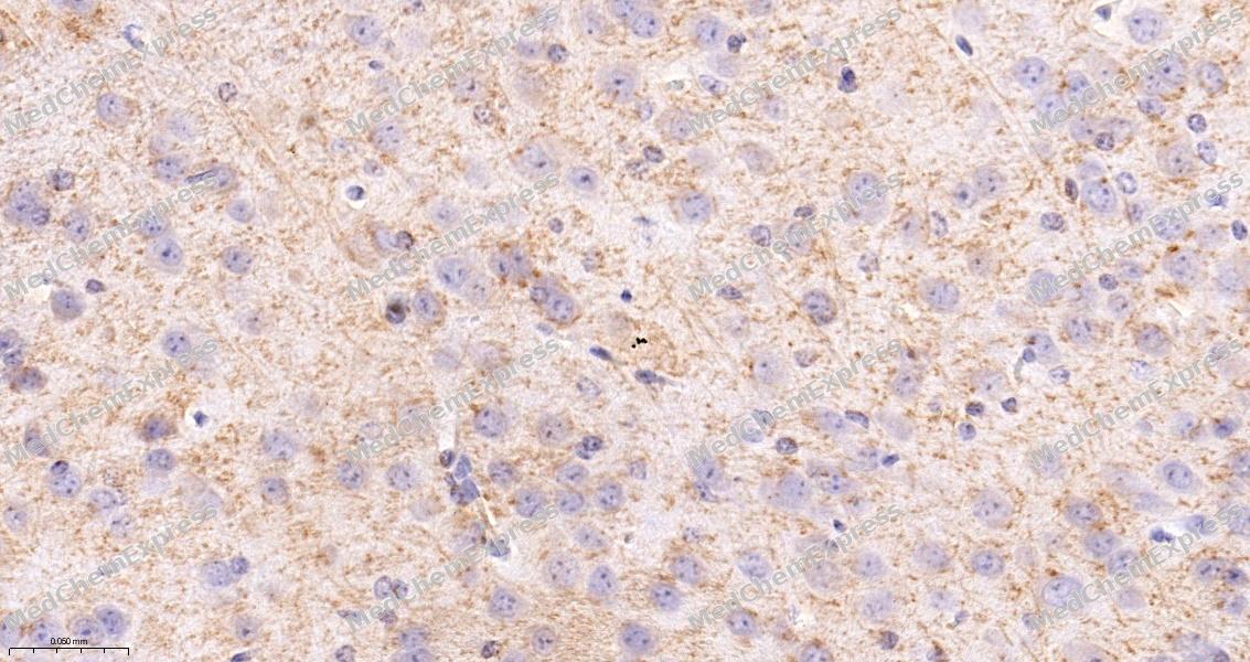 Immunohistochemical analysis of paraffin-embedded mouse caudate tissue using Myelin Basic Protein Antibody (HY-P85921, 1/1000). The section was pretreated using heat mediated antigen retrieval with sodium citrate buffer (pH 6.0) for 8 minutes. The tissues were blocked with quick block buffer for 0.5 hours at room temperature, washed with PBS and PBST, and then incubated with the primary antibody overnight at 4℃. The detection was performed using an HRP conjugated compact polymer system. DAB was used as the chromogen. Tissues were counterstained with hematoxylin and mounted with DPX.
 -
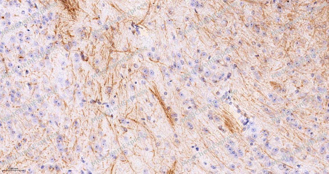 Immunohistochemical analysis of paraffin-embedded mouse cerebellum tissue using Myelin Basic Protein Antibody (HY-P85921, 1/1000). The section was pretreated using heat mediated antigen retrieval with sodium citrate buffer (pH 6.0) for 8 minutes. The tissues were blocked with quick block buffer for 0.5 hours at room temperature, washed with PBS and PBST, and then incubated with the primary antibody overnight at 4℃. The detection was performed using an HRP conjugated compact polymer system. DAB was used as the chromogen. Tissues were counterstained with hematoxylin and mounted with DPX.
 -
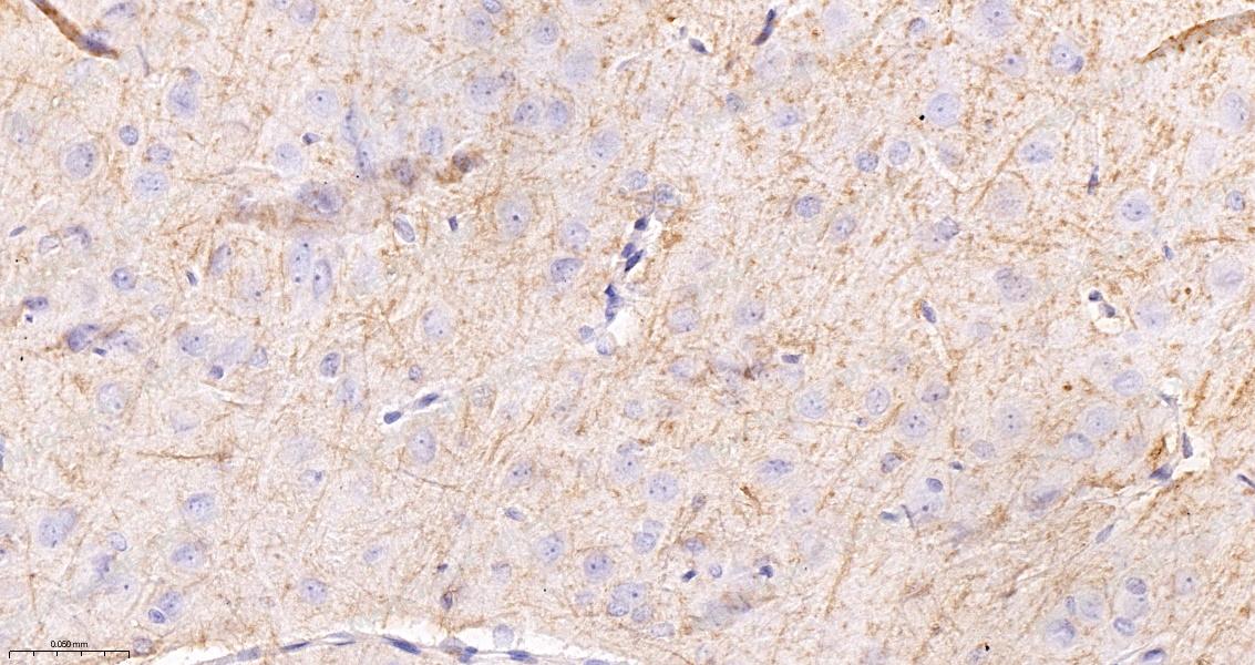 Immunohistochemical analysis of paraffin-embedded rat hippocampus tissue using Myelin Basic Protein Antibody (HY-P85921, 1/1000). The section was pretreated using heat mediated antigen retrieval with sodium citrate buffer (pH 6.0) for 8 minutes. The tissues were blocked with quick block buffer for 0.5 hours at room temperature, washed with PBS and PBST, and then incubated with the primary antibody overnight at 4℃. The detection was performed using an HRP conjugated compact polymer system. DAB was used as the chromogen. Tissues were counterstained with hematoxylin and mounted with DPX.
 -
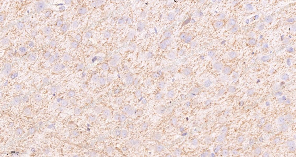 Immunohistochemical analysis of paraffin-embedded rat caudate tissue using Myelin Basic Protein Antibody (HY-P85921, 1/1000). The section was pretreated using heat mediated antigen retrieval with sodium citrate buffer (pH 6.0) for 8 minutes. The tissues were blocked with quick block buffer for 0.5 hours at room temperature, washed with PBS and PBST, and then incubated with the primary antibody overnight at 4℃. The detection was performed using an HRP conjugated compact polymer system. DAB was used as the chromogen. Tissues were counterstained with hematoxylin and mounted with DPX.
 -
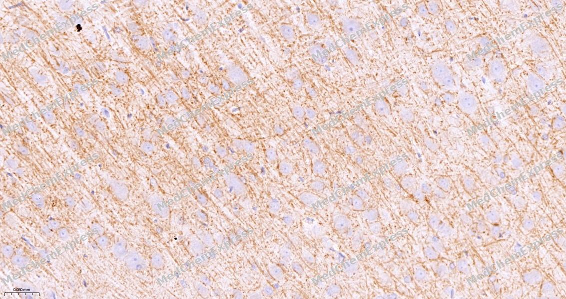 Immunohistochemical analysis of paraffin-embedded rat cerebral cortex tissue using Myelin Basic Protein Antibody (HY-P85921, 1/1000). The section was pretreated using heat mediated antigen retrieval with sodium citrate buffer (pH 6.0) for 8 minutes. The tissues were blocked with quick block buffer for 0.5 hours at room temperature, washed with PBS and PBST, and then incubated with the primary antibody overnight at 4℃. The detection was performed using an HRP conjugated compact polymer system. DAB was used as the chromogen. Tissues were counterstained with hematoxylin and mounted with DPX.
 -
 Immunocytochemistry analysis of PC-3 cells labeling Myelin Basic Protein with Myelin Basic Protein Antibody (HY-P85921) at 1/200 dilution. Cells were fixed in 4% paraformaldehyde for 15 minutes at room temperature, permeabilized with 0.1% Triton X-100 in PBS for 15 minutes at room temperature, then blocked with quick block buffer for 10 minutes at room temperature. Cells were then incubated with Myelin Basic Protein Antibody (HY-P85921) at 1/200 dilution in quick block buffer overnight at 4 ℃.AF488-conjugated Goat Anti-Mouse IgG H&L (HY-P8005, Green) was used as the secondary antibody at 1/1,000 dilution. PBS instead of the primary antibody was used as the secondary antibody only control. The Nuclear counterstain was DAPI (Blue).
 -
 Immunocytochemistry analysis of SH-SY5Y cells labeling Myelin Basic Protein with Myelin Basic Protein Antibody (HY-P85921) at 1/200 dilution. Cells were fixed in 4% paraformaldehyde for 15 minutes at room temperature, permeabilized with 0.1% Triton X-100 in PBS for 15 minutes at room temperature, then blocked with quick block buffer for 10 minutes at room temperature. Cells were then incubated with Myelin Basic Protein Antibody (HY-P85921) at 1/200 dilution in quick block buffer overnight at 4 ℃.AF488-conjugated Goat Anti-Mouse IgG H&L (HY-P8005, Green) was used as the secondary antibody at 1/1,000 dilution. PBS instead of the primary antibody was used as the secondary antibody only control. The Nuclear counterstain was DAPI (Blue).


Background
-
Function
Myelin Basic Protein is a The classic group of MBP isoforms (isoform 4-isoform 14) are with PLP the most abundant protein components of the myelin membrane in the CNS. They have a role in both its formation and stabilization. The smaller isoforms might have an important role in remyelination of denuded axons in multiple sclerosis. The non-classic group of MBP isoforms (isoform 1-isoform 3/Golli-MBPs) may preferentially have a role in the early developing brain long before myelination, maybe as components of transcriptional complexes, and may also be involved in signaling pathways in T-cells and neural cells. Differential splicing events combined with optional post-translational modifications give a wide spectrum of isomers, with each of them potentially having a specialized function. Induces T-cell proliferation
-
Subcellular Localization
Myelin membrane; Peripheral membrane protein; Cytoplasmic side; Nucleus
-
Expression
Tissue_specificity:MBP isoforms are found in the central and peripheral nervous systems, while the Golli-MBP isoform is expressed in the fetal thymus, spleen, spinal cord, and cell lines derived from the immune system. -
Isoforms & Post-Translational Modification
P02686 has 6 isomers: P02686-1: 33117 Da (predicted); P02686-2: 21522 Da (predicted); P02686-3: 21493 Da (predicted); P02686-4: 20246 Da (predicted); P02686-5: 18591 Da (predicted); P02686-6: 17343 Da (predicted).
Several charge isomers of MBP; C1 (the most cationic, least modified, and most abundant form), C2, C3, C4, C5, C6, C7, C8-A and C8-B (the least cationic form); are produced as a result of optional PTM, such as phosphorylation, deamidation of glutamine or asparagine, arginine citrullination and methylation. C8-A and C8-B contain each two mass isoforms termed C8-A(H), C8-A(L), C8-B(H) and C8-B(L), (H) standing for higher and (L) for lower molecular weight. C3, C4 and C5 are phosphorylated. The ratio of methylated arginine residues decreases during aging, making the protein more cationic;The N-terminal alanine is acetylated (isoform 3, isoform 4, isoform 5 and isoform 6);Arg-241 was found to be 6% monomethylated and 60% symmetrically dimethylated;Proteolytically cleaved in B cell lysosomes by cathepsin CTSG which degrades the major immunogenic MBP epitope and prevents the activation of MBP-specific autoreactive T cells;Phosphorylated by TAOK2, VRK2, MAPK11, MAPK12, MAPK14 and MINK1 -
Subunit
Homodimer. Isoform 3 exists as a homodimer
-
SwissProt ID
-
Synonyms
GDB; Golli MBP; Golli MBP; myelin basic protein; Hemopoietic MBP; HMBPR; HUGO; MBP; MBP_CAVPO; MBP_HUMAN; MGC99675; MLD; Myelin A1 protein; Myelin A1 Protein, basic; Myelin basic protein; Myelin Deficient; Myelin membrane encephalitogenic protein; OTTHUMP00000163776; OTTHUMP00000174387; OTTHUMP00000174388; SHI; Shiverer; SP


Documentation
-
Data Sheet (234 KB)
-
SDS (252 KB)
- English - EN (252 KB)
- Français - FR (252 KB)
- Deutsch - DE (252 KB)
- Norwegian - NO (252 KB)
- Español - ES (252 KB)
- Swedish - SV (252 KB)
- Italian - IT (252 KB)
- Korean - KR (252 KB)
- Portuguese - PT (252 KB)
-
User Guide for Antibodies (1077 KB)

 Powered by Bioz
