



P4HB Antibody (YA1503)
(Synonyms: P4HB; DSI; ERBA2L; GIT; P4Hbeta; PDI; PDIA1; PHDB; PO4DB; PO4HB; PROHB)
Based on 1 Customer Validation
P4HB Antibody (YA1503) is a Rabbit-derived and non-conjugated IgG monoclonal antibody, targeting to P4HB.
-
Host:
Rabbit
-
Isotype:
IgG
-
Application:
WB, IHC-F, IHC-P, ICC/IF
-
Reactivity :
Human, Mouse, Rat
-
Formulation:
Supplied in 50mM Tris-Glycine(pH 7.4), 0.15M NaCl, 40% Glycerol, 0.01% Sodium azide and 0.05% BSA
-
Conjugation:
Non-conjugated


To place orders, for customer services and technical support, please contact: MedChemExpress USA
Tel: 609-228-6898 E-mail: [email protected] [email protected]





Applications
| Application |
WB

WB: Western Blot
|
IHC-P
IHC-P: Immunohistochemistry-Paraffin
|
IHC-F
IHC-F: Immunohistochemistry-Frozen
|
ICC/IF
ICC/IF: Immunocytochemistry/
Immunofluorescence |
|---|---|---|---|---|
| Dilution Ratio | 1:500-1:1000 | 1:50-1:100 | 1:50-1:100 | 1:50-1:200 |


Product Details
P4HB Antibody (YA1503) is a Rabbit-derived and non-conjugated IgG monoclonal antibody, targeting to P4HB.
-
Host Rabbit

-
Clonality Recombinant,Monoclonal

-
Species ReactivityHuman, Mouse, Rat
-
Observed Molecular WeightObserved band size: 57 kDa
Note: Due to possible protein modifications or aggregation, the molecular weight should be confirmed by actual measurement, and the predicted value is for reference only.

-
Calculated Molecular Weight Predicted band size: 57 kDa

Entrez Gene: 5034 Human ; 18453 Mouse ; 25506 Rat
SwissProt: P07237 Human ; P09103 Mouse ; P04785 Rat
OMIM: 112240 Human
A synthetic peptide of human P4HB
Endogenous
Affinity Purified
Non-conjugated
Unmodified
IgG


Product Properties
-
Appearance
Solution
-
Formulation
Supplied in 50mM Tris-Glycine(pH 7.4), 0.15M NaCl, 40% Glycerol, 0.01% Sodium azide and 0.05% BSA
-
Concentration
Batch-dependent, Please check the COA for the concentration of each lot. Check Lot Concentration
-
Storage & Stability
Stored at -20°C for 1 year. Avoid repeated freeze / thaw cycles.
-
Shipping
Shipping with blue ice.


Verification Images
-
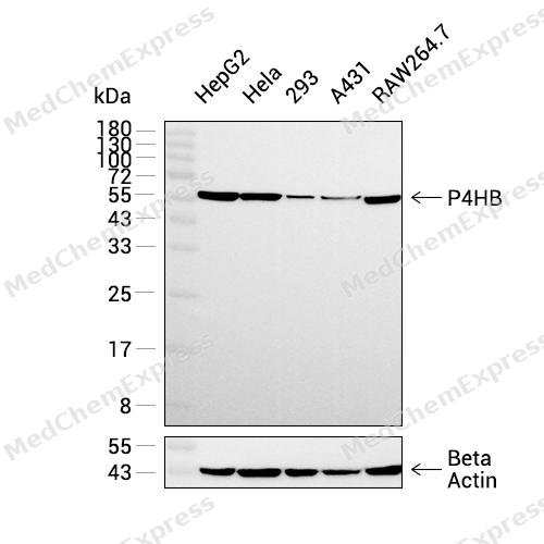 Western blot analysis was performed on extracts from HepG2 (lane 1, 15 μg), HeLa (lane 2, 15 μg), 293 (lane 3, 15 μg), A431 (lane 4, 15 μg), and RAW264.7 (lane 5, 15 μg) using P4HB Rabbit mAb.Proteins were transferred to a PVDF membrane and blocked with 5% non - fat milk in TBST at 4°C overnight.The primary antibody (1:1000 dilution) and the loading control antibody (beta-Actin, HY-P83730, 1:20000 dilution) were incubated in 5% non-fat milk in TBST for 1 hour at 37°C.Goat Anti - Rabbit IgG - HRP Secondary Antibody (1:20000 dilution) was then applied for 40 minutes at 37°C.
 -
 Immunohistochemical analysis of paraffin-embedded mouse epididymis tissue using P4HB Antibody (HY-P81758, 1/100). The section was pretreated using heat mediated antigen retrieval with sodium citrate buffer (pH 6.0) for 8 minutes. The tissues were blocked with quick block buffer for 0.5 hours at room temperature, washed with PBS and PBST, and then incubated with the primary antibody overnight at 4℃. The detection was performed using an HRP conjugated compact polymer system. DAB was used as the chromogen. Tissues were counterstained with hematoxylin and mounted with DPX.
 -
 Immunohistochemical analysis of paraffin-embedded mouse liver tissue using P4HB Antibody (HY-P81758, 1/100). The section was pretreated using heat mediated antigen retrieval with sodium citrate buffer (pH 6.0) for 8 minutes. The tissues were blocked with quick block buffer for 0.5 hours at room temperature, washed with PBS and PBST, and then incubated with the primary antibody overnight at 4℃. The detection was performed using an HRP conjugated compact polymer system. DAB was used as the chromogen. Tissues were counterstained with hematoxylin and mounted with DPX.
 -
 Immunohistochemical analysis of paraffin-embedded mouse small intestine tissue using P4HB Antibody (HY-P81758, 1/100). The section was pretreated using heat mediated antigen retrieval with sodium citrate buffer (pH 6.0) for 8 minutes. The tissues were blocked with quick block buffer for 0.5 hours at room temperature, washed with PBS and PBST, and then incubated with the primary antibody overnight at 4℃. The detection was performed using an HRP conjugated compact polymer system. DAB was used as the chromogen. Tissues were counterstained with hematoxylin and mounted with DPX.
 -
 Immunohistochemical analysis of paraffin-embedded human tonsil tissue using P4HB Antibody (HY-P81758, 1/100). The section was pretreated using heat mediated antigen retrieval with sodium citrate buffer (pH 6.0) for 8 minutes. The tissues were blocked with quick block buffer for 0.5 hours at room temperature, washed with PBS and PBST, and then incubated with the primary antibody overnight at 4℃. The detection was performed using an HRP conjugated compact polymer system. DAB was used as the chromogen. Tissues were counterstained with hematoxylin and mounted with DPX.
 -
 Immunohistochemical analysis of paraffin-embedded mouse colon tissue using P4HB Antibody (HY-P81758, 1/100). The section was pretreated using heat mediated antigen retrieval with sodium citrate buffer (pH 6.0) for 8 minutes. The tissues were blocked with quick block buffer for 0.5 hours at room temperature, washed with PBS and PBST, and then incubated with the primary antibody overnight at 4℃. The detection was performed using an HRP conjugated compact polymer system. DAB was used as the chromogen. Tissues were counterstained with hematoxylin and mounted with DPX.
 -
 Immunohistochemical analysis of paraffin-embedded mouse duodenum tissue using P4HB Antibody (HY-P81758, 1/100). The section was pretreated using heat mediated antigen retrieval with sodium citrate buffer (pH 6.0) for 8 minutes. The tissues were blocked with quick block buffer for 0.5 hours at room temperature, washed with PBS and PBST, and then incubated with the primary antibody overnight at 4℃. The detection was performed using an HRP conjugated compact polymer system. DAB was used as the chromogen. Tissues were counterstained with hematoxylin and mounted with DPX.
 -
 Immunohistochemical analysis of paraffin-embedded human endometrium tissue using P4HB Antibody (HY-P81758, 1/100). The section was pretreated using heat mediated antigen retrieval with sodium citrate buffer (pH 6.0) for 8 minutes. The tissues were blocked with quick block buffer for 0.5 hours at room temperature, washed with PBS and PBST, and then incubated with the primary antibody overnight at 4℃. The detection was performed using an HRP conjugated compact polymer system. DAB was used as the chromogen. Tissues were counterstained with hematoxylin and mounted with DPX.
 -
 Immunohistochemical analysis of paraffin-embedded human thyroid gland tissue using P4HB Antibody (HY-P81758, 1/100). The section was pretreated using heat mediated antigen retrieval with sodium citrate buffer (pH 6.0) for 8 minutes. The tissues were blocked with quick block buffer for 0.5 hours at room temperature, washed with PBS and PBST, and then incubated with the primary antibody overnight at 4℃. The detection was performed using an HRP conjugated compact polymer system. DAB was used as the chromogen. Tissues were counterstained with hematoxylin and mounted with DPX.
 -
 Immunohistochemical analysis of paraffin-embedded human adrenal gland tissue using P4HB Antibody (HY-P81758, 1/100). The section was pretreated using heat mediated antigen retrieval with sodium citrate buffer (pH 6.0) for 8 minutes. The tissues were blocked with quick block buffer for 0.5 hours at room temperature, washed with PBS and PBST, and then incubated with the primary antibody overnight at 4℃. The detection was performed using an HRP conjugated compact polymer system. DAB was used as the chromogen. Tissues were counterstained with hematoxylin and mounted with DPX.
 -
 Immunohistochemical analysis of paraffin-embedded human small intestine tissue using P4HB Antibody (HY-P81758, 1/100). The section was pretreated using heat mediated antigen retrieval with sodium citrate buffer (pH 6.0) for 8 minutes. The tissues were blocked with quick block buffer for 0.5 hours at room temperature, washed with PBS and PBST, and then incubated with the primary antibody overnight at 4℃. The detection was performed using an HRP conjugated compact polymer system. DAB was used as the chromogen. Tissues were counterstained with hematoxylin and mounted with DPX.
 -
 Immunocytochemistry analysis of Hela cells labeling P4HB with P4HB Antibody (HY-P81758) at 1/100 dilution. Cells were fixed in 4% paraformaldehyde for 15 minutes at room temperature, permeabilized with 0.1% Triton X-100 in PBS for 15 minutes at room temperature, then blocked with quick block buffer for 10 minutes at room temperature. Cells were then incubated with P4HB Antibody (HY-P81758) at 1/100 dilution in quick block buffer overnight at 4 ℃. AF488-conjugated Goat Anti-Rabbit IgG H&L (HY-P8002, Green) was used as the secondary antibody at 1/1,000 dilution. PBS instead of the primary antibody was used as the secondary antibody only control. The Nuclear counterstain was DAPI (Blue).
 -
 Immunocytochemistry analysis of PC-3 cells labeling P4HB with P4HB Antibody (HY-P81758) at 1/100 dilution. Cells were fixed in 4% paraformaldehyde for 15 minutes at room temperature, permeabilized with 0.1% Triton X-100 in PBS for 15 minutes at room temperature, then blocked with quick block buffer for 10 minutes at room temperature. Cells were then incubated with P4HB Antibody (HY-P81758) at 1/100 dilution in quick block buffer overnight at 4 ℃. AF488-conjugated Goat Anti-Rabbit IgG H&L (HY-P8002, Green) was used as the secondary antibody at 1/1,000 dilution. PBS instead of the primary antibody was used as the secondary antibody only control. The Nuclear counterstain was DAPI (Blue).


Background
-
Function
P4HB is a This multifunctional protein catalyzes the formation, breakage and rearrangement of disulfide bonds. At the cell surface, seems to act as a reductase that cleaves disulfide bonds of proteins attached to the cell. May therefore cause structural modifications of exofacial proteins. Inside the cell, seems to form/rearrange disulfide bonds of nascent proteins. At high concentrations and following phosphorylation by FAM20C, functions as a chaperone that inhibits aggregation of misfolded proteins. At low concentrations, facilitates aggregation (anti-chaperone activity). May be involved with other chaperones in the structural modification of the TG precursor in hormone biogenesis. Also acts as a structural subunit of various enzymes such as prolyl 4-hydroxylase and microsomal triacylglycerol transfer protein MTTP. Receptor for LGALS9; the interaction retains P4HB at the cell surface of Th2 T helper cells, increasing disulfide reductase activity at the plasma membrane, altering the plasma membrane redox state and enhancing cell migration[1][2].
-
Subcellular Localization
Endoplasmic reticulum; Endoplasmic reticulum lumen; Melanosome; Cell membrane; Peripheral membrane protein
-
Subunit
Heterodimer; heterodimerizes with the protein microsomal triglyceride transfer MTTP (PubMed:16478722, PubMed:23475612, PubMed:26224785). Homodimer. Monomers and homotetramers may also occur. Interacts with P4HA2, forming a heterotetramer consisting of 2 alpha subunits (P4HA2) and 2 beta (P4HB), where P4HB plays the role of a structural subunit; this tetramer catalyzes the formation of 4-hydroxyproline in collagen (PubMed:7753822). Also constitutes the structural subunit of the microsomal triacylglycerol transfer protein MTTP in mammalian cells. Stabilizes both enzymes and retain them in the ER without contributing to the catalytic activity (By similarity). Binds UBQLN1 (PubMed:12095988). Interacts with ERO1B (PubMed:11707400). Binds to CD4, and upon HIV-1 binding to the cell membrane, is part of a P4HB/PDI-CD4-CXCR4-gp120 complex. Interacts with ILDR2 (By similarity). Interacts with ERN1/IRE1A (via N-terminus); the interaction is enhanced by phosphorylation of P4HB by FAM20C in response to endoplasmic reticulum stress and results in attenuation of ERN1 activity (PubMed:32149426)
-
SwissProt ID
-
Synonyms
P4HB; DSI; ERBA2L; GIT; P4Hbeta; PDI; PDIA1; PHDB; PO4DB; PO4HB; PROHB
-
Research Field
Tags & Cell Markers


Documentation
-
Data Sheet (235 KB)
-
SDS (251 KB)
- English - EN (251 KB)
- Français - FR (251 KB)
- Deutsch - DE (251 KB)
- Norwegian - NO (251 KB)
- Español - ES (251 KB)
- Swedish - SV (251 KB)
- Italian - IT (251 KB)
- Korean - KR (251 KB)
- Portuguese - PT (251 KB)
-
User Guide for Antibodies (1077 KB)

 Powered by Bioz
