



PDPK1 Antibody (YA1569)
(Synonyms: PDPK1; PDK1; 3-phosphoinositide-dependent protein kinase 1; hPDK1)
Based on 1 Customer Validation
PDPK1 Antibody (YA1569) is a Rabbit-derived and non-conjugated IgG monoclonal antibody, targeting to PDPK1.
-
Host:
Rabbit
-
Isotype:
IgG
-
Application:
WB
-
Reactivity :
Human, Mouse, Rat
-
Formulation:
Supplied in 50mM Tris-Glycine(pH 7.4), 0.15M NaCl, 40% Glycerol, 0.01% Sodium azide and 0.05% BSA
-
Conjugation:
Non-conjugated


To place orders, for customer services and technical support, please contact: MedChemExpress USA
Tel: 609-228-6898 E-mail: [email protected] [email protected]





Applications
| Application |
WB

WB: Western Blot
|
|---|---|
| Dilution Ratio | 1:500-1:1000 |


Product Details
PDPK1 Antibody (YA1569) is a Rabbit-derived and non-conjugated IgG monoclonal antibody, targeting to PDPK1.
-
Host Rabbit

-
Clonality Recombinant,Monoclonal

-
Species ReactivityHuman, Mouse, Rat
-
Observed Molecular WeightObserved band size: 63 kDa
Note: Due to possible protein modifications or aggregation, the molecular weight should be confirmed by actual measurement, and the predicted value is for reference only.

-
Calculated Molecular Weight Predicted band size: 63 kDa

Entrez Gene: 5170 Human ; 18607 Mouse ; 81745 Rat
SwissProt: O15530 Human ; Q9Z2A0 Mouse ; O55173 Rat
OMIM: 605213 Human
A synthetic peptide of human PDPK1
Endogenous
Affinity Purified
Non-conjugated
Unmodified
IgG


Product Properties
-
Appearance
Liquid
-
Formulation
Supplied in 50mM Tris-Glycine(pH 7.4), 0.15M NaCl, 40% Glycerol, 0.01% Sodium azide and 0.05% BSA
-
Concentration
Batch-dependent, Please check the COA for the concentration of each lot. Check Lot Concentration
-
Storage & Stability
Stored at -20°C for 1 year. Avoid repeated freeze / thaw cycles.
-
Shipping
Shipping with blue ice.


Verification Images
-
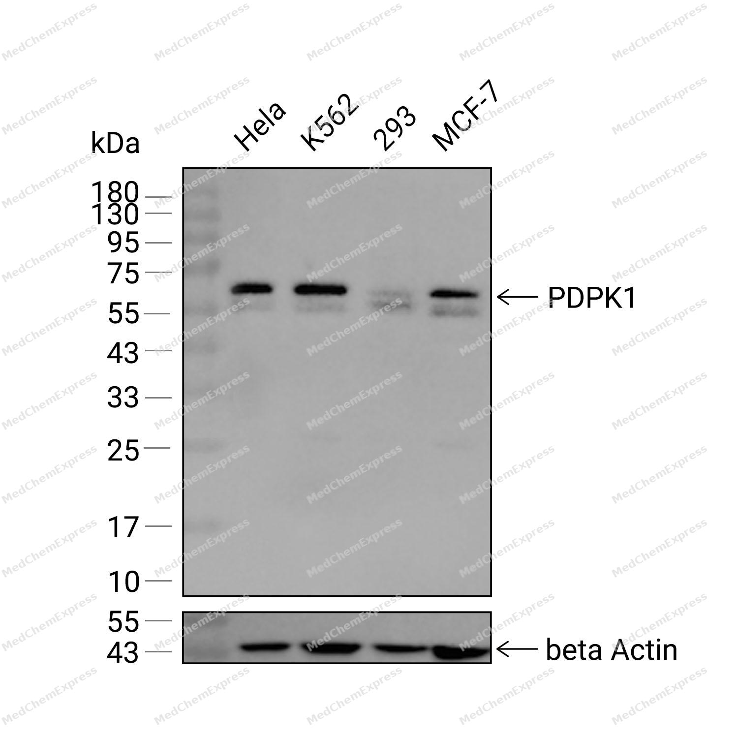 Western blot analysis was performed on extracts from Hela (lane 1, 15 μg), K562 (lane 2, 15 μg), 293 (lane 3, 15 μg), and MCF-7 (lane 4, 15 μg) using PDPK1 Rabbit mAb.Proteins were transferred to a PVDF membrane and blocked with 5% non-fat milk in TBST at 4°C overnight.The primary antibody (1:1000 dilution) and the loading control antibody (beta-Actin, HY-P80438, 1:20000 dilution) were incubated in 5% non-fat milk in TBST for 1 hour at 37°C.Goat Anti-Rabbit IgG-HRP Secondary Antibody (1:20000 dilution) was then applied for 40 minutes at 37°C.
 -
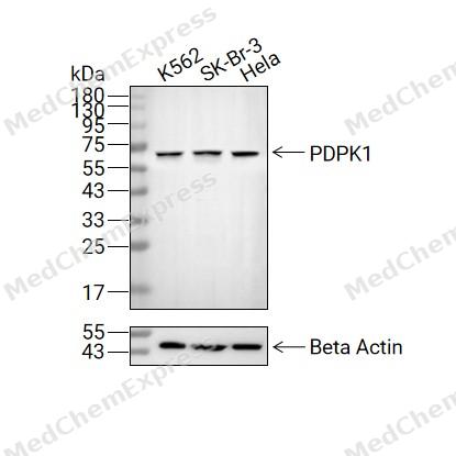 Western blot analysis of extracts from K562 (lane 2(20μg), SK-Br-3 (lane 3(20μg), and Hela (lane 4(20μg) using PDPK1 (HY-P81824) Rabbit mAb. Proteins were transferred to a PVDF membrane and blocked with 5% non-fat milk in TBST for 2 hour at room temperature. The primary antibody (1/1000) and Loading control antibody (Beta Actin, HY-P80438, 1/10000) was used in 5% non-fat milk in TBST at 4°C overnight. Goat Anti-Rabbit IgG-HRP Secondary Antibody (1/10000) was used for 1 hour at room temperature.


Background
-
Function
PDPK1 is a Serine/threonine kinase which acts as a master kinase, phosphorylating and activating a subgroup of the AGC family of protein kinases. Its targets include: protein kinase B (PKB/AKT1, PKB/AKT2, PKB/AKT3), p70 ribosomal protein S6 kinase (RPS6KB1), p90 ribosomal protein S6 kinase (RPS6KA1, RPS6KA2 and RPS6KA3), cyclic AMP-dependent protein kinase (PRKACA), protein kinase C (PRKCD and PRKCZ), serum and glucocorticoid-inducible kinase (SGK1, SGK2 and SGK3), p21-activated kinase-1 (PAK1), TSSK3, protein kinase PKN (PKN1 and PKN2). Plays a central role in the transduction of signals from insulin by providing the activating phosphorylation to PKB/AKT1, thus propagating the signal to downstream targets controlling cell proliferation and survival, as well as glucose and amino acid uptake and storage. Negatively regulates the TGF-beta-induced signaling by: modulating the association of SMAD3 and SMAD7 with TGF-beta receptor, phosphorylating SMAD2, SMAD3, SMAD4 and SMAD7, preventing the nuclear translocation of SMAD3 and SMAD4 and the translocation of SMAD7 from the nucleus to the cytoplasm in response to TGF-beta. Activates PPARG transcriptional activity and promotes adipocyte differentiation. Activates the NF-kappa-B pathway via phosphorylation of IKKB. The tyrosine phosphorylated form is crucial for the regulation of focal adhesions by angiotensin II. Controls proliferation, survival, and growtH2O2 developing pancreatic cells. Participates in the regulation of Ca(2+) entry and Ca(2+)-activated K(+) channels of mast cells. Essential for the motility of vascular endothelial cells (ECs) and is involved in the regulation of their chemotaxis. Plays a critical role in cardiac homeostasis by serving as a dual effector for cell survival and beta-adrenergic response. Plays an important role during thymocyte development by regulating the expression of key nutrient receptors on the surface of pre-T cells and mediating Notch-induced cell growth and proliferative responses. Provides negative feedback inhibition to toll-like receptor-mediated NF-kappa-B activation in macrophages; Catalytically inactive[1][2][3][4][5][6][7][8][9][10][11][12][13][14][15][16][17].
-
Subcellular Localization
Cytoplasm; Nucleus; Cell membrane; Peripheral membrane protein; Cell junction, focal adhesion
-
Expression
Tissue_specificity:This protein appears to be universally expressed. Compared to normal tissues of the lung, liver, colon, and breast, the phosphorylated form of Tyr-9 protein was significantly increased in diseased tissues.
Induction:Stimulated by insulin, and the oxidants hydrogen peroxide and peroxovanadate -
Isoforms & Post-Translational Modification
O15530 has 5 isomers: O15530-1: 63152 Da (predicted); O15530-2: 58035 Da (predicted); O15530-3: 60396 Da (predicted); O15530-4: 48201 Da (predicted); O15530-5: 50838 Da (predicted).
Phosphorylation on Ser-241 in the activation loop is required for full activity. PDPK1 itself can autophosphorylate Ser-241, leading to its own activation. Autophosphorylation is inhibited by the apoptotic C-terminus cleavage product of PKN2 (By similarity). Tyr-9 phosphorylation is critical for stabilization of both PDPK1 and the PDPK1/SRC complex via HSP90-mediated protection of PDPK1 degradation. Angiotensin II stimulates the tyrosine phosphorylation of PDPK1 in vascular smooth muscle in a calcium- and SRC-dependent manner. Phosphorylated on Tyr-9, Tyr-373 and Tyr-376 by INSR in response to insulin. Palmitate negatively regulates autophosphorylation at Ser-241 and palmitate-induced phosphorylation at Ser-529 and Ser-501 by PKC/PRKCQ negatively regulates its ability to phosphorylate PKB/AKT1. Phosphorylation at Thr-354 by MELK partially inhibits kinase activity, the inhibition is cooperatively enhanced by phosphorylation at Ser-394 and Ser-398 by MAP3K5;Autophosphorylated; autophosphorylation is inhibited by the apoptotic C-terminus cleavage product of PKN2;Monoubiquitinated in the kinase domain, deubiquitinated by USP4 -
Subunit
Homodimer in its autoinhibited state. Active as monomer. Interacts with NPRL2, PPARG, PAK1, PTK2B, GRB14, PKN1 (via C-terminus), STRAP and IKKB. The Tyr-9 phosphorylated form interacts with SRC, RASA1 and CRK (via their SH2 domains). Interacts with SGK3 in a phosphorylation-dependent manner. The tyrosine-phosphorylated form interacts with PTPN6. The Ser-241 phosphorylated form interacts with YWHAH and YWHAQ. Binds INSR in response to insulin. Interacts (via PH domain) with SMAD3, SMAD4 and SMAD7. Interacts with PKN2; the interaction stimulates PDPK1 autophosphorylation, its PI(3,4,5)P3-dependent kinase activity toward 'Ser-473' of AKT1 but also activates its kinase activity toward PRKCD and PRKCZ
-
SwissProt ID
-
Synonyms
PDPK1; PDK1; 3-phosphoinositide-dependent protein kinase 1; hPDK1
-
Research Field
Signal Transduction


Documentation
-
Data Sheet (232 KB)
-
SDS (251 KB)
- English - EN (251 KB)
- Français - FR (251 KB)
- Deutsch - DE (251 KB)
- Norwegian - NO (251 KB)
- Español - ES (251 KB)
- Swedish - SV (251 KB)
- Italian - IT (251 KB)
- Korean - KR (251 KB)
- Portuguese - PT (251 KB)
-
User Guide for Antibodies (1077 KB)

 Powered by Bioz
