



SAMHD1 Antibody (YA1542)
(Synonyms: SAMHD1; AGS5; CHBL2; DCIP; HDDC1; MOP-5; MOP5; SBBI88; Mg11)
Based on 1 Customer Validation
SAMHD1 Antibody (YA1542) is a Rabbit-derived and non-conjugated IgG monoclonal antibody, targeting to SAMHD1.
-
Host:
Rabbit
-
Isotype:
IgG
-
Application:
WB, IHC-P, ICC/IF, FC
-
Reactivity :
Human
-
Formulation:
Supplied in 50mM Tris-Glycine(pH 7.4), 0.15M NaCl, 40%Glycerol, 0.01% sodium azide and 0.05% BSA.
-
Conjugation:
Non-conjugated


To place orders, for customer services and technical support, please contact: MedChemExpress USA
Tel: 609-228-6898 E-mail: [email protected] [email protected]





Applications
| Application |
WB

WB: Western Blot
|
IHC-P
IHC-P: Immunohistochemistry-Paraffin
|
ICC/IF
ICC/IF: Immunocytochemistry/
Immunofluorescence |
FC
FC: Flow Cytometry
|
|---|---|---|---|---|
| Dilution Ratio | 1:500-1:1000 | 1:50-1:100 | 1:50-1:200 | 1:50-1:100 |


Product Details
SAMHD1 Antibody (YA1542) is a Rabbit-derived and non-conjugated IgG monoclonal antibody, targeting to SAMHD1.
-
Host Rabbit

-
Clonality Recombinant,Monoclonal

-
Species ReactivityHuman
-
Observed Molecular WeightObserved band size: 72 kDa
Note: Due to possible protein modifications or aggregation, the molecular weight should be confirmed by actual measurement, and the predicted value is for reference only.

-
Calculated Molecular Weight Predicted band size: 72 kDa

A synthesized peptide derived from human SAMHD1 aa580-626/626.
Endogenous
Affinity Chromatography
Non-conjugated
Unmodified
IgG


Product Properties
-
Appearance
Solution
-
Formulation
Supplied in 50mM Tris-Glycine(pH 7.4), 0.15M NaCl, 40%Glycerol, 0.01% sodium azide and 0.05% BSA.
-
Concentration
Batch-dependent, Please check the COA for the concentration of each lot. Check Lot Concentration
-
Storage & Stability
Stored at -20°C for 1 year. Avoid repeated freeze / thaw cycles.
-
Shipping
Shipping with blue ice.


Verification Images
-
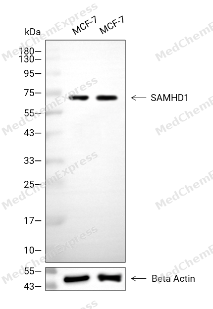 Western blot analysis of extracts from MCF-7 (lane 2(20μg), MCF-7 (lane 3(40μg), using SAMHD1 Antibody. Proteins were transferred to a PVDF membrane and blocked with 5% BSA in TBST for 2 hour at room temperature. The primary antibody and Loading control antibody (Beta Actin, HY-P80438, 1/3000) was used in 5% BSA in TBST at 4°C overnight. Goat Anti-Mouse/Rabbit IgG-HRP Secondary Antibody (HY-P8004/HY-P8001, 1/10,000) was used for 1 hour at room temperature.
 -
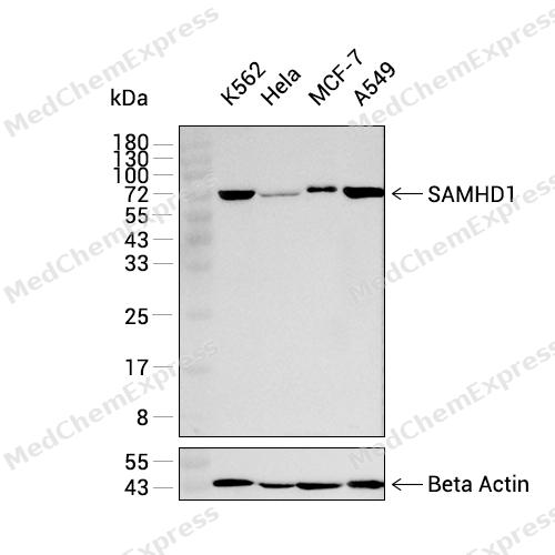 Western blot analysis was performed on extracts from K562 (lane 1, 15 μg), Hela (lane 2, 15 μg), MCF-7 (lane 3, 15 μg), and A549 (lane 4, 15 μg) using SAMHD1 Rabbit mAb.Proteins were transferred to a PVDF membrane and blocked with 5% non-fat milk in TBST at 4°C overnight.The primary antibody (1:1000 dilution) and the loading control antibody (beta-Actin, HY-P83730, 1:20000 dilution) were incubated in 5% non-fat milk in TBST for 1 hour at 37°C.Goat Anti-Rabbit IgG-HRP Secondary Antibody (1:20000 dilution) was then applied for 40 minutes at 37°C.
 -
 Immunohistochemical analysis of paraffin-embedded human cervix tissue using SAMHD1 Antibody (HY-P81797, 1/100). The section was pretreated using heat mediated antigen retrieval with sodium citrate buffer (pH 6.0) for 8 minutes. The tissues were blocked with quick block buffer for 0.5 hours at room temperature, washed with PBS and PBST, and then incubated with the primary antibody overnight at 4℃. The detection was performed using an HRP conjugated compact polymer system. DAB was used as the chromogen. Tissues were counterstained with hematoxylin and mounted with DPX.
 -
 Immunohistochemical analysis of paraffin-embedded human endometrium tissue using SAMHD1 Antibody (HY-P81797, 1/100). The section was pretreated using heat mediated antigen retrieval with sodium citrate buffer (pH 6.0) for 8 minutes. The tissues were blocked with quick block buffer for 0.5 hours at room temperature, washed with PBS and PBST, and then incubated with the primary antibody overnight at 4℃. The detection was performed using an HRP conjugated compact polymer system. DAB was used as the chromogen. Tissues were counterstained with hematoxylin and mounted with DPX.
 -
 Immunohistochemical analysis of paraffin-embedded human adrenal gland tissue using SAMHD1 Antibody (HY-P81797, 1/100). The section was pretreated using heat mediated antigen retrieval with sodium citrate buffer (pH 6.0) for 8 minutes. The tissues were blocked with quick block buffer for 0.5 hours at room temperature, washed with PBS and PBST, and then incubated with the primary antibody overnight at 4℃. The detection was performed using an HRP conjugated compact polymer system. DAB was used as the chromogen. Tissues were counterstained with hematoxylin and mounted with DPX.
 -
 Immunohistochemical analysis of paraffin-embedded human tonsil tissue using SAMHD1 Antibody (HY-P81797, 1/100). The section was pretreated using heat mediated antigen retrieval with sodium citrate buffer (pH 6.0) for 8 minutes. The tissues were blocked with quick block buffer for 0.5 hours at room temperature, washed with PBS and PBST, and then incubated with the primary antibody overnight at 4℃. The detection was performed using an HRP conjugated compact polymer system. DAB was used as the chromogen. Tissues were counterstained with hematoxylin and mounted with DPX.
 -
 Immunohistochemical analysis of paraffin-embedded human small intestine tissue using SAMHD1 Antibody (HY-P81797, 1/100). The section was pretreated using heat mediated antigen retrieval with sodium citrate buffer (pH 6.0) for 8 minutes. The tissues were blocked with quick block buffer for 0.5 hours at room temperature, washed with PBS and PBST, and then incubated with the primary antibody overnight at 4℃. The detection was performed using an HRP conjugated compact polymer system. DAB was used as the chromogen. Tissues were counterstained with hematoxylin and mounted with DPX.
 -
 Immunohistochemical analysis of paraffin-embedded human gallbladder tissue using SAMHD1 Antibody (HY-P81797, 1/100). The section was pretreated using heat mediated antigen retrieval with sodium citrate buffer (pH 6.0) for 8 minutes. The tissues were blocked with quick block buffer for 0.5 hours at room temperature, washed with PBS and PBST, and then incubated with the primary antibody overnight at 4℃. The detection was performed using an HRP conjugated compact polymer system. DAB was used as the chromogen. Tissues were counterstained with hematoxylin and mounted with DPX.
 -
 Immunocytochemistry analysis of Hela cells labeling SAMHD1 with SAMHD1 Antibody (HY-P81797) at 1/100 dilution. Cells were fixed in 4% paraformaldehyde for 15 minutes at room temperature, permeabilized with 0.1% Triton X-100 in PBS for 15 minutes at room temperature, then blocked with quick block buffer for 10 minutes at room temperature. Cells were then incubated with SAMHD1 Antibody (HY-P81797) at 1/100 dilution in quick block buffer overnight at 4 ℃. AF488-conjugated Goat Anti-Rabbit IgG H&L(HY-P8002, Green) was used as the secondary antibody at 1/1,000 dilution. PBS instead of the primary antibody was used as the secondary antibody only control. The Nuclear counterstain was DAPI (Blue).
 -
 Immunocytochemistry analysis of Hela cells labeling SAMHD1 with SAMHD1 Antibody (HY-P81797) at 1/200 dilution. Cells were fixed in 4% paraformaldehyde for 15 minutes at room temperature, permeabilized with 0.1% Triton X-100 in PBS for 15 minutes at room temperature, then blocked with quick block buffer for 10 minutes at room temperature. Cells were then incubated with SAMHD1 Antibody (HY-P81797) at 1/200 dilution in quick block buffer overnight at 4 ℃. AF488-conjugated Goat Anti-Rabbit IgG H&L(HY-P8002, Green) was used as the secondary antibody at 1/1,000 dilution. PBS instead of the primary antibody was used as the secondary antibody only control. The Nuclear counterstain was DAPI (Blue).


Background
-
Function
SAMHD1 is a Protein that acts both as a host restriction factor involved in defense response to virus and as a regulator of DNA end resection at stalled replication forks. Has deoxynucleoside triphosphate (dNTPase) activity, which is required to restrict infection by viruses, such as HIV-1: dNTPase activity reduces cellular dNTP levels to levels too low for retroviral reverse transcription to occur, blocking early-stage virus replication in dendritic and other myeloid cells. Likewise, suppresses LINE-1 retrotransposon activity. Not able to restrict infection by HIV-2 virus; because restriction activity is counteracted by HIV-2 viral protein Vpx. In addition to virus restriction, dNTPase activity acts as a regulator of DNA precursor pools by regulating dNTP pools. Phosphorylation at Thr-592 acts as a switch to control dNTPase-dependent and -independent functions: it inhibits dNTPase activity and ability to restrict infection by viruses, while it promotes DNA end resection at stalled replication forks. Functions during S phase at stalled DNA replication forks to promote the resection of gapped or reversed forks: acts by stimulating the exonuclease activity of MRE11, activating the ATR-CHK1 pathway and allowing the forks to restart replication. Its ability to promote degradation of nascent DNA at stalled replication forks is required to prevent induction of type I interferons, thereby preventing chronic inflammation. Ability to promote DNA end resection at stalled replication forks is independent of dNTPase activity. Enhances immunoglobulin hypermutation in B-lymphocytes by promoting transversion mutation[1][2][3][4][5][6][7][8][9][10][11][12][13][14][15][16][17][18][19][20].
-
Subcellular Localization
Nucleus; Chromosome
-
Expression
Tissue_specificity:Expressed in heart, skeletal muscle, spleen, liver, small intestine, placenta, lung and peripheral blood leukocytes (PubMed:11064105) . No expression is seen in brain and thymus (PubMed:11064105)
Induction:By IFNG/IFN-gamma. Up-regulated in TNF treated lung fibroblasts -
Isoforms & Post-Translational Modification
Q9Y3Z3 has 3 isomers: Q9Y3Z3-1: 72201 Da (predicted); Q9Y3Z3-3: 63676 Da (predicted); Q9Y3Z3-4: 68122 Da (predicted).
Phosphorylation at Thr-592 by CDK1 acts as a switch to control deoxynucleoside triphosphate (dNTPase)-dependent and -independent functions (PubMed:29670289). Phosphorylation at Thr-592 takes place in cycling cells: it reduces the stability of the homotetramer, impairing the dNTPase activity and subsequent ability to restrict infection by viruses (PubMed:23601106, PubMed:23602554, PubMed:26294762, PubMed:26431200, PubMed:31291580). It also inhibits ability to suppress LINE-1 retrotransposon activity (PubMed:29610582). In contrast, phosphorylation at Thr-592 promotes DNA end resection at stalled replication forks in response to DNA damage (PubMed:29670289);(Microbial infection) Phosphorylation at Thr-592 by Epstein-Barr virus kinase BGLF4 and human cytomegalovirus/HCMV UL97 leads to a reduced level of dCTPase and dTTPase activity and the loss of viral restriction;(Microbial infection) Ubiquitinated following interaction with HIV-2 viral protein Vpx; Vpx promotes interaction and with a DCX (DDB1-CUL4-X-box) E3 ubiquitin ligase, leading to proteasomal degradation -
Subunit
Homodimer; in absence of GTP and dNTP (PubMed:24141705, PubMed:24217394, PubMed:25760601, PubMed:28229507). Homotetramer; in GTP- and dNTP-bound form (PubMed:23601106, PubMed:24141705, PubMed:24217394, PubMed:25267621, PubMed:25288794, PubMed:25760601, PubMed:26101257, PubMed:26294762, PubMed:26431200, PubMed:28229507). Interacts with MRE11; leading to stimulate the exonuclease activity of MRE11 (PubMed:28834754, PubMed:29670289). Interacts with RBBP8/CtIP (PubMed:28834754). Interacts (via its C-terminus) with CD81
-
SwissProt ID
-
Synonyms
SAMHD1; AGS5; CHBL2; DCIP; HDDC1; MOP-5; MOP5; SBBI88; Mg11
-
Research Field
Microbiology


Documentation
-
Data Sheet (234 KB)
-
SDS (251 KB)
- English - EN (251 KB)
- Français - FR (251 KB)
- Deutsch - DE (251 KB)
- Norwegian - NO (251 KB)
- Español - ES (251 KB)
- Swedish - SV (251 KB)
- Italian - IT (251 KB)
- Korean - KR (251 KB)
- Portuguese - PT (251 KB)
-
User Guide for Antibodies (1077 KB)

 Powered by Bioz
