



alpha 1 Antitrypsin Antibody (YA3592)
(Synonyms: PI; A1A; AAT; PI1; A1AT; PRO2275; alpha1AT)Based on 1 Customer Validation
alpha 1 Antitrypsin Antibody (YA3592) is a Mouse-derived and non-conjugated IgG1 monoclonal antibody, targeting to alpha 1 Antitrypsin.
-
Host:
Mouse
-
Isotype:
IgG1
-
Application:
IHC-P, ICC/IF, FC, ELISA
-
Reactivity :
Human
-
Formulation:
Supplied in PBS with 0.05% sodium azide
-
Conjugation:
Non-conjugated


To place orders, for customer services and technical support, please contact: MedChemExpress USA
Tel: 609-228-6898 E-mail: [email protected] [email protected]





Applications
| Application |
IHC-P

IHC-P: Immunohistochemistry-Paraffin
|
ICC/IF
ICC/IF: Immunocytochemistry/
Immunofluorescence |
FC
FC: Flow Cytometry
|
ELISA
ELISA: Enzyme Linked Immunosorbent Assay
|
|---|---|---|---|---|
| Dilution Ratio | 1:200-1:1000 | 1:200-1:1000 | 1:200-1:400 | 1:10000 |


Product Details
alpha 1 Antitrypsin Antibody (YA3592) is a Mouse-derived and non-conjugated IgG1 monoclonal antibody, targeting to alpha 1 Antitrypsin.
-
Host Mouse

-
Clonality Monoclonal

-
Species ReactivityHuman
-
Observed Molecular WeightObserved band size: 55 kDa
Note: Due to possible protein modifications or aggregation, the molecular weight should be confirmed by actual measurement, and the predicted value is for reference only.

-
Calculated Molecular Weight Predicted band size: 47 kDa

Purified recombinant fragment of human SERPINA1 (AA: 23-237) expressed in E. Coli.
affinity purified.
Non-conjugated
Unmodified
IgG1


Product Properties
-
Appearance
Liquid
-
Formulation
Supplied in PBS with 0.05% sodium azide
-
Concentration
Batch-dependent, Please check the COA for the concentration of each lot. Check Lot Concentration
-
Storage & Stability
Stored at -20°C for 1 year. Avoid repeated freeze / thaw cycles.
-
Shipping
Shipping with blue ice.


Verification Images
-
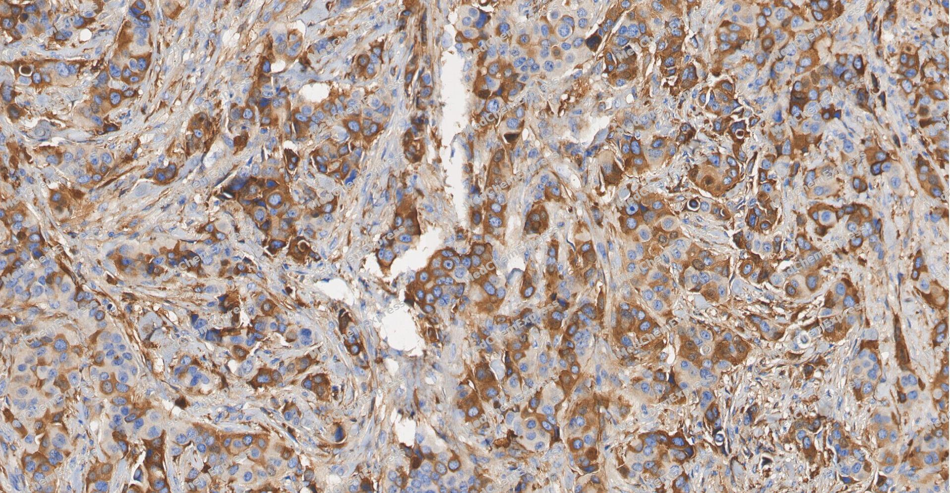 Immunohistochemical analysis of paraffin-embedded human Breast Cancer tissue using alpha 1 Antitrypsin antibody was performed. The section was pretreated using high-temperature and high-pressure mediated EDTA antigen retrieval buffer (pH 9.0), for 5 minutes. The tissues were incubated with primary antibody (HY-P83895, 1:2000 dilution) at room temperature for Leave overnight at 4°C. Detection was performed using an HRP conjugated compact polymer system. DAB was used as the chromogen. The tissues were counterstained with hematoxylin and mounted with neutral balsam mounting medium.
 -
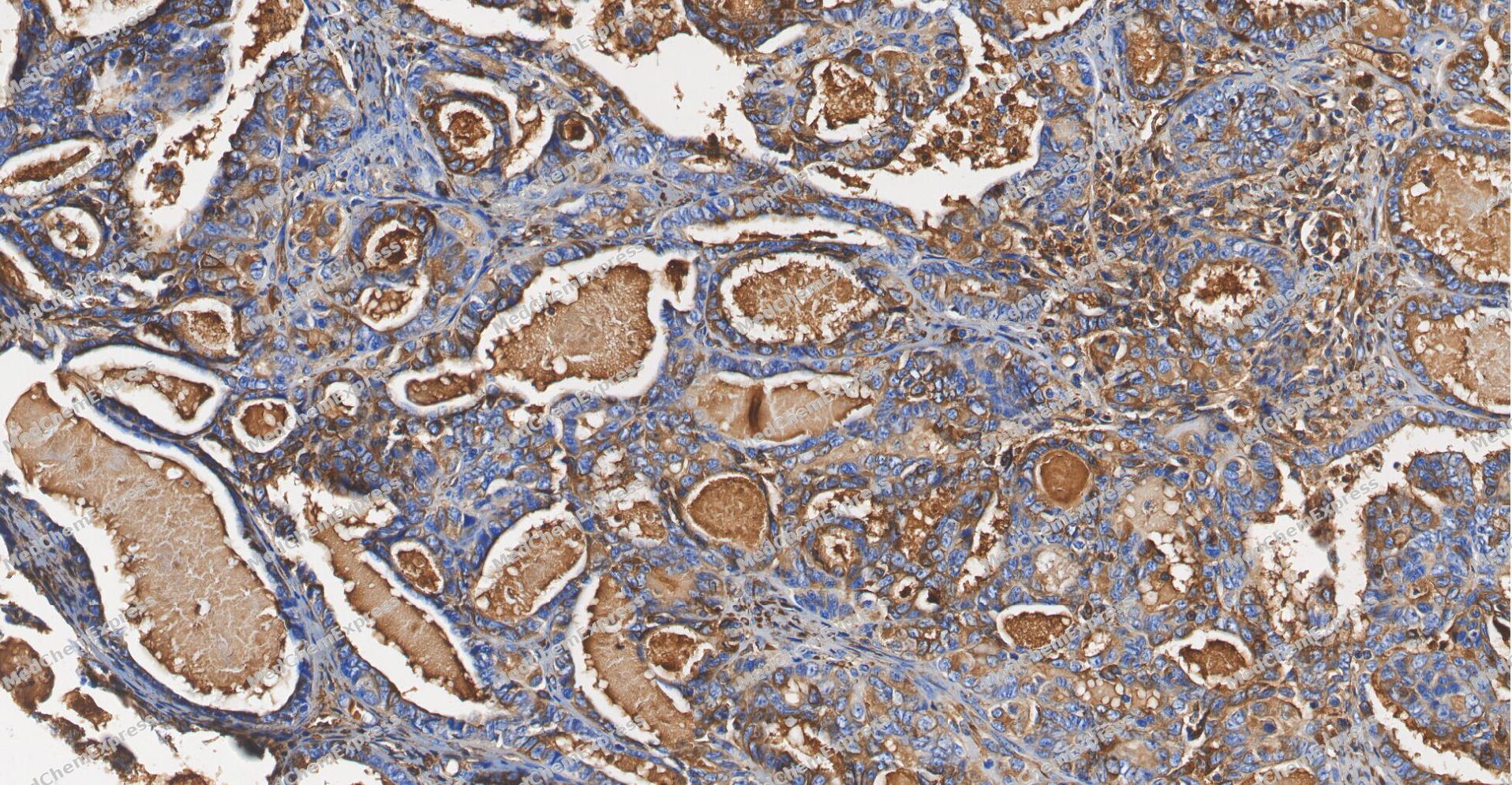 Immunohistochemical analysis of paraffin-embedded human ovarian carcinoma tissue using alpha 1 Antitrypsin antibody was performed. The section was pretreated using high-temperature and high-pressure mediated EDTA antigen retrieval buffer (pH 9.0), for 5 minutes. The tissues were incubated with primary antibody (HY-P83895, 1:2000 dilution) at room temperature for Leave overnight at 4°C. Detection was performed using an HRP conjugated compact polymer system. DAB was used as the chromogen. The tissues were counterstained with hematoxylin and mounted with neutral balsam mounting medium.
 -
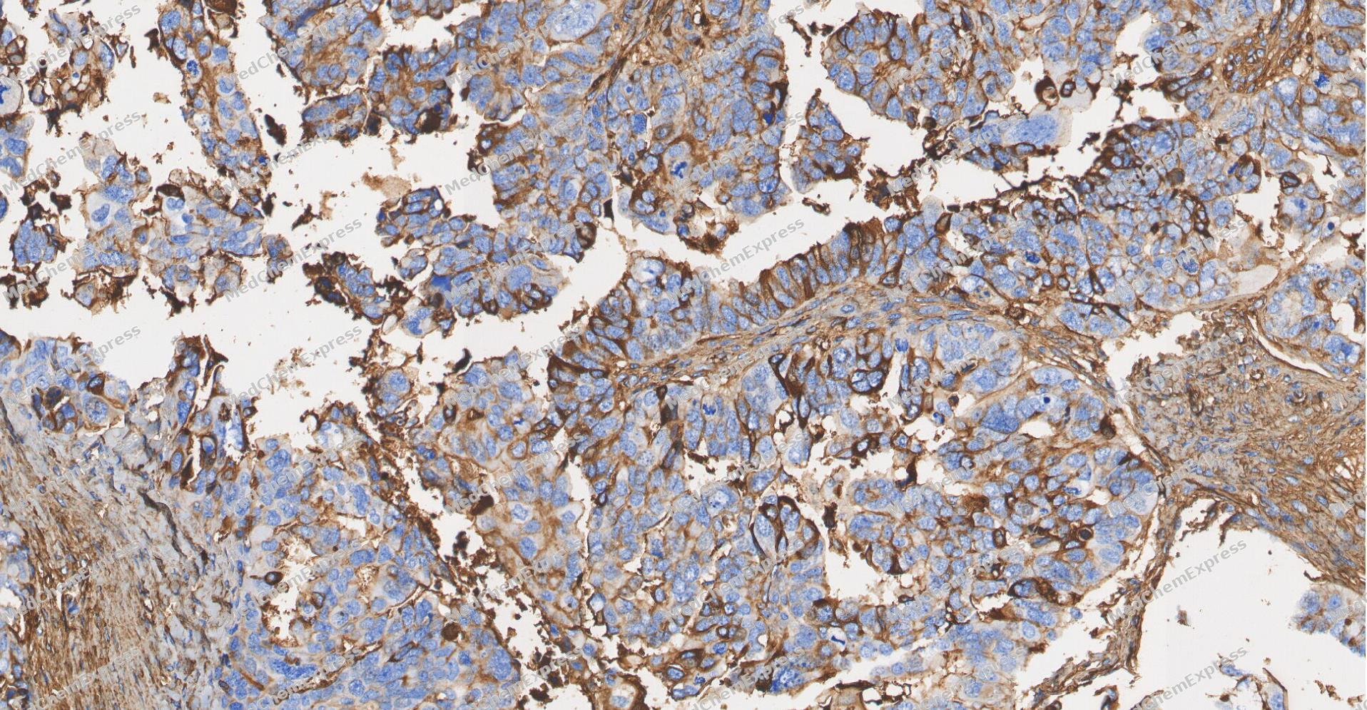 Immunohistochemical analysis of paraffin-embedded human ovarian carcinoma tissue using alpha 1 Antitrypsin antibody was performed. The section was pretreated using high-temperature and high-pressure mediated EDTA antigen retrieval buffer (pH 9.0), for 5 minutes. The tissues were incubated with primary antibody (HY-P83895, 1:2000 dilution) at room temperature for Leave overnight at 4°C. Detection was performed using an HRP conjugated compact polymer system. DAB was used as the chromogen. The tissues were counterstained with hematoxylin and mounted with neutral balsam mounting medium.
 -
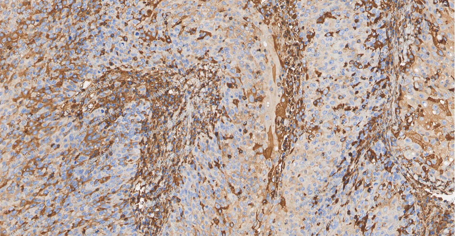 Immunohistochemical analysis of paraffin-embedded human Cervical cancer tissue using alpha 1 Antitrypsin antibody was performed. The section was pretreated using high-temperature and high-pressure mediated EDTA antigen retrieval buffer (pH 9.0), for 5 minutes. The tissues were incubated with primary antibody (HY-P83895, 1:2000 dilution) at room temperature for Leave overnight at 4°C. Detection was performed using an HRP conjugated compact polymer system. DAB was used as the chromogen. The tissues were counterstained with hematoxylin and mounted with neutral balsam mounting medium.
 -
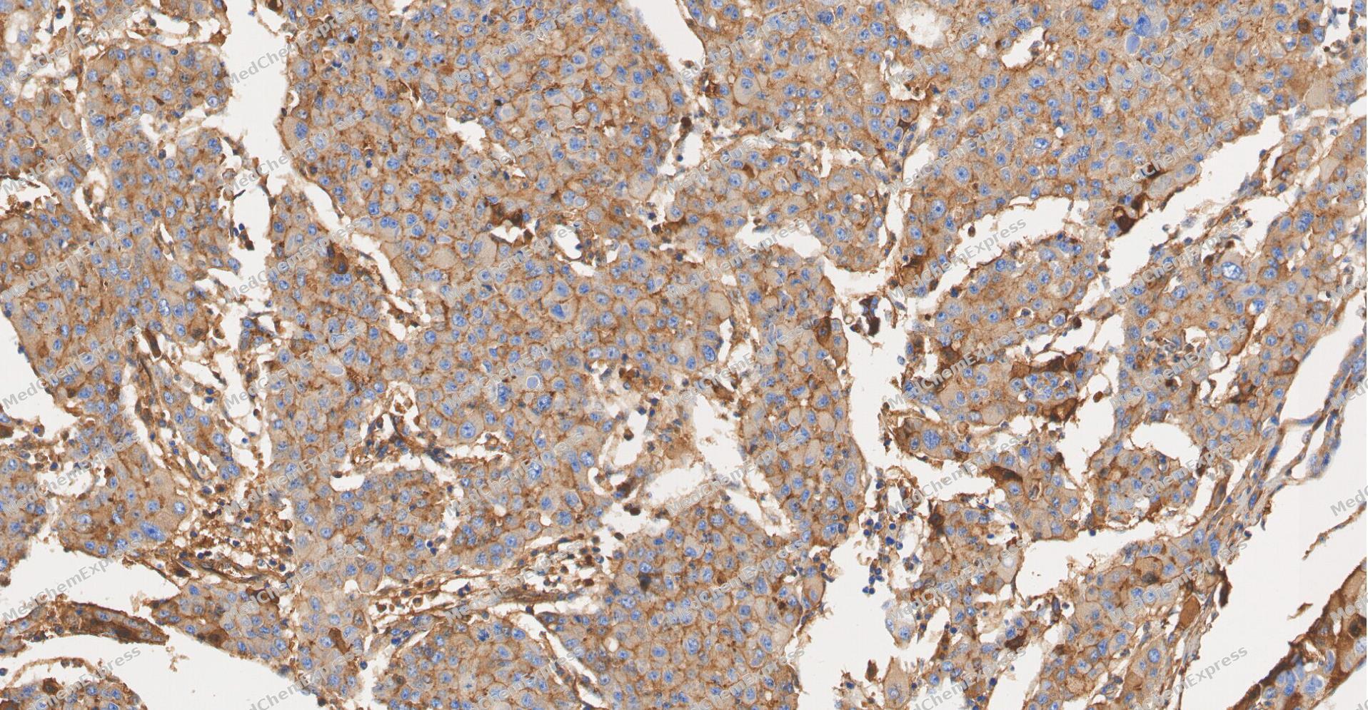 Immunohistochemical analysis of paraffin-embedded human Liver cancer tissue using alpha 1 Antitrypsin antibody was performed. The section was pretreated using high-temperature and high-pressure mediated EDTA antigen retrieval buffer (pH 9.0), for 5 minutes. The tissues were incubated with primary antibody (HY-P83895, 1:2000 dilution) at room temperature for Leave overnight at 4°C. Detection was performed using an HRP conjugated compact polymer system. DAB was used as the chromogen. The tissues were counterstained with hematoxylin and mounted with neutral balsam mounting medium.
 -
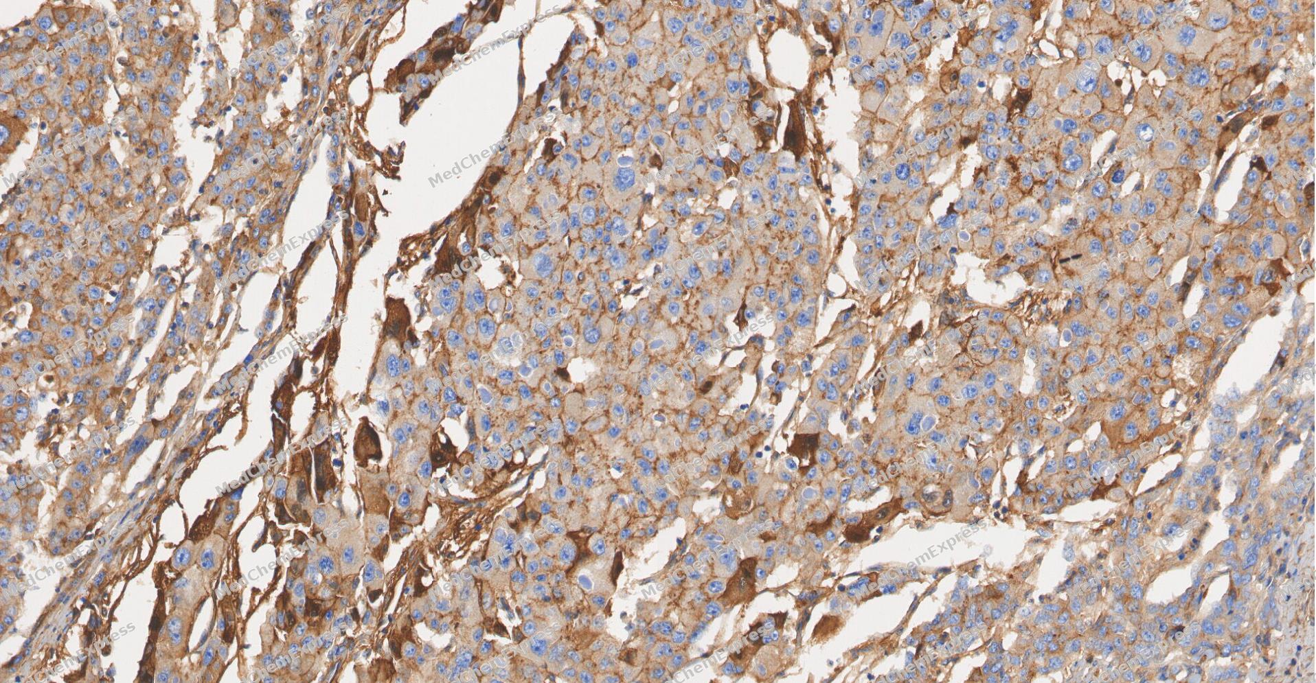 Immunohistochemical analysis of paraffin-embedded human Liver cancer tissue using alpha 1 Antitrypsin antibody was performed. The section was pretreated using high-temperature and high-pressure mediated EDTA antigen retrieval buffer (pH 9.0), for 5 minutes. The tissues were incubated with primary antibody (HY-P83895, 1:2000 dilution) at room temperature for Leave overnight at 4°C. Detection was performed using an HRP conjugated compact polymer system. DAB was used as the chromogen. The tissues were counterstained with hematoxylin and mounted with neutral balsam mounting medium.


Background
-
Function
alpha 1 Antitrypsin is an Inhibitor of serine proteases. Its primary target is elastase, but it also has a moderate affinity for plasmin and thrombin. Irreversibly inhibits trypsin, chymotrypsin and plasminogen activator. The aberrant form inhibits insulin-induced NO synthesis in platelets, decreases coagulation time and has proteolytic activity against insulin and plasmin; Reversible chymotrypsin inhibitor. It also inhibits elastase, but not trypsin. Its major physiological function is the protection of the lower respiratory tract against proteolytic destruction by human leukocyte elastase (HLE)
-
Subcellular Localization
Secreted; Endoplasmic reticulum; Secreted, extracellular space, extracellular matrix
-
Expression
Tissue_specificity:It is ubiquitous. It is expressed in both leukocytes and plasma. -
Isoforms & Post-Translational Modification
P01009 has 3 isomers: P01009-1: 46737 Da (predicted); P01009-2: 40263 Da (predicted); P01009-3: 34755 Da (predicted).
N-glycosylated. Differential glycosylation produces a number of isoforms. N-linked glycan at Asn-107 is alternatively di-antennary, tri-antennary or tetra-antennary. The glycan at Asn-70 is di-antennary with trace amounts of tri-antennary. Glycan at Asn-271 is exclusively di-antennary. Structure of glycans at Asn-70 and Asn-271 is Hex5HexNAc4. The structure of the antennae is Neu5Ac(alpha1-6)Gal(beta1-4)GlcNAc attached to the core structure Man(alpha1-6)[Man(alpha1-3)]Man(beta1-4)GlcNAc(beta1-4)GlcNAc. Some antennae are fucosylated, which forms a Lewis-X determinant;Proteolytic processing may yield the truncated form that ranges from Asp-30 to Lys-418;(Microbial infection) Proteolytically processed by Staphylococcus aureus seryl, cysteinyl, and metallo-proteases -
Subunit
Interacts with CELA2A (PubMed:31358993). Interacts with ERGIC3 and LMAN1/ERGIC53 (PubMed:31142615). Interacts with PRSS1/trypsin (PubMed:11057674). Interacts with PRSS1/Trypsin (PubMed:11057674). The variants S and Z interact with CANX and PDIA3 (PubMed:23826168)
-
SwissProt ID
-
Synonyms
PI; A1A; AAT; PI1; A1AT; PRO2275; alpha1AT


Documentation
-
Data Sheet (234 KB)
-
SDS (251 KB)
- English - EN (251 KB)
- Français - FR (251 KB)
- Deutsch - DE (251 KB)
- Norwegian - NO (251 KB)
- Español - ES (251 KB)
- Swedish - SV (251 KB)
- Italian - IT (251 KB)
- Korean - KR (251 KB)
- Portuguese - PT (251 KB)
-
User Guide for Antibodies (1077 KB)

 Powered by Bioz
