



SOCS-3 Antibody (YA3498)
(Synonyms: CIS3; SSI3; ATOD4; Cish3; SSI-3; SOCS-3)Based on 1 Customer Validation
SOCS-3 Antibody (YA3498) is a Mouse-derived and non-conjugated IgG1 monoclonal antibody, targeting to SOCS-3.
-
Host:
Mouse
-
Isotype:
IgG
-
Application:
WB, IHC-P, ICC/IF, FC, ELISA
-
Reactivity :
Human
-
Formulation:
Supplied in PBS with 0.05% sodium azide
-
Conjugation:
Non-conjugated


To place orders, for customer services and technical support, please contact: MedChemExpress USA
Tel: 609-228-6898 E-mail: [email protected] [email protected]





Applications
| Application |
WB

WB: Western Blot
|
IHC-P
IHC-P: Immunohistochemistry-Paraffin
|
ICC/IF
ICC/IF: Immunocytochemistry/
Immunofluorescence |
FC
FC: Flow Cytometry
|
ELISA
ELISA: Enzyme Linked Immunosorbent Assay
|
|---|---|---|---|---|---|
| Dilution Ratio | 1:500-1:2000 | 1:200-1:1000 | 1:200-1:1000 | 1:200-1:400 | 1:10000 |


Product Details
SOCS-3 Antibody (YA3498) is a Mouse-derived and non-conjugated IgG1 monoclonal antibody, targeting to SOCS-3.
-
Host Mouse

-
Clonality Monoclonal

-
Species ReactivityHuman
-
Observed Molecular WeightObserved band size: 30 kDa
Note: Due to possible protein modifications or aggregation, the molecular weight should be confirmed by actual measurement, and the predicted value is for reference only.

-
Calculated Molecular Weight Predicted band size: 25 kDa

Purified recombinant fragment of human SOCS3 (AA: full(1-225)) expressed in E. Coli.
affinity purified.
Non-conjugated
Unmodified
IgG


Product Properties
-
Appearance
Solution
-
Formulation
Supplied in PBS with 0.05% sodium azide
-
Concentration
Batch-dependent, Please check the COA for the concentration of each lot. Check Lot Concentration
-
Storage & Stability
Stored at -20°C for 1 year. Avoid repeated freeze / thaw cycles.
-
Shipping
Shipping with blue ice.


Verification Images
-
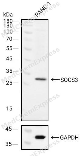 Western blot analysis of extracts from PANC-1(lane 1(40μg) ) using SOCS3 antibody. Proteins were transferred to a NC membrane and blocked with 5% Skim milk in TBST for 2 hour at room temperature. The primary antibody ( 1/1000) and Loading control antibody (GAPDH, 1/1000) was used in 5% Skim milk in TBST at 4°C overnight. Goat Anti-Mouse IgG-HRP Secondary Antibody (1/10,000) was used for 1 hour at room temperature.
 -
 Immunohistochemical analysis of paraffin-embedded human bronchus tissue using SOCS-3 Antibody (HY-P83801, 1/1000). The section was pretreated using heat mediated antigen retrieval with sodium citrate buffer (pH 6.0) for 8 minutes. The tissues were blocked with quick block buffer for 0.5 hours at room temperature, washed with PBS and PBST, and then incubated with the primary antibody overnight at 4℃. The detection was performed using an HRP conjugated compact polymer system. DAB was used as the chromogen. Tissues were counterstained with hematoxylin and mounted with DPX.
 -
 Immunohistochemical analysis of paraffin-embedded human testis tissue using SOCS-3 Antibody (HY-P83801, 1/1000). The section was pretreated using heat mediated antigen retrieval with sodium citrate buffer (pH 6.0) for 8 minutes. The tissues were blocked with quick block buffer for 0.5 hours at room temperature, washed with PBS and PBST, and then incubated with the primary antibody overnight at 4℃. The detection was performed using an HRP conjugated compact polymer system. DAB was used as the chromogen. Tissues were counterstained with hematoxylin and mounted with DPX.
 -
 Immunohistochemical analysis of paraffin-embedded human epididymis tissue using SOCS-3 Antibody (HY-P83801, 1/1000). The section was pretreated using heat mediated antigen retrieval with sodium citrate buffer (pH 6.0) for 8 minutes. The tissues were blocked with quick block buffer for 0.5 hours at room temperature, washed with PBS and PBST, and then incubated with the primary antibody overnight at 4℃. The detection was performed using an HRP conjugated compact polymer system. DAB was used as the chromogen. Tissues were counterstained with hematoxylin and mounted with DPX.
 -
 Immunohistochemical analysis of paraffin-embedded human salivary gland tissue using SOCS-3 Antibody (HY-P83801, 1/1000). The section was pretreated using heat mediated antigen retrieval with sodium citrate buffer (pH 6.0) for 8 minutes. The tissues were blocked with quick block buffer for 0.5 hours at room temperature, washed with PBS and PBST, and then incubated with the primary antibody overnight at 4℃. The detection was performed using an HRP conjugated compact polymer system. DAB was used as the chromogen. Tissues were counterstained with hematoxylin and mounted with DPX.
 -
 Immunohistochemical analysis of paraffin-embedded human kidney tissue using SOCS-3 Antibody (HY-P83801, 1/1000). The section was pretreated using heat mediated antigen retrieval with sodium citrate buffer (pH 6.0) for 8 minutes. The tissues were blocked with quick block buffer for 0.5 hours at room temperature, washed with PBS and PBST, and then incubated with the primary antibody overnight at 4℃. The detection was performed using an HRP conjugated compact polymer system. DAB was used as the chromogen. Tissues were counterstained with hematoxylin and mounted with DPX.
 -
 Immunohistochemical analysis of paraffin-embedded human duodenum tissue using SOCS-3 Antibody (HY-P83801, 1/1000). The section was pretreated using heat mediated antigen retrieval with sodium citrate buffer (pH 6.0) for 8 minutes. The tissues were blocked with quick block buffer for 0.5 hours at room temperature, washed with PBS and PBST, and then incubated with the primary antibody overnight at 4℃. The detection was performed using an HRP conjugated compact polymer system. DAB was used as the chromogen. Tissues were counterstained with hematoxylin and mounted with DPX.
 -
 Immunohistochemical analysis of paraffin-embedded human seminal vesicle tissue using SOCS-3 Antibody (HY-P83801, 1/1000). The section was pretreated using heat mediated antigen retrieval with sodium citrate buffer (pH 6.0) for 8 minutes. The tissues were blocked with quick block buffer for 0.5 hours at room temperature, washed with PBS and PBST, and then incubated with the primary antibody overnight at 4℃. The detection was performed using an HRP conjugated compact polymer system. DAB was used as the chromogen. Tissues were counterstained with hematoxylin and mounted with DPX.
 -
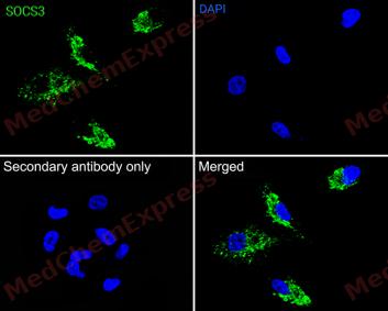 Immunofluorescence analysis of A549 cells labeling SOCS3 antibody at 1/50 dilution. Cells were fixed in 4% paraformaldehyde for 15 minutes at room temperature, permeabilized with 0.1% Triton X-100 in PBS for 15 minutes at room temperature, then blocked with 1% BSA in 10% negative goat serum for 1 hour at room temperature.Cells were then incubated with SOCS3 antibody at 1/50 dilution in 1% BSA in PBST overnight at 4 ℃. AF 488 Goat Anti-mouse IgG H&L (green) was used as the secondary antibody at 1/500 dilution. PBS instead of the primary antibody was used as the secondary antibody only control. The Nuclear counterstain was DAPI (Blue)


Background
-
Function
SOCS-3 is a SOCS family proteins form part of a classical negative feedback system that regulates cytokine signal transduction. SOCS3 is involved in negative regulation of cytokines that signal through the JAK/STAT pathway. Inhibits cytokine signal transduction by binding to tyrosine kinase receptors including IL6ST/gp130, LIF, erythropoietin, insulin, IL12, GCSF and leptin receptors. Binding to JAK2 inhibits its kinase activity and regulates IL6 signaling. Suppresses fetal liver erythropoiesis. Regulates onset and maintenance of allergic responses mediated by T-helper type 2 cells. Probable substrate recognition component of a SCF-like ECS (Elongin BC-CUL2/5-SOCS-box protein) E3 ubiquitin-protein ligase complex which mediates the ubiquitination and subsequent proteasomal degradation of target proteins[1].
-
Expression
Tissue_specificity:It is widely expressed, with high expression levels in the heart, placenta, skeletal muscle, peripheral blood leukocytes, human lungs, and fetal liver and kidneys. Expression levels are lower in the thymus. -
Subunit
Interacts with multiple activated proteins of the tyrosine kinase signaling pathway including IGF1 receptor, insulin receptor and JAK2. Binding to JAK2 is mediated through the KIR and SH2 domains to a phosphorylated tyrosine residue within the JAK2 JH1 domain (PubMed:10421843). Binds specific activated tyrosine residues of the leptin, EPO, IL12, GSCF and gp130 receptors (PubMed:12027890, PubMed:14559241). Interaction with CSNK1E stabilizes SOCS3 protein (PubMed:15070676). Component of the probable ECS(SOCS3) E3 ubiquitin-protein ligase complex which contains CUL5, RNF7/RBX2, Elongin BC complex and SOCS3 (PubMed:15601820). Interacts with CUL5, RNF7, ELOB and ELOC (PubMed:15601820). Interacts with CUL2 (PubMed:15601820). Interacts with FGFR3 (PubMed:16410555). Interacts with INSR (By similarity). Interacts with BCL10; this interaction may interfere with BCL10-binding with PELI2 (By similarity). Interacts with NOD2 (via CARD domain); the interaction promotes NOD2 degradation (PubMed:23019338)
-
SwissProt ID
-
Synonyms
CIS3; SSI3; ATOD4; Cish3; SSI-3; SOCS-3


Documentation

 Powered by Bioz
