



STK3 Antibody (YA7494)

Based on 1 Customer Validation
STK3 Antibody (YA7494) is a Rabbit-derived and non-conjugated IgG, Kappa monoclonal antibody, targeting to STK3.
-
Host:
Rabbit
-
Isotype:
IgG,Kappa
-
Application:
WB, IHC-P, ICC/IF, IP, ELISA
-
Reactivity :
Human, Mouse, Rat
-
Formulation:
Supplied in PBS (pH7.4) containing 50% glycerol, 0.05% Proclin 300, 0.05%BSA
-
Conjugation:
Non-conjugated


To place orders, for customer services and technical support, please contact: MedChemExpress USA
Tel: 609-228-6898 E-mail: [email protected] [email protected]





Applications
| Application |
IHC-P

IHC-P: Immunohistochemistry-Paraffin
|
WB
WB: Western Blot
|
ICC/IF
ICC/IF: Immunocytochemistry/
Immunofluorescence |
ELISA
ELISA: Enzyme Linked Immunosorbent Assay
|
IP
IP: Immunoprecipitation
|
|---|---|---|---|---|---|
| Dilution Ratio | 1:100-1:500 | 1:2000-1:10000 | 1:200-1:1000 | 1:5000-1:20000 | 1:50-1:200 |


Product Details
STK3 Antibody (YA7494) is a Rabbit-derived and non-conjugated IgG, Kappa monoclonal antibody, targeting to STK3.
-
Host Rabbit

-
Clonality Monoclonal,Recombinant

-
Species ReactivityHuman, Mouse, Rat
-
Observed Molecular WeightObserved band size: 56 kDa
Note: Due to possible protein modifications or aggregation, the molecular weight should be confirmed by actual measurement, and the predicted value is for reference only.

-
Calculated Molecular Weight Predicted band size: 56 kDa

The exact sequence is proprietary to MCE.
Endogenous
Protein A affinity purified
Non-conjugated
Unmodified
IgG,Kappa


Product Properties
-
Appearance
Liquid
-
Formulation
Supplied in PBS (pH7.4) containing 50% glycerol, 0.05% Proclin 300, 0.05%BSA
-
Concentration
Batch-dependent, Please check the COA for the concentration of each lot. Check Lot Concentration
-
Storage & Stability
Stored at -20°C for 1 year. Avoid repeated freeze / thaw cycles.
-
Shipping
Shipping with blue ice.


Verification Images
-
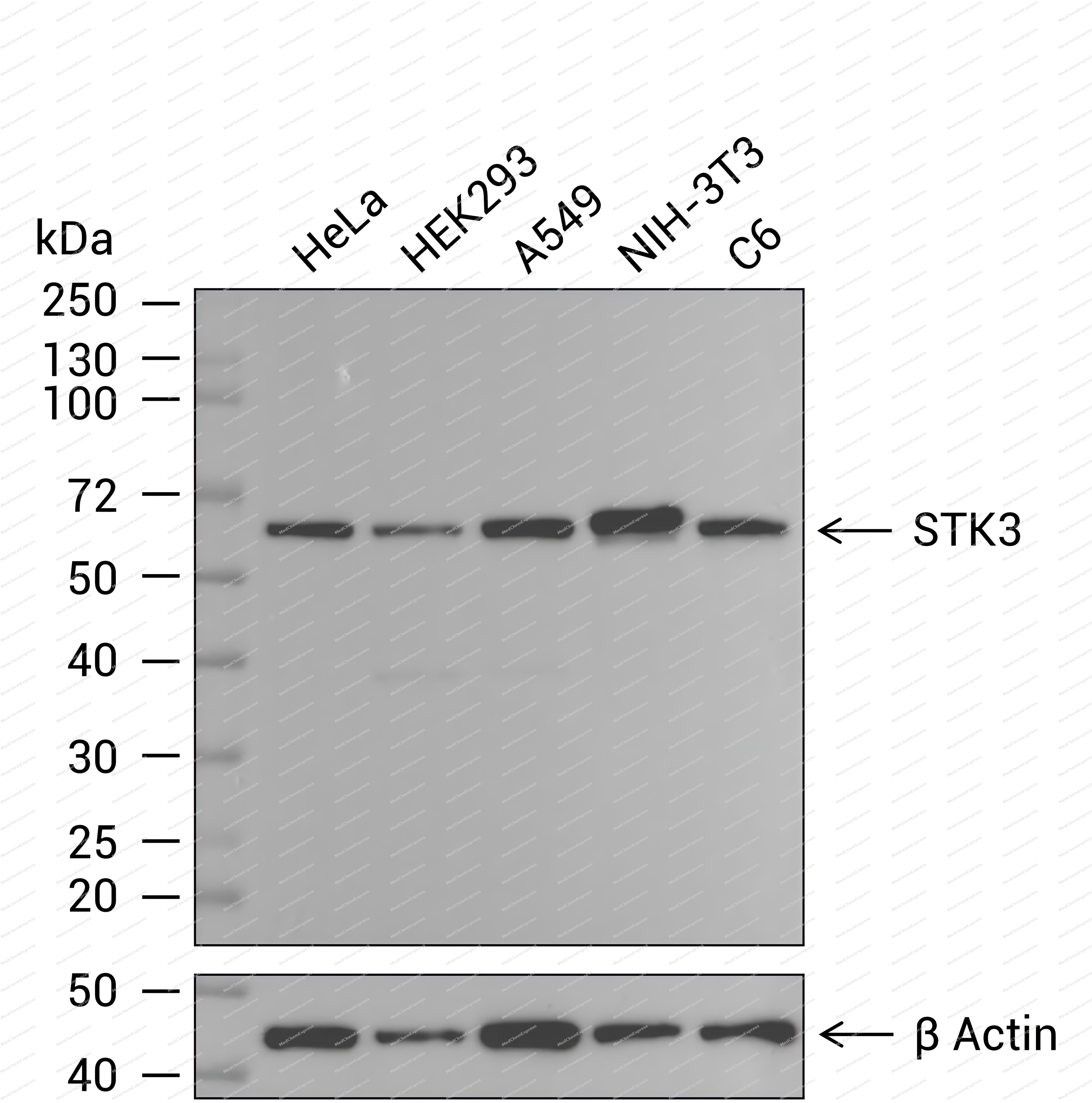 Western blot analysis of extracts from HeLa (lane 1(20μg)) 、HEK293 (lane 2(20μg)) 、A549 (lane 3(20μg)) 、NIH-3T3 (lane 4(20μg)) and C6 (lane 5(20μg)) using STK3 Antibody (HY-P87809) . Proteins were transferred to a PVDF membrane and blocked with 5% nonfat dry milk in TBST for 1.5 hour at room temperature. The primary antibody (1/2000) and Loading control antibody (β Actin, 1/10000) was used in 5% nonfat dry milk in TBST at 4℃ overnight. Goat Anti-Rabbit IgG-HRP Secondary Antibody (1/10,000) was used for 1 hour at room temperature.


Background
-
Function
STK3 is a Stress-activated, pro-apoptotic kinase which, following caspase-cleavage, enters the nucleus and induces chromatin condensation followed by internucleosomal DNA fragmentation. Key component of the Hippo signaling pathway which plays a pivotal role in organ size control and tumor suppression by restricting proliferation and promoting apoptosis. The core of this pathway is composed of a kinase cascade wherein STK3/MST2 and STK4/MST1, in complex with its regulatory protein SAV1, phosphorylates and activates LATS1/2 in complex with its regulatory protein MOB1, which in turn phosphorylates and inactivates YAP1 oncoprotein and WWTR1/TAZ. Phosphorylation of YAP1 by LATS2 inhibits its translocation into the nucleus to regulate cellular genes important for cell proliferation, cell death, and cell migration. STK3/MST2 and STK4/MST1 are required to repress proliferation of mature hepatocytes, to prevent activation of facultative adult liver stem cells (oval cells), and to inhibit tumor formation. Phosphorylates NKX2-1. Phosphorylates NEK2 and plays a role in centrosome disjunction by regulating the localization of NEK2 to centrosome, and its ability to phosphorylate CROCC and CEP250. In conjunction with SAV1, activates the transcriptional activity of ESR1 through the modulation of its phosphorylation. Positively regulates RAF1 activation via suppression of the inhibitory phosphorylation of RAF1 on 'Ser-259'. Phosphorylates MOBKL1A and RASSF2. Phosphorylates MOBKL1B on 'Thr-74'. Acts cooperatively with MOBKL1B to activate STK38[1][2][3][4][5][6][7][8][9][10][11][12][13][14][15][16].
-
Subcellular Localization
Cytoplasm,Nucleus,Cytoplasm, cytoskeleton, microtubule organizing center, centrosome
-
Expression
Tissue_Specificity: Expressed at high levels in adult kidney, skeletal and placenta tissues and at very low levels in adult heart, lung and brain tissues
Induction: Activity increases during mitosis -
Isoforms & Post-Translational Modification
Q13188 has two isomers: Q13188-1: 56301 Da (predicted); Q13188-2: 59462 Da (predicted).
Autophosphorylated on two residues Thr-174 and Thr-180, leading to activation (PubMed:23972470, PubMed:29063833)丨Proteolytically cleaved by caspase-3 during apoptosis丨Ubiquitinated by TRIM69; leading to its redistribution to the perinuclear cytoskeleton, where it is phosphorylated by PLK1 and subsequently activated -
Subunit
Homodimer; mediated via the coiled-coil region
-
SwissProt ID


Documentation

 Powered by Bioz
