



XBP1 Antibody (YA6235)
(Synonyms: XBP1; TREB5; XBP2; X-box-binding protein 1; XBP-1; Tax-responsive element-binding protein 5)
Based on 1 Customer Validation
XBP1 Antibody (YA6235) is a Rabbit-derived and non-conjugated IgG monoclonal antibody, targeting to XBP1.
-
Host:
Rabbit
-
Isotype:
IgG
-
Application:
WB, IHC-P, ICC/IF, IP, ELISA
-
Reactivity :
Human, Mouse, Rat
-
Formulation:
Supplied in PBS, 50% glycerol, 0.05% Proclin 300, 0.05%BSA
-
Conjugation:
Non-conjugated


To place orders, for customer services and technical support, please contact: MedChemExpress USA
Tel: 609-228-6898 E-mail: [email protected] [email protected]





Applications
| Application |
IHC-P

IHC-P: Immunohistochemistry-Paraffin
|
WB
WB: Western Blot
|
ICC/IF
ICC/IF: Immunocytochemistry/
Immunofluorescence |
ELISA
ELISA: Enzyme Linked Immunosorbent Assay
|
IP
IP: Immunoprecipitation
|
|---|---|---|---|---|---|
| Dilution Ratio | 1:200-1:1000 | 1:1000-1:5000 | 1:200-1:1000 | 1:5000-1:20000 | 1:50-1:200 |


Product Details
XBP1 Antibody (YA6235) is a Rabbit-derived and non-conjugated IgG monoclonal antibody, targeting to XBP1.
-
Host Rabbit

-
Clonality Monoclonal

-
Species ReactivityHuman, Mouse, Rat
-
Observed Molecular WeightObserved band size: 35 kDa,60 kDa
Note: Due to possible protein modifications or aggregation, the molecular weight should be confirmed by actual measurement, and the predicted value is for reference only.

-
Calculated Molecular Weight Predicted band size: 29 kDa

Protein A
Non-conjugated
Unmodified
IgG


Product Properties
-
Appearance
Liquid
-
Formulation
Supplied in PBS, 50% glycerol, 0.05% Proclin 300, 0.05%BSA
-
Concentration
Batch-dependent, Please check the COA for the concentration of each lot. Check Lot Concentration
-
Storage & Stability
Stored at -20°C for 1 year. Avoid repeated freeze / thaw cycles.
-
Shipping
Shipping with blue ice.


Verification Images
-
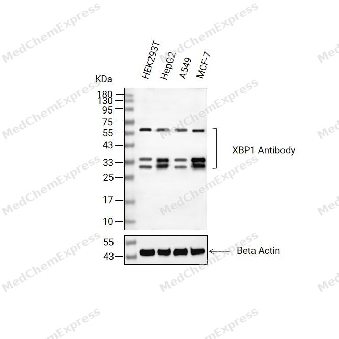 Western blot analysis of extracts from HEK293T(lane 2(20ug) , HepG2(lane 3(20ug) and A549(lane 4(20ug),MCF-7(lane 5(20ug) using XBP1 Antibody (HY-P86543) Rabbit mAb. Proteins were transferred to a PVDF membrane and blocked with 5% non-fat milk in TBST for 2 hour at room temperature. The primary antibody (1/1000) and Loading control antibody (Beta Actin, HY-P83730, 1/10000) was used in 5% non-fat milk in TBST at 4°C overnight. Goat Anti-Mouse/Rabbit IgG-HRP Secondary Antibody (1/10000) was used for 1 hour at room temperature.
 -
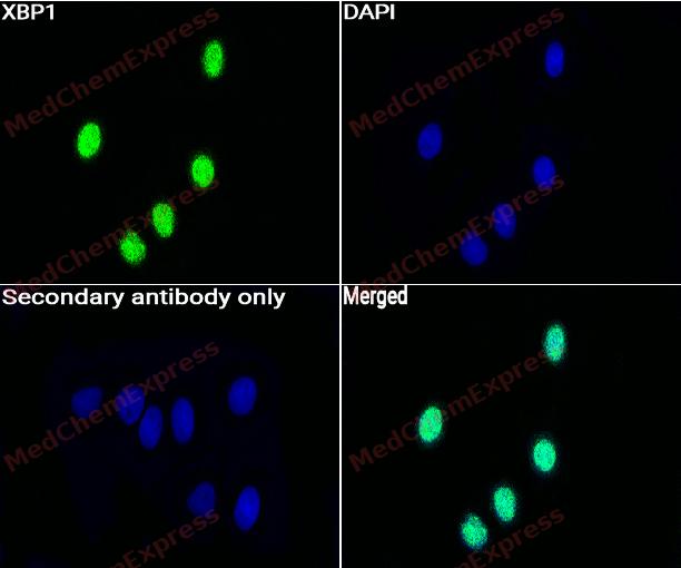 Immunocytochemistry analysis of HeLa cells labeling XBP1 with XBP1 Antibody (HY-P86543) at 1/50 dilution. Cells were fixed in 4% paraformaldehyde for 15 minutes at room temperature, permeabilized with 0.1% Triton X-100 for 10 minutes at room temperature, then blocked with BSA for Immunol Staining for 10 min at room temperature. Cells were then incubated with XBP1 Antibody (HY-P86543) at 1/50 dilution in BSA for Immunol Staining at 4 ℃ overnight. AF488-conjugated AffiniPure Goat Anti-Rabbit IgG H&L(HY-P8002, Green) was used as the secondary antibody at 1/1,000 dilution. PBS instead of the primary antibody was used as the secondary antibody only control. The Nuclear counterstain was DAPI (Blue).
 -
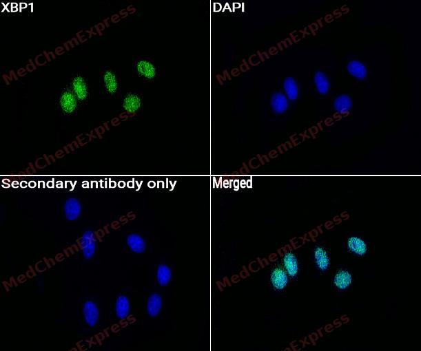 Immunocytochemistry analysis of HepG2 cells labeling XBP1 with XBP1 Antibody (HY-P86543) at 1/50 dilution. Cells were fixed in 4% paraformaldehyde for 15 minutes at room temperature, permeabilized with 0.1% Triton X-100 for 10 minutes at room temperature, then blocked with BSA for Immunol Staining for 10 min at room temperature. Cells were then incubated with XBP1 Antibody (HY-P86543) at 1/50 dilution in BSA for Immunol Staining at 4 ℃ overnight. AF488-conjugated AffiniPure Goat Anti-Rabbit IgG H&L(HY-P8002,Green) was used as the secondary antibody at 1/1,000 dilution. PBS instead of the primary antibody was used as the secondary antibody only control. The Nuclear counterstain was DAPI (Blue).


Background
-
Function
XBP1 functions as a transcription factor during endoplasmic reticulum (ER) stress by regulating the unfolded protein response (UPR). Required for cardiac myogenesis and hepatogenesis during embryonic development, and the development of secretory tissues such as exocrine pancreas and salivary gland. Involved in terminal differentiation of B lymphocytes to plasma cells and production of immunoglobulins. Modulates the cellular response to ER stress in a PIK3R-dependent manner. Binds to the cis-acting X box present in the promoter regions of major histocompatibility complex class II genes. Involved in VEGF-induced endothelial cell (EC) proliferation and retinal blood vessel formation during embryonic development but also for angiogenesis in adult tissues under ischemic conditions. Also functions as a major regulator of the UPR in obesity-induced insulin resistance and type 2 diabetes for the management of obesity and diabetes prevention; Plays a role in the unconventional cytoplasmic splicing processing of its own mRNA triggered by the endoplasmic reticulum (ER) transmembrane endoribonuclease ERN1: upon ER stress, the emerging XBP1 polypeptide chain, as part of a mRNA-ribosome-nascent chain (R-RNC) complex, cotranslationally recruits its own unprocessed mRNA through transient docking to the ER membrane and translational pausing, therefore facilitating efficient IRE1-mediated XBP1 mRNA isoform 2 production. In endothelial cells (EC), associated with KDR, promotes IRE1-mediated XBP1 mRNA isoform 2 productions in a vascular endothelial growth factor (VEGF)-dependent manner, leading to EC proliferation and angiogenesis. Functions as a negative feed-back regulator of the potent transcription factor XBP1 isoform 2 protein levels through proteasome-mediated degradation, thus preventing the constitutive activation of the ER stress response signaling pathway. Inhibits the transactivation activity of XBP1 isoform 2 in myeloma cells. Acts as a weak transcriptional factor. Together with HDAC3, contributes to the activation of NFE2L2-mediated HMOX1 transcription factor gene expression in a PI(3)K/mTORC2/Akt-dependent signaling pathway leading to EC survival under disturbed flow/oxidative stress. Binds to the ER stress response element (ERSE) upon ER stress. Binds to the consensus 5'-GATGACGTG[TG]N(3)[AT]T-3' sequence related to cAMP responsive element (CRE)-like sequences. Binds the Tax-responsive element (TRE) present in the long terminal repeat (LTR) of T-cell leukemia virus type 1 (HTLV-I) and to the TPA response elements (TRE). Associates preferentially to the HDAC3 gene promoter region in a static flow-dependent manner. Binds to the CDH5/VE-cadherin gene promoter region; Functions as a stress-inducible potent transcriptional activator during endoplasmic reticulum (ER) stress by inducing unfolded protein response (UPR) target genes via binding to the UPR element (UPRE). Up-regulates target genes encoding ER chaperones and ER-associated degradation (ERAD) components to enhance the capacity of productive folding and degradation mechanism, respectively, in order to maintain the homeostasis of the ER under ER stress. Plays a role in the production of immunoglobulins and interleukin-6 in the presence of stimuli required for plasma cell differentiation. Induces phospholipid biosynthesis and ER expansion. Contributes to the VEGF-induced endothelial cell (EC) growth and proliferation in a Akt/GSK-dependent and/or -independent signaling pathway, respectively, leading to beta-catenin nuclear translocation and E2F2 gene expression. Promotes umbilical vein EC apoptosis and atherosclerotisis development in a caspase-dependent signaling pathway, and contributes to VEGF-induced EC proliferation and angiogenesis in adult tissues under ischemic conditions. Involved in the regulation of endostatin-induced autophagy in EC through BECN1 transcriptional activation. Plays a role as an oncogene by promoting tumor progression: stimulates zinc finger protein SNAI1 transcription to induce epithelial-to-mesenchymal (EMT) transition, cell migration and invasion of breast cancer cells. Involved in adipocyte differentiation by regulating lipogenic gene expression during lactation. Plays a role in the survival of both dopaminergic neurons of the substantia nigra pars compacta (SNpc), by maintaining protein homeostasis and of myeloma cells. Increases insulin sensitivity in the liver as a response to a high carbohydrate diet, resulting in improved glucose tolerance. Also improves glucose homeostasis in an ER stress- and/or insulin-independent manner through both binding and proteasome-induced degradation of the transcription factor FOXO1, hence resulting in suppression of gluconeogenic genes expression and in a reduction of blood glucose levels. Controls the induction of de novo fatty acid synthesis in hepatocytes by regulating the expression of a subset of lipogenic genes in an ER stress- and UPR-independent manner. Associates preferentially to the HDAC3 gene promoter region in a disturbed flow-dependent manner. Binds to the BECN1 gene promoter region. Binds to the CDH5/VE-cadherin gene promoter region. Binds to the ER stress response element (ERSE) upon ER stress. Binds to the 5'-CCACG-3' motif in the PPARG promoter[1][2][3][4][5][6][7][8][9][10][11][12][13][14][15][16][17][18].
-
Subcellular Localization
Endoplasmic reticulum; Nucleus; Cytoplasm; Endoplasmic reticulum membrane; Single-pass type II membrane protein; Endoplasmic reticulum membrane; Peripheral membrane protein; Membrane; Peripheral membrane protein; Nucleus; Cytoplasm; Cytoplasm; Nucleus
-
Expression
Tissue_specificity:It is expressed in plasma cells of the synovium in rheumatoid arthritis (PubMed:11460154) . It is overexpressed in primary and metastatic breast cancer cells (PubMed:25280941) . Compared with fused quiescent endothelial cells, proliferating endothelial cells show higher expression levels of isoform 1 and isoform 2 (PubMed:19416856) .
Induction:Isoform 1 is up-regulated at the recovery phase of the endoplasmic reticulum (ER) stress response and isoform 2 is up-regulated early during the ER stress response and gradually decreased at later phase of ER stress (PubMed:16461360) . -
Isoforms & Post-Translational Modification
P17861 has 2 isomers: P17861-1: 28695 Da (predicted); P17861-2: 40148 Da (predicted).
Acetylated by EP300; acetylation positively regulates the transcriptional activity of XBP1 isoform 2 (PubMed:20955178). Isoform 2 is deacetylated by SIRT1; deacetylation negatively regulates the transcriptional activity of XBP1 isoform 2 (PubMed:20955178);Ubiquitinated, leading to proteasome-mediated degradation in response to ER stress (PubMed:11779464, PubMed:16461360, PubMed:25239945);X-box-binding protein 1, cytoplasmic form and luminal form are produced by intramembrane proteolytic cleavage of ER membrane-anchored isoform 1 triggered by HM13/SPP in a DERL1-RNF139-dependent and VCP/p97-independent manner. X-box-binding protein 1, luminal form is ubiquitinated leading to proteasomal degradation (PubMed:25239945) -
Subunit
Isoform 2 interacts with SIRT1. Isoform 2 interacts with PIK3R1 and PIK3R2; the interactions are direct and induce translocation of XBP1 isoform 2 into the nucleus and the unfolded protein response (UPR) XBP1-dependent target genes activation in a ER stress- and/or insulin-dependent but PI3K-independent manner. Isoform 2 interacts with FOXO1; the interaction is direct and leads to FOXO1 ubiquitination and degradation via the proteasome pathway in hepatocytes (By similarity). Isoform 1 interacts with HM13 (PubMed:25239945). Isoform 1 interacts with RNF139; the interaction induces ubiquitination and degradation of isoform 1 (PubMed:25239945). Isoform 1 interacts (via luminal domain) with DERL1; the interaction obviates the need for ectodomain shedding prior HM13/SPP-mediated XBP1 isoform 1 cleavage (PubMed:25239945). Isoform 1 interacts with isoform 2; the interaction sequesters isoform 2 from the nucleus and enhances isoform 2 degradation in the cytoplasm (PubMed:16461360, PubMed:25239945). Isoform 1 interacts with HDAC3 and AKT1; the interactions occur in endothelial cell (EC) under disturbed flow (PubMed:25190803). Isoform 1 interacts with the oncoprotein FOS (PubMed:1903538). Isoform 2 interacts with ATF6; the interaction occurs in a ER stress-dependent manner and is required for DNA binding to the unfolded protein response element (UPRE) (PubMed:17765680). Isoform 2 interacts with PIK3R1; the interaction is direct and induces translocation of XBP1 isoform 2 into the nucleus and the unfolded protein response (UPR) XBP1-dependent target genes activation in a ER stress- and/or insulin-dependent but PI3K-independent manner (PubMed:20348923)
-
SwissProt ID
-
Synonyms
XBP1; TREB5; XBP2; X-box-binding protein 1; XBP-1; Tax-responsive element-binding protein 5


Documentation
-
Data Sheet (235 KB)
-
SDS (252 KB)
- English - EN (252 KB)
- Français - FR (252 KB)
- Deutsch - DE (252 KB)
- Norwegian - NO (252 KB)
- Español - ES (252 KB)
- Swedish - SV (252 KB)
- Italian - IT (252 KB)
- Korean - KR (252 KB)
- Portuguese - PT (252 KB)
-
User Guide for Antibodies (1077 KB)

 Powered by Bioz
