



Chiauranib

Based on 1 Customer Validation
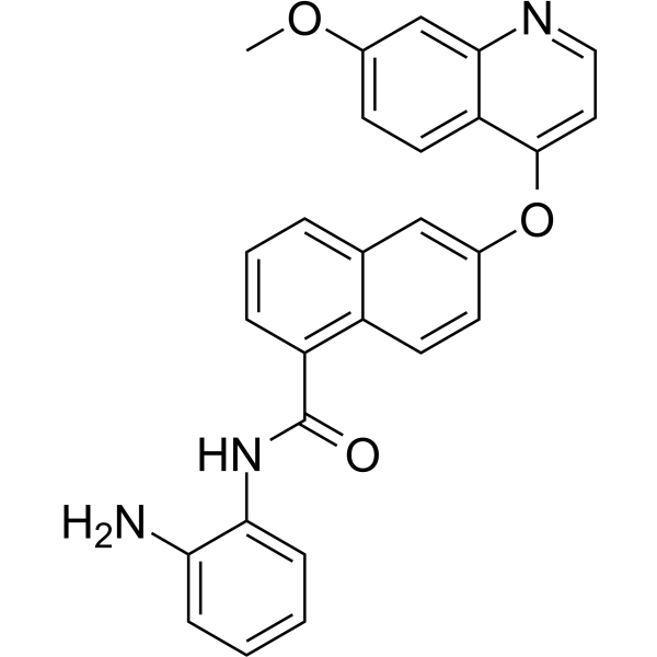
Chiauranib (CS2164) is an orally active multi-target inhibitor against tumor angiogenesis. Chiauranib potently inhibits the angiogenesis-related kinases (VEGFR1, VEGFR2, VEGFR3, PDGFRα and c-Kit), mitosis-related kinase Aurora B, and chronic inflammation-related kinase CSF-1R, with IC50 values ranging from 1-9 nM. Chiauranib has strongly anticancer effects.
Nos produits utilisent uniquement pour la recherche. Nous ne vendons pas aux patients.
- Pureté : 98.97%
- CAS No.: 1256349-48-0
- Formule: C27H21N3O3
- Masse moléculaire:435.47
-
Stockage:
4°C, protect from light
* In solvent : -80°C, 6 months; -20°C, 1 month (protect from light)

To place orders, for customer services and technical support, please contact: MedChemExpress USA
Tel: 609-228-6898 E-mail: [email protected] [email protected]


-
 Activité biologique
-
Chemical Information
-
Solvant et solubilité
- Protocole
- Pureté et documentation
- Références
-
 Help & FAQs


-
HY-10331
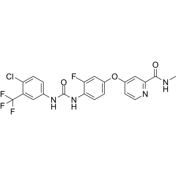
RegorafenibMulti-kinase Inhibitor -
HY-12768
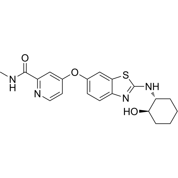
SotuletinibCSF-1R Inhibitor -
HY-16018
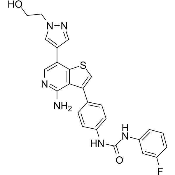
IlorasertibAurora/VEGF/PDGF Inhibitor -
HY-10208
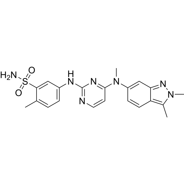
PazopanibVEGFR Inhibitor -
HY-50751
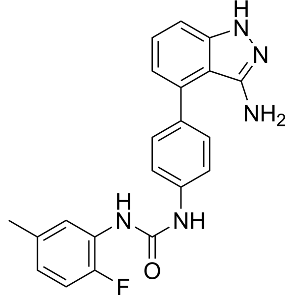
LinifanibMulti-Targeted RTK Inhibitor -
HY-10873
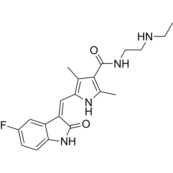
N-Desethyl SunitinibSunitinib Metabolite -
HY-10126
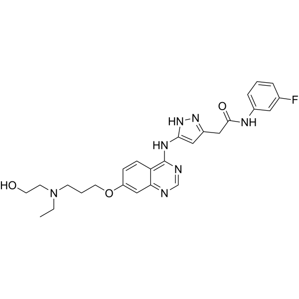
Barasertib-HQPAAurora B Inhibitor -
HY-10917
GW2580c-Fms Inhibitor -
HY-15391
LucitanibVEGFR/FGFR Inhibitor -
HY-114258
LY3295668Aurora A Kinase Inhibitor
Voir tous les produits spécifiques à Isoform VEGFR
MoreVoir tous les produits spécifiques à Isoform Aurora Kinase
More

Activité biologique
Description
IC50 & Target
[1]|
Flt-1 8 nM (IC50) |
KDR 7 nM (IC50) |
Flt-4 9 nM (IC50) |
PDGFRα 1 nM (IC50) |
c-Kit 4 nM (IC50) |
Aurora B 9 nM (IC50) |
PDGFRβ 93 nM (IC50) |
CSF-1R 7 nM (IC50) |
In Vitro
Chiauranib (CS2164; 3 μM; 24 hours) shows induction of G2/M cell cycle arrest and suppression of cell proliferation in tumor tissues through the inhibition of Aurora B-mediated H3 phosphorylation[1].
In HUVEC and PDGFRβ phosphorylation in PDGFRβ overexpressed NIH3T3 cells, Chiauranib (CS2164; 0.03-3 μM) displays anti-angiogenic activities through suppression of VEGFR/PDGFR phosphorylation, inhibition of ligand-dependent cell proliferation and capillary tube formation, and prevention of vasculature formation in tumor tissues[1].
.
Chiauranib (CS2164) inhibits CSF-1R phosphorylation that leads to the suppression of ligand-stimulated monocyte-to-macrophage differentiation and reduces CSF-1R+ cells in tumor tissues[1].
MedChemExpress (MCE) has not independently confirmed the accuracy of these methods. They are for reference only.
-
Cell Line:Molt-4 cells
-
Concentration:3 μM
-
Incubation Time:24 hours
-
Result:Induced the pronounced cell cycle arrest in the G2/M phase at 3 μM.
-
Cell Line:Molt-4 cells
-
Concentration:1.5 μM, 3 μM, 6 μM
-
Incubation Time:24 hours
-
Result:Yielded a substantial reduction in the level of p-H3 in Molt‐4 cells in a concentration-dependent fashion.
In Vivo
MedChemExpress (MCE) has not independently confirmed the accuracy of these methods. They are for reference only.
-
Animal Model:Female BALB/c athymic (nu+/nu+) mice (6-week old) bearing HCT-8, SMMC-7721, MGC‐803 or A549 cells[1]
-
Dosage:2.5 mg/kg, 5 mg/kg, 10 mg/kg, 20 mg/kg, 40 mg/kg
-
Administration:Oral administration; once daily; for 33 days or 43 days
-
Result:Induced remarkable regression or complete inhibition of tumor growth in several human tumor xenograft models.
Essai clinique
| NCT Number | Sponsor | Condition | Start Date |
Phase

|
|---|---|---|---|---|
| NCT01329991 | Plexxikon| | 2011-05 | PHASE1 |


Chemical Information
-
CAS No. 1256349-48-0
-
Appearance Solid
-
Masse moléculaire 435.47
-
Formule C27H21N3O3
-
Color White to light brown
-
SMILES
O=C(NC1=CC=CC=C1N)C2=C3C=CC(OC4=CC=NC5=CC(OC)=CC=C54)=CC3=CC=C2
-
Synonyms
Ibcasertib; CS2164
-
Livraison
Room temperature in continental US; may vary elsewhere.
-
Stockage
4°C, protect from light
* In solvent : -80°C, 6 months; -20°C, 1 month (protect from light)


Solvant et solubilité
In Vitro:
DMSO : 62.5 mg/mL (143.52 mM; Need ultrasonic; Hygroscopic DMSO has a significant impact on the solubility of product, please use newly opened DMSO)
Please refer to the solubility information to select the appropriate solvent. Once prepared, please aliquot and store the solution to prevent product inactivation from repeated freeze-thaw cycles.
Storage method and period of stock solution: -80°C, 6 months; -20°C, 1 month (protect from light). When stored at -80°C, please use it within 6 months. When stored at -20°C, please use it within 1 month.
Please refer to the solubility information to select the appropriate solvent. Once prepared, please aliquot and store the solution to prevent product inactivation from repeated freeze-thaw cycles.
Storage method and period of stock solution: -80°C, 6 months; -20°C, 1 month (protect from light). When stored at -80°C, please use it within 6 months. When stored at -20°C, please use it within 1 month.
Concentration (start) × Volume (start) = Concentration (final) × Volume (final)
In Vivo:
Select the appropriate dissolution method based on your experimental animal and administration route.
- For the following dissolution methods, please ensure to first prepare a clear stock solution using an In Vitro approach and then sequentially add co-solvents:
- To ensure reliable experimental results, the clarified stock solution can be appropriately stored based on storage conditions. As for the working solution for In Vivo experiments, it is recommended to prepare freshly and use it on the same day.
- The percentages shown for the solvents indicate their volumetric ratio in the final prepared solution. If precipitation or phase separation occurs during preparation, heat and/or sonication can be used to aid dissolution.
Add each solvent one by one: 10% DMSO 90% (20% SBE-β-CD in Saline)
Solubility: ≥ 2.08 mg/mL (4.78 mM); Clear solution
This protocol yields a clear solution of ≥ 2.08 mg/mL (saturation unknown).
Taking 1 mL working solution as an example, add 100 μL DMSO stock solution (20.8 mg/mL) to 900 μL 20% SBE-β-CD in Saline, and mix evenly.
Preparation of 20% SBE-β-CD in Saline (4°C, storage for one week): 2 g SBE-β-CD powder is dissolved in 10 mL Saline, completely dissolve until clear.
In Vivo Dissolution Calculator
Please enter the basic information of animal experiments:
-
-
-
-
Recommended: Prepare an additional quantity of animals to account for potential losses during experiments.
Please enter your animal formula composition:
-
%DMSO +
Recommended: Keep the proportion of DMSO in working solution below 2% if your animal is weak.
-
%+
-
+%Tween-80 + +
-
%Saline +
The co-solvents required include: DMSO, . All of co-solvents are available by MedChemExpress (MCE). , Tween 80. All of co-solvents are available by MedChemExpress (MCE).
Working solution concentration: 0.22 mg/mL
Method for preparing stock solution: mg drug dissolved in μL DMSO. Stock solution concentration: mg/mL. * In solvent : -80°C, 6 months; -20°C, 1 month (protect from light)
1. Take μL DMSO stock solution;
2. Add μL .
μL , mix evenly;
3. Then add μL Tween 80, mix evenly;
4. Then add μL
Please ensure that the stock solution in the first step is dissolved to a clear state, and add co-solvents in sequence. You can use ultrasonic heating (ultrasonic cleaner, recommended frequency 20-40 kHz), vortexing, etc. to assist dissolution.


Pureté et documentation
-
Fiche technique (279 KB)
-
SDS (394 KB)
- English - EN (394 KB)
- Français - FR (394 KB)
- Deutsch - DE (394 KB)
- Norwegian - NO (394 KB)
- Español - ES (394 KB)
- Swedish - SV (394 KB)
- Italian - IT (394 KB)
- Korean - KR (394 KB)
- Portuguese - PT (394 KB)
-
Instruction de manipulation (2659 KB)
Références


Complete Stock Solution Preparation Table
Please refer to the solubility information to select the appropriate solvent. Once prepared, please aliquot and store the solution to prevent product inactivation from repeated freeze-thaw cycles.
Storage method and period of stock solution: -80°C, 6 months; -20°C, 1 month (protect from light). When stored at -80°C, please use it within 6 months. When stored at -20°C, please use it within 1 month.
| Optional Solvent | Concentration Solvent Mass | 1 mg | 5 mg | 10 mg | 25 mg |
|---|---|---|---|---|---|
| DMSO | 1 mM | 2.2964 mL | 11.4818 mL | 22.9637 mL | 57.4092 mL |
| 5 mM | 0.4593 mL | 2.2964 mL | 4.5927 mL | 11.4818 mL | |
| 10 mM | 0.2296 mL | 1.1482 mL | 2.2964 mL | 5.7409 mL | |
| 15 mM | 0.1531 mL | 0.7655 mL | 1.5309 mL | 3.8273 mL | |
| 20 mM | 0.1148 mL | 0.5741 mL | 1.1482 mL | 2.8705 mL | |
| 25 mM | 0.0919 mL | 0.4593 mL | 0.9185 mL | 2.2964 mL | |
| 30 mM | 0.0765 mL | 0.3827 mL | 0.7655 mL | 1.9136 mL | |
| 40 mM | 0.0574 mL | 0.2870 mL | 0.5741 mL | 1.4352 mL | |
| 50 mM | 0.0459 mL | 0.2296 mL | 0.4593 mL | 1.1482 mL | |
| 60 mM | 0.0383 mL | 0.1914 mL | 0.3827 mL | 0.9568 mL | |
| 80 mM | 0.0287 mL | 0.1435 mL | 0.2870 mL | 0.7176 mL | |
| 100 mM | 0.0230 mL | 0.1148 mL | 0.2296 mL | 0.5741 mL |


Chiauranib Related Classifications
HY-124526 Related Classifications
 Powered by Bioz