



Diethylcarbamazine
Based on 1 publication(s) in Google Scholar
Diethylcarbamazine is an orally active microfilaricidal agent used originally in onchocerciasis and lymphatic filiariasis. Diethylcarbamazine reduces eosinophil trafficking to the lung tissue and exerts anti-allergic effects. Diethylcarbamazine reduces serum levels of leptin, TNF-α, IL-6, MCP-1, glucose, insulin, and triglycerides, and ameliorates insulin resistance without altering body, liver, or adipose tissue weights. Diethylcarbamazine enhances reactive oxygen intermediate expression by polymorphonuclear neutrophils, increases lymphocyte proliferation, and inhibits actinomycetoma lesion development. Diethylcarbamazine can be used for the researches of bronchial asthma, insulin resistance and infection.
For research use only. We do not sell to patients.
- Purity : 99.86%
- CAS No.: 90-89-1
- Formula: C10H21N3O
- Molecular Weight:199.29
-
Storage:
RT, sealed storage, away from moisture and light.
In solvent -80°C, 1 year , -20°C, 6 months

To place orders, for customer services and technical support, please contact: MedChemExpress USA
Tel: 609-228-6898 E-mail: [email protected] [email protected]


-
 Biological Activity
-
Chemical Information
-
Solvent & Solubility
- Protocols
- Purity & Documentation
- References
-
 Help & FAQs


-

HY-12642AR
Diethylcarbamazine (Standard)Prasite Inhibitor -

HY-W037893
CHMA1004 dihydrochlorideMETTL3/METTL14/WTAP Activator -

HY-109659
1-(3-Aminopropyl)-4-methylpiperazineCapping Agent -

HY-75117A
1-(Cyclopropylcarbonyl)piperazine hydrochlorideDrug Intermediate -
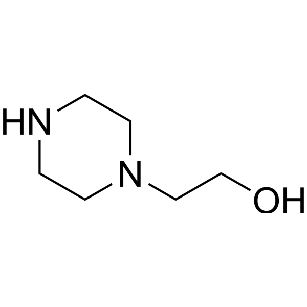
HY-41939
1-(2-Hydroxyethyl)piperazineBiochemical Reagent -

HY-W027208
1,4-Bis(3-aminopropyl)piperazineBiochemical Assay Reagent -
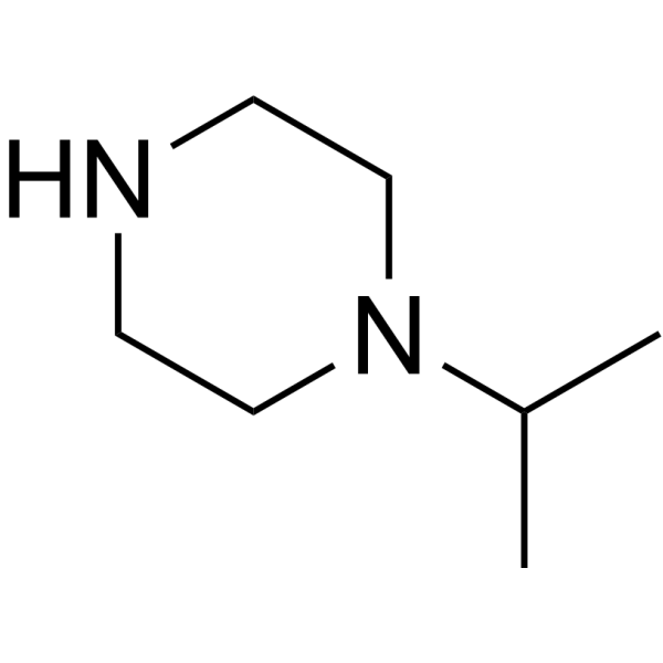
HY-W012744
IsopropylpiperazineBiochemical Reagent -
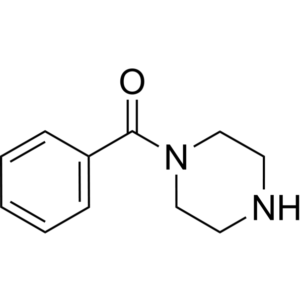
HY-W002698
1-BenzoylpiperazineBiochemical Reagent -
HY-40040
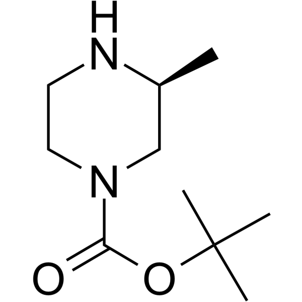
(S)-1-Boc-3-methylpiperazineSynthetic Intermediate -

HY-W042668
2-(4-(tert-Butoxycarbonyl)-2-oxopiperazin-1-yl)acetic acidPROTAC Linker -

HY-W370953
Piperazine-CH2-propargylPROTAC Linker -

HY-W015158
1-Benzyl-3-methylpiperazinePiperazine
Publications Citing Use of MedChemExpress (MCE) Diethylcarbamazine
MoreAll Parasite Isoforms
More

Biological Activity
Description
IC50 & Target
[1]|
IL-6 |
In Vitro
In Vivo
Diethylcarbamazine (12-200 mg/kg; p.o.; twice weekly; 12 weeks) significantly ameliorates high-fat diet-induced insulin resistance in male Swiss mice via suppression of adipose tissue inflammation at the optimal 50 mg/kg dose, without affecting body or tissue weights, with significant reductions in serum glucose, insulin, triglycerides, proinflammatory mediators, liver cyclooxygenase activity, and NF-κBp65 nuclear translocation[2].
Diethylcarbamazine (6 mg/kg; p.o.; daily; 1 week) inhibits actinomycetoma development in BALB/c mice, with a statistically significant reduction in lesion size, and enhances cellular immune responses including neutrophil reactive oxygen intermediate production and lymphocyte proliferation[3].
MedChemExpress (MCE) has not independently confirmed the accuracy of these methods. They are for reference only.
-
Animal Model:white albino mice (6 to 8-week-old female, 18-20 g, intradermal sensitization + aerosol exposure Ovalbumin-induced asthma)[1]
-
Dosage:12 mg/kg
-
Administration:p.o.;
-
Result:Reduced serum IL-4 and IL-5, total BALF IgE and specific anti-ovalbumin IgE, lung tissue EPO and eotaxin2, and reduced inflammatory cell infiltration and thickened alveolar walls compared to untreated asthmatic mice.
Showed no significant changes in serum IL-4, IL-5, BALF IgE, lung EPO, or eotaxin2, and similar histopathological improvement compared to DEC alone when preceded by anti-DEC antibody.
Showed no significant changes in serum IL-4, IL-5, lung EPO, or eotaxin2, and partial reduction in inflammatory cell infiltration and alveolar wall thickening compared to DEC alone when preceded by 10 mg/kg Quercetin.
Significantly reduced serum IL-4 and IL-5, increased IFN-γ/IL-4 ratio to near control levels, decreased total BALF IgE, and reduced inflammatory cell infiltration compared to DEC alone when preceded by 20 mg/kg and 40 mg/kg Quercetin.
-
Animal Model:Swiss mice (male, 5 weeks old, initial 10 g body weight, high-fat diet-induced insulin resistance)[2]
-
Dosage:12 mg/kg; 50 mg/kg; 200 mg/kg
-
Administration:p.o.; twice weekly; 12 weeks (from 6 to 18 week)
-
Result:Reduced serum glucose levels at all oral glucose tolerance test time points (0, 30, 60, 120 min) with a statistically significant reduction in area under the curve at 50 mg/kg.
Reduced serum triglyceride levels at 50 mg/kg.
Reduced serum insulin levels and HOMA-IR score at 50 mg/kg.
Reduced serum leptin levels at 50 mg/kg.
Reduced serum levels of TNF-α, IL-6, and MCP-1 at 50 mg/kg.
Significantly reduced liver cyclooxygenase activity at 50 mg/kg.
Significantly inhibited nuclear localization of NF-κBp65 with increased cytoplasmic retention at 50 mg/kg.
Showed no statistically significant reductions in serum glucose, triglycerides, insulin, HOMA-IR, leptin, TNF-α, IL-6, MCP-1, or liver cyclooxygenase activity relative to untreated high-fat diet-fed mice at 12 mg/kg and 200 mg/kg.
Exhibited less potent effects on NF-κBp65 nuclear localization at 12 mg/kg and 200 mg/kg compared to 50 mg/kg.
Caused no significant changes to body, adipose tissue, or liver weights at 50 mg/kg.
-
Animal Model:BALB/c (male, 10-12 weeks old, 37-45 g, actinomycetoma model via footpad inoculation with Nocardia brasiliensis)[3]
-
Dosage:6 mg/kg
-
Administration:p.o.; daily; 1 week
-
Result:Reduced actinomycetoma lesion size with statistical significance compared to untreated infected controls.
Resolved fistulae, granule discharge, hyperemia, and erythema by day 20 post-infection, and prevented mycetoma establishment through day 90.
Reduced cellular infiltrate, eliminated granuloma layer formation, restored epidermal integrity, and reorganized damaged striated muscle fibers by day 28 post-infection.
Enhanced polymorphonuclear neutrophil reactive oxygen intermediate production with statistical significance at day 3 post-infection.
Increased lymphocyte proliferation in response to N.
brasiliensis cellular crude extract with statistical significance at day 21 post-infection.
Increased lymphocyte proliferation in response to concanavalin A with statistical significance at day 28 post-infection.
Caused no significant changes in IgG or IgM antibody production against N.
brasiliensis P24 antigen.


Chemical Information
-
CAS No. 90-89-1
-
Appearance Solid-Liquid Mixture
-
Molecular Weight 199.29
-
Formula C10H21N3O
-
Color Off-white to light yellow
-
SMILES
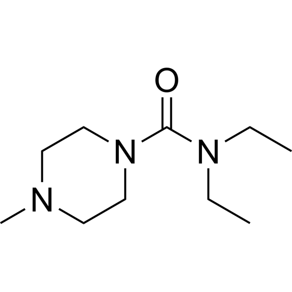
O=C(N1CCN(C)CC1)N(CC)CC
-
Shipping
Room temperature in continental US; may vary elsewhere.
-
Storage
RT, sealed storage, away from moisture and light
In solvent -80°C 1 year -20°C 6 months


Publications (1)
-
Journal Impact Factor
-
Most Recent
-
Cancer Sci
PSMD14/E2F1 Axis-Mediated CENPF Promotes the Metastasis of Triple-Negative Breast Cancer Through Inhibiting Ferroptosis. [Abstract]2025 Aug;116(8):2281-2295. PMID: 40365861


Solvent & Solubility
In Vitro:
DMSO : 200 mg/mL (1003.56 mM; Need ultrasonic; Hygroscopic DMSO has a significant impact on the solubility of product, please use newly opened DMSO)
Please refer to the solubility information to select the appropriate solvent. Once prepared, please aliquot and store the solution to prevent product inactivation from repeated freeze-thaw cycles.
Storage method and period of stock solution: -80°C, 1 year; -20°C, 6 months. When stored at -80°C, please use it within 1 year. When stored at -20°C, please use it within 6 months.
Please refer to the solubility information to select the appropriate solvent. Once prepared, please aliquot and store the solution to prevent product inactivation from repeated freeze-thaw cycles.
Storage method and period of stock solution: -80°C, 1 year; -20°C, 6 months. When stored at -80°C, please use it within 1 year. When stored at -20°C, please use it within 6 months.
Concentration (start) × Volume (start) = Concentration (final) × Volume (final)
In Vivo:
Select the appropriate dissolution method based on your experimental animal and administration route.
- For the following dissolution methods, please ensure to first prepare a clear stock solution using an In Vitro approach and then sequentially add co-solvents:
- To ensure reliable experimental results, the clarified stock solution can be appropriately stored based on storage conditions. As for the working solution for In Vivo experiments, it is recommended to prepare freshly and use it on the same day.
- The percentages shown for the solvents indicate their volumetric ratio in the final prepared solution. If precipitation or phase separation occurs during preparation, heat and/or sonication can be used to aid dissolution.
Add each solvent one by one: 10% DMSO 40% PEG300 5% Tween-80 45% Saline
Solubility: ≥ 5 mg/mL (25.09 mM); Clear solution
This protocol yields a clear solution of ≥ 5 mg/mL (saturation unknown).
Taking 1 mL working solution as an example, add 100 μL DMSO stock solution (50.0 mg/mL) to 400 μL PEG300, and mix evenly; then add 50 μL Tween-80 and mix evenly; then add 450 μL Saline to adjust the volume to 1 mL.
Preparation of Saline: Dissolve 0.9 g sodium chloride in ddH₂O and dilute to 100 mL to obtain a clear Saline solution.
Add each solvent one by one: 10% DMSO 90% (20% SBE-β-CD in Saline)
Solubility: ≥ 5 mg/mL (25.09 mM); Clear solution
This protocol yields a clear solution of ≥ 5 mg/mL (saturation unknown).
Taking 1 mL working solution as an example, add 100 μL DMSO stock solution (50.0 mg/mL) to 900 μL 20% SBE-β-CD in Saline, and mix evenly.
Preparation of 20% SBE-β-CD in Saline (4°C, storage for one week): 2 g SBE-β-CD powder is dissolved in 10 mL Saline, completely dissolve until clear.
In Vivo Dissolution Calculator
Please enter the basic information of animal experiments:
-
-
-
-
Recommended: Prepare an additional quantity of animals to account for potential losses during experiments.
Please enter your animal formula composition:
-
%DMSO +
Recommended: Keep the proportion of DMSO in working solution below 2% if your animal is weak.
-
%+
-
+%Tween-80 + +
-
%Saline +
The co-solvents required include: DMSO, . All of co-solvents are available by MedChemExpress (MCE). , Tween 80. All of co-solvents are available by MedChemExpress (MCE).
Working solution concentration: 0.22 mg/mL
Method for preparing stock solution: mg drug dissolved in μL DMSO. Stock solution concentration: mg/mL.
1. Take μL DMSO stock solution;
2. Add μL .
μL , mix evenly;
3. Then add μL Tween 80, mix evenly;
4. Then add μL
Please ensure that the stock solution in the first step is dissolved to a clear state, and add co-solvents in sequence. You can use ultrasonic heating (ultrasonic cleaner, recommended frequency 20-40 kHz), vortexing, etc. to assist dissolution.


Purity & Documentation
-
Data Sheet (281 KB)
-
SDS (394 KB)
- English - EN (394 KB)
- Français - FR (394 KB)
- Deutsch - DE (394 KB)
- Norwegian - NO (394 KB)
- Español - ES (394 KB)
- Swedish - SV (394 KB)
- Italian - IT (394 KB)
- Korean - KR (394 KB)
- Portuguese - PT (394 KB)
-
Handling Instructions (2659 KB)
References
[1]. Abdul-Razek N, et al. Enhancement of Anti-allergic Effect of Diethylcarbamazine Citrate in Asthmatic Mouse Model: Testing of Anti-drug Antibodies and Quercetin. Iran J Allergy Asthma Immunol. 2020;19(4):373-385. Published 2020 Aug 25. [Content Brief]
[2]. Abdel-Latif M, et al. Diethylcarbamazine citrate ameliorates insulin resistance in high-fat diet-induced obese mice via modulation of adipose tissue inflammation. Int Immunopharmacol. 2015;29(2):607-612. [Content Brief]
[3]. García-Hernández M, et al. Immunomodulatory effect of diethylcarbamazine in mice infected with Nocardia brasiliensis. Int Immunopharmacol. 2014;23(1):113-120. [Content Brief]


Complete Stock Solution Preparation Table
Please refer to the solubility information to select the appropriate solvent. Once prepared, please aliquot and store the solution to prevent product inactivation from repeated freeze-thaw cycles.
Storage method and period of stock solution: -80°C, 1 year; -20°C, 6 months. When stored at -80°C, please use it within 1 year. When stored at -20°C, please use it within 6 months.
| Optional Solvent | Concentration Solvent Mass | 1 mg | 5 mg | 10 mg | 25 mg |
|---|---|---|---|---|---|
| DMSO | 1 mM | 5.0178 mL | 25.0891 mL | 50.1781 mL | 125.4453 mL |
| 5 mM | 1.0036 mL | 5.0178 mL | 10.0356 mL | 25.0891 mL | |
| 10 mM | 0.5018 mL | 2.5089 mL | 5.0178 mL | 12.5445 mL | |
| 15 mM | 0.3345 mL | 1.6726 mL | 3.3452 mL | 8.3630 mL | |
| 20 mM | 0.2509 mL | 1.2545 mL | 2.5089 mL | 6.2723 mL | |
| 25 mM | 0.2007 mL | 1.0036 mL | 2.0071 mL | 5.0178 mL | |
| 30 mM | 0.1673 mL | 0.8363 mL | 1.6726 mL | 4.1815 mL | |
| 40 mM | 0.1254 mL | 0.6272 mL | 1.2545 mL | 3.1361 mL | |
| 50 mM | 0.1004 mL | 0.5018 mL | 1.0036 mL | 2.5089 mL | |
| 60 mM | 0.0836 mL | 0.4182 mL | 0.8363 mL | 2.0908 mL | |
| 80 mM | 0.0627 mL | 0.3136 mL | 0.6272 mL | 1.5681 mL | |
| 100 mM | 0.0502 mL | 0.2509 mL | 0.5018 mL | 1.2545 mL |


Diethylcarbamazine Related Classifications
HY-12642A Related Classifications
 Powered by Bioz