



Ibuprofen sodium
Based on 23 publication(s) in Google Scholar
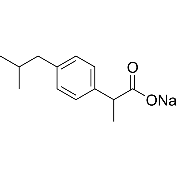
Ibuprofen ((±)-Ibuprofen) sodium is an orally active, selective COX-1 inhibitor with an IC50 value of 13 μM. Ibuprofen sodium inhibits cell proliferation, angiogenesis, and induces cell apoptosis. Ibuprofen sodium is a nonsteroidal anti-inflammatory agent and a nitric oxide (NO) donor. Ibuprofen sodium can be used in the research of pain, swelling, inflammation, infection, immunology, cancers.
For research use only. We do not sell to patients.
- Purity : 99.92%
- CAS No.: 31121-93-4
- Formula: C13H17NaO2
- Molecular Weight:228.26
-
Storage:
4°C, sealed storage, away from moisture
* In solvent : -80°C, 6 months; -20°C, 1 month (sealed storage, away from moisture)

To place orders, for customer services and technical support, please contact: MedChemExpress USA
Tel: 609-228-6898 E-mail: [email protected] [email protected]


-
 Biological Activity
-
Chemical Information
-
Solvent & Solubility
- Protocols
- Purity & Documentation
- References
-
 Help & FAQs


-
HY-B1756
RotenoneMitochondrial Complex I Inhibitor -
HY-66005
AcetaminophenCOX-2 Inhibitor -
HY-B0246
CarbamazepineAnticonvulsant -
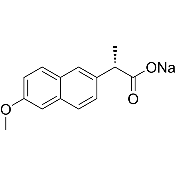
HY-15030A
Naproxen sodiumCOX Inhibitor -
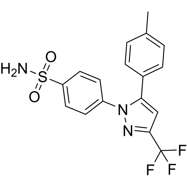
HY-14398
CelecoxibCOX-2inhibitor -
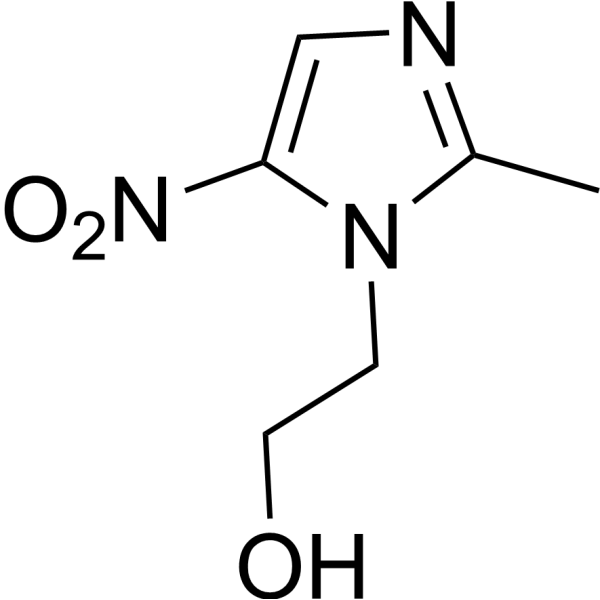
HY-B0318
MetronidazoleAntibiotic -
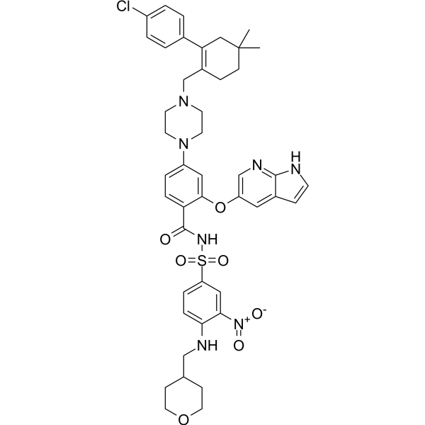
HY-15531
VenetoclaxBCL-2 Inhibitor -
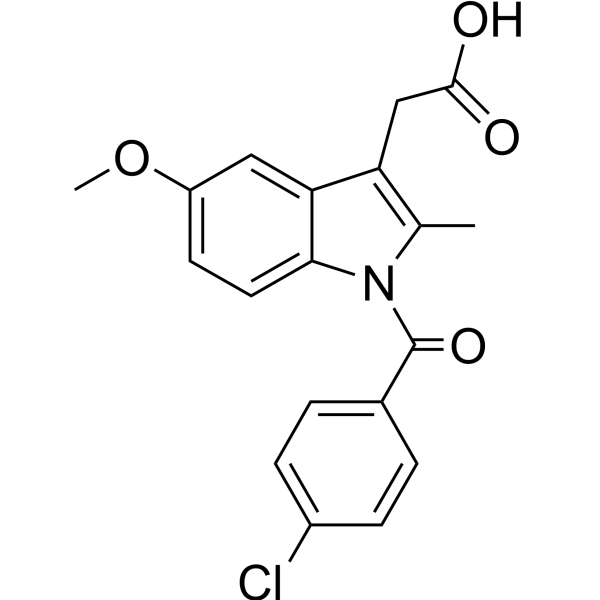
HY-14397
IndomethacinCOX1/2 Inhibitor -
HY-D1056
Lipopolysaccharides, from E. coli O55:B5Endotoxin
Publications Citing Use of MedChemExpress (MCE) Ibuprofen sodium
More- Biomaterials. 2025 Nov:322:123402. [Abstract]
- Phytomedicine. 2022 Nov:106:154427. [Abstract]
- J Hazard Mater Lett. 2025 Nov.
- Int J Biol Macromol. 2025 Dec 9;337(Pt 2):149613. [Abstract]
- Plant Physiol. 2026 Mar 2;200(3):kiag108. [Abstract]
- Cell Rep. 2025 Apr 2;44(4):115489. [Abstract]
- Cell Rep. 2019 Dec 17;29(12):3847-3858.e5. [Abstract]
- Biochem Pharmacol. 2024 Nov:229:116478. [Abstract]
- Inflammopharmacology. 2024 Feb;32(1):733-745. [Abstract]
- Cells. 2022 Jun 9;11(12):1870. [Abstract]
- EMBO Rep. 2022 Jun 7;23(6):e53932. [Abstract]
- Cell Rep Methods. 2023 Oct 23;3(10):100599. [Abstract]
- Int Immunopharmacol. 2026 Aug 15:183:116873. [Abstract]
- Int J Mol Sci. 2021 Nov 22;22(22):12597. [Abstract]
- Bioresour Technol Rep. 2023 Nov 10, 101696.
- Bioresour Technol Rep. 2023 Dec, 24, 101619.
- Sci Rep. 2020 Oct 2;10(1):16383. [Abstract]
- Neuropharmacology. 2022 Oct 1:217:109191. [Abstract]
- ACS Chem Neurosci. 2025 Sep 3;16(17):3257-3266. [Abstract]
- J Neurosci. 2024 Aug 21;44(34):e0740242024. [Abstract]
- Reprod Toxicol. 2025 May:134:108895. [Abstract]
- J Appl Toxicol. 2024 Oct;44(10):1528-1539. [Abstract]
- Chemosphere. 2019 Jun:225:378-387. [Abstract]
 Customer Validation & Images

-
Cell Proliferation/Viability Assay
-
Cell Migration/Invasion Assay
-
Cell Migration/Invasion Assay
-
Flow Cytometry
-
Bio/Physico-chemical Assay
All Parasite Isoforms
More

Biological Activity
Description
IC50 & Target
[1]|
COX-1 13 μM (IC50) |
COX-2 370 μM (IC50) |
In Vitro
Ibuprofen sodium (24 h) inhibits COX-1 and COX-2 activity with IC50 values of 13 μM and 370 μM[1].
Ibuprofen sodium (500 μM, 48 h) inhibits cell proliferation and angiogenesis, and induces apoptosis in AGS cells (Adenocarcinoma gastric cell line)[2].
Ibuprofen sodium (500 μM, 48 h) downregulates transcription of Akt, VEGF-A, PCNA, Bcl2, OCT3/4 and CD44 genes, but upregulates RNA levels of wild type P53 and Bax genes in AGS cell[2].
Ibuprofen sodium (500 μM, 24 h) restores microtubule reformation, microtubule-dependent intracellular cholesterol transport, and induces extension of microtubules to the cell periphery in both cystic fibrosis (CF) cell models and primary CF nasal epithelial cells[3].
Ibuprofen sodium (500 μM, 24 h) enhances UV-induced cell death in MCF-7 cells and MDA-MB-231 cells by a photosensitization process[4].
MedChemExpress (MCE) has not independently confirmed the accuracy of these methods. They are for reference only.
-
Cell Line:AGS cells
-
Concentration:100-1000 μM
-
Incubation Time:24 h, 48 h
-
Result:Inhibited AGS cell viability with IC50 values of 630 μM (trypan blue staining, 24 h), 456 μM (neutral red assay, 24 h), 549 μM (trypan blue staining , 48 h) and 408 μM (neutral red assay, 48 h).
In Vivo
Ibuprofen sodium (subcutaneous injection, 60 mg/kg, every second day for 15 days) reduces the risk of neuropathy in a rat model of chronic Oxaliplatin induced peripheral neuropathy[6].
Ibuprofen sodium (oral administration, 20 mg/kg, every 12 hours, 5 doses total) decreases muscle growth (average muscle fiber cross-sectional area) without affecting regulation of supraspinatus tendon adaptions to exercise[7].
Ibuprofen sodium (oral administration, 35 mg/kg, twice daily) attenuates the Inflammatory response to pseudomonas aeruginosa in a rat model of chronic pulmonary infection[8].
MedChemExpress (MCE) has not independently confirmed the accuracy of these methods. They are for reference only.
-
Animal Model:Syngeneic (D2A1) orthotopic Balb/c mouse model of PPBC (postpartum)[5]
-
Dosage:300 mg/kg, daily for 14 days
-
Administration:Fed in animal feedings (added to pulverized standard chow and mixed dry, then mixed with water, made into chow pellets and dried thoroughly)
-
Result:Suppresed tumor growth, reduced presence of immature monocytes and increased numbers of T cells.
Enhanced Th1 associated cytokines as well as promoted tumor border accumulation of T cells.
-
Animal Model:Oxaliplatin‑induced peripheral neuropathy[6]
-
Dosage:60 mg/kg, every second day for 15 days
-
Administration:Subcutaneous injection
-
Result:Lowered sensory nerve conduction velocity (SNCV).
- Research Protocol for Infectious Diseases
- Human pluripotent stem cell endothelial-cell differentiation
- Vascular/Branching Fractal Analysis
- Annexin V plus membrane-impermeant dye apoptosis staining
- BrdU Incorporation Assay
- Somatic Cell Culture
- CFSE Dye Dilution Proliferation Assay
- Dye-dilution cell tracking and proliferation staining
- CCK-8/WST-8 Cell Proliferation Assay
- Protocol for Cell Cycle
- Cell Counting-Based Growth Curve Assay
- Research Protocol for Inflammation-related Diseases
- Colony Formation (Clonogenic) Assay
- Cell Viability Determination by MTT Colorimetric Assay
- EdU Incorporation Assay (Click Chemistry-Based DNA Synthesis Measurement)
- Ki-67 Immunostaining Proliferation Assay
- Detection of Apoptosis
- TUNEL staining for apoptotic DNA fragmentation
- PCNA Immunodetection Proliferation Assay
- Protocol for Cell Counting and Cell Density Analysis
- Endothelial Tube Formation Assay
- Apoptosis Solutions
- MTT Cell Proliferation Assay
- Endothelial Cell Migration/Angiogenic Sprouting Assay


Chemical Information
-
CAS No. 31121-93-4
-
Appearance Solid
-
Molecular Weight 228.26
-
Formula C13H17NaO2
-
Color White to off-white
-
SMILES
O=C(O[Na])C(C)C1=CC=C(CC(C)C)C=C1
-
Synonyms
(±)-Ibuprofen sodium
-
Shipping
Room temperature in continental US; may vary elsewhere.
-
Storage
4°C, sealed storage, away from moisture
* In solvent : -80°C, 6 months; -20°C, 1 month (sealed storage, away from moisture)


Publications (23)
-
Journal Impact Factor
-
Most Recent
-
Biomaterials
GSH-activable and cytolytic iPep-coupled immune nanoagonist for cancer synergetic therapy. [Abstract]2025 Nov:322:123402. PMID: 40373515 -
Phytomedicine
Prim-O-glucosycimifugin attenuates liver injury in septic mice by inhibiting NLRP3 inflammasome/caspase-1 signaling cascades in macrophages. [Abstract]2022 Nov:106:154427. PMID: 36088791 -
-
Int J Biol Macromol
Molecular interaction of abemaciclib with human serum albumin: Insights from spectroscopy, microscopy, and computational approaches. [Abstract]2025 Dec 9;337(Pt 2):149613. PMID: 41380878 -
Plant Physiol
The MdSP1-MdANK immune module activated by effector SyCD1 enhances Malus domestica resistance to Valsa mali. [Abstract]2026 Mar 2;200(3):kiag108. PMID: 41757702 -
Cell Rep
LAMP2A-mediated neuronal hyperexcitability by enhancing NKAβ1 degradation underlies depression-induced allodynia. [Abstract]2025 Apr 2;44(4):115489. PMID: 40178973 -
Cell Rep
A Central Amygdala Input to the Parafascicular Nucleus Controls Comorbid Pain in Depression. [Abstract]2019 Dec 17;29(12):3847-3858.e5. PMID: 31851918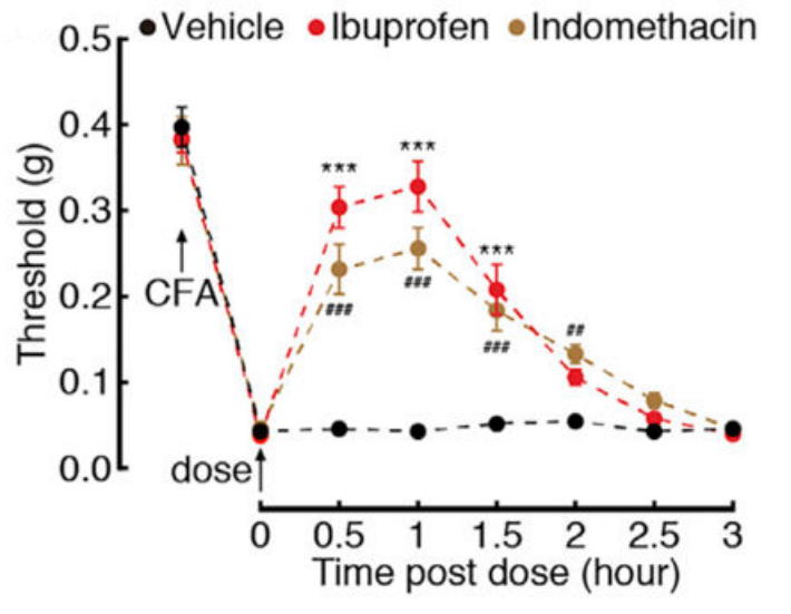
Ibuprofen sodium purchased from MedChemExpress. Usage Cited in: Cell Rep. 2019 Dec 17;29(12):3847-3858.e5. [Abstract]
Effects of Ibuprofen (10 mg/kg, i.p.) or indomethacin (10 mg/kg, i.p.) on pain thresholds at day 3 after CFA treatment.
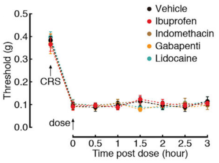
Ibuprofen sodium purchased from MedChemExpress. Usage Cited in: Cell Rep. 2019 Dec 17;29(12):3847-3858.e5. [Abstract]
Effects of Ibuprofen (10 mg/kg, i.p.), Indomethacin, Gabapentin, or Lidocaine on pain thresholds of CRS 3W mice.
-
Biochem Pharmacol
The 'ABC' of split-nanoluciferase HIF heterodimerization bioassays: Applications, Benefits & Considerations. [Abstract]2024 Nov:229:116478. PMID: 39128589 -
Inflammopharmacology
Ibuprofen inhibits anaplastic thyroid cells in vivo and in vitro by triggering NLRP3-ASC-GSDMD-dependent pyroptosis. [Abstract]2024 Feb;32(1):733-745. PMID: 37999895
Ibuprofen sodium purchased from MedChemExpress. Usage Cited in: Inflammopharmacology. 2024 Feb;32(1):733-745. [Abstract]
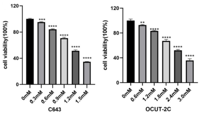
The inhibitory effect of Ibuprofen (0-3 mM, 48 h) on the proliferation of C643 and OCUT-2C cells was detected by CCK8 assay.
Ibuprofen sodium purchased from MedChemExpress. Usage Cited in: Inflammopharmacology. 2024 Feb;32(1):733-745. [Abstract]
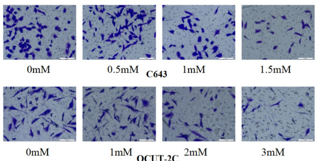
Transwell assay, cells were treated with various concentrations of Ibuprofen (C643 at 0, 0.5, 1, and 1.5 mM; OCUT-2C at 0, 1, 2, and 3 mM) for 48 h view cell invasion abilities.
Ibuprofen sodium purchased from MedChemExpress. Usage Cited in: Inflammopharmacology. 2024 Feb;32(1):733-745. [Abstract]
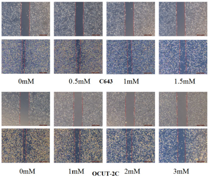
Wound healing assay, cells were treated with different concentrations of Ibuprofen (C643 at 0, 0.5, 1, and 1.5 mM; OCUT-2C at 0, 1, 2, and 3 mM) for 48 h view cell migration abilities.
Ibuprofen sodium purchased from MedChemExpress. Usage Cited in: Inflammopharmacology. 2024 Feb;32(1):733-745. [Abstract]
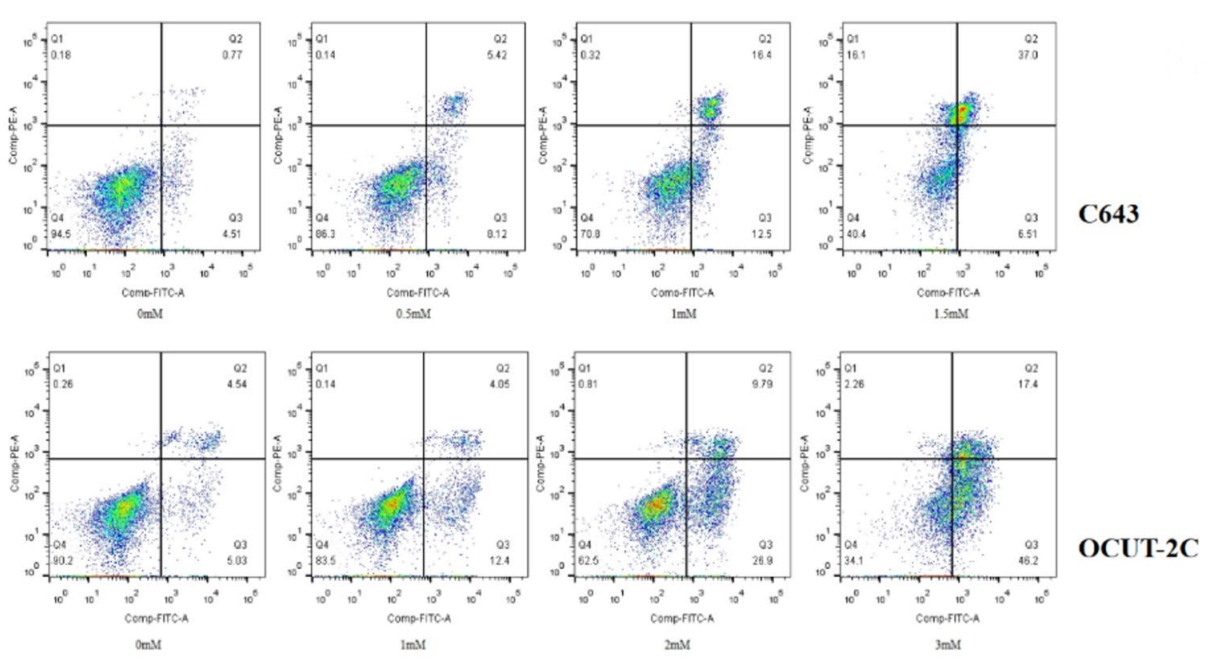
Flow cytometry analysis of ATC cells treated with different Ibuprofen (0-3 mM) concentrations for 48 h stained with Annexin V-FITC and PI.
Ibuprofen sodium purchased from MedChemExpress. Usage Cited in: Inflammopharmacology. 2024 Feb;32(1):733-745. [Abstract]
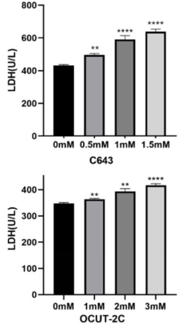
The effect of Ibuprofen (0-1.5 mM) on LDH activity in C643 and OCUT-2C cells.
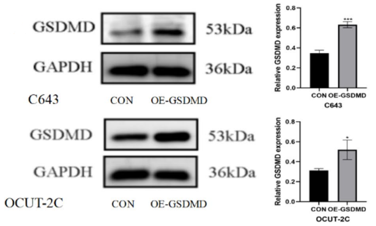
Ibuprofen sodium purchased from MedChemExpress. Usage Cited in: Inflammopharmacology. 2024 Feb;32(1):733-745. [Abstract]
Ibuprofen-induced expression of GSDMD protein in C643 and OCUT-2C cells.
-
Cells
Prostaglandin 2α Promotes Autophagy and Mitochondrial Energy Production in Fish Hepatocytes. [Abstract]2022 Jun 9;11(12):1870. PMID: 35740999 -
EMBO Rep
2022 Jun 7;23(6):e53932. PMID: 35403787 -
Cell Rep Methods
RECOVER identifies synergistic drug combinations in vitro through sequential model optimization. [Abstract]2023 Oct 23;3(10):100599. PMID: 37797618 -
Int Immunopharmacol
Oxyphenbutazone suppresses non-canonical inflammasome activation and protects against LPS-induced sepsis. [Abstract]2026 Aug 15:183:116873. PMID: 42150290 -
Int J Mol Sci
Singapore Grouper Iridovirus Disturbed Glycerophospholipids Homeostasis: Cytosolic Phospholipase A2 Was Essential for Virus Replication. [Abstract]2021 Nov 22;22(22):12597. PMID: 34830477 -
-
-
Sci Rep
Characterization of five novel vasopressin V2 receptor mutants causing nephrogenic diabetes insipidus reveals a role of tolvaptan for M272R-V2R mutation. [Abstract]2020 Oct 2;10(1):16383. PMID: 33009446 -
Neuropharmacology
Malfunction of astrocyte and cholinergic input is involved in postoperative impairment of hippocampal synaptic plasticity and cognitive function. [Abstract]2022 Oct 1:217:109191. PMID: 35835213 -
ACS Chem Neurosci
Identification of a Nonelectrophilic and Selective TRPA1 Agonist for Alleviation of Inflammatory Pain through Channel Desensitization. [Abstract]2025 Sep 3;16(17):3257-3266. PMID: 40846466 -
J Neurosci
Acute Ongoing Nociception Delays Recovery of Consciousness from Sevoflurane Anesthesia via a Midbrain Circuit. [Abstract]2024 Aug 21;44(34):e0740242024. PMID: 39019613 -
Reprod Toxicol
2025 May:134:108895. PMID: 40097051 -
J Appl Toxicol
Differential impacts of nonsteroidal anti-inflammatory drugs on lifespan and healthspan in aged Caenorhabditis elegans. [Abstract]2024 Oct;44(10):1528-1539. PMID: 38840409 -
Chemosphere
Mass-balance-model-based evaluation of sewage treatment plant contribution to residual pharmaceuticals in environmental waters. [Abstract]2019 Jun:225:378-387. PMID: 30884299


Solvent & Solubility
In Vitro:
H2O : ≥ 100 mg/mL (438.10 mM)
DMSO : 20.83 mg/mL (91.26 mM; ultrasonic and warming and heat to 60°C; Hygroscopic DMSO has a significant impact on the solubility of product, please use newly opened DMSO)
* "≥" means soluble, but saturation unknown.
Please refer to the solubility information to select the appropriate solvent. Once prepared, please aliquot and store the solution to prevent product inactivation from repeated freeze-thaw cycles.
Storage method and period of stock solution: -80°C, 6 months; -20°C, 1 month (sealed storage, away from moisture). When stored at -80°C, please use it within 6 months. When stored at -20°C, please use it within 1 month.
* Note: If you choose water as the stock solution, please dilute it to the working solution, then filter and sterilize it with a 0.22 μm filter before use.
Please refer to the solubility information to select the appropriate solvent. Once prepared, please aliquot and store the solution to prevent product inactivation from repeated freeze-thaw cycles.
Storage method and period of stock solution: -80°C, 6 months; -20°C, 1 month (sealed storage, away from moisture). When stored at -80°C, please use it within 6 months. When stored at -20°C, please use it within 1 month.
* Note: If you choose water as the stock solution, please dilute it to the working solution, then filter and sterilize it with a 0.22 μm filter before use.
Concentration (start) × Volume (start) = Concentration (final) × Volume (final)


Purity & Documentation
-
Data Sheet (278 KB)
-
SDS (393 KB)
- English - EN (393 KB)
- Français - FR (393 KB)
- Deutsch - DE (393 KB)
- Norwegian - NO (393 KB)
- Español - ES (393 KB)
- Swedish - SV (393 KB)
- Italian - IT (393 KB)
- Korean - KR (393 KB)
- Portuguese - PT (393 KB)
-
Handling Instructions (2659 KB)
References
[1]. Noreen Y, et al. Development of a radiochemical cyclooxygenase-1 and -2 in vitro assay for identification of natural products as inhibitors of prostaglandin biosynthesis. J Nat Prod. 1998 Jan;61(1):2-7. [Content Brief]
[2]. Hassan Akrami, et al. Inhibitory effect of ibuprofen on tumor survival and angiogenesis in gastric cancer cell. Tumour Biol. 2015 May;36(5):3237-43. [Content Brief]
[3]. Sharon M Rymut, et al. Ibuprofen regulation of microtubule dynamics in cystic fibrosis epithelial cells. Am J Physiol Lung Cell Mol Physiol. 2016 Aug 1;311(2):L317-27. [Content Brief]
[4]. Emmanuelle Bignon, et al. Ibuprofen and ketoprofen potentiate UVA-induced cell death by a photosensitization process. Sci Rep. 2017 Aug 21;7(1):8885. [Content Brief]
[5]. Nathan D Pennock, et al. Ibuprofen supports macrophage differentiation, T cell recruitment, and tumor suppression in a model of postpartum breast cancer. J Immunother Cancer. 2018 Oct 1;6(1):98. [Content Brief]
[6]. Thomas Krøigård, et al. Protective effect of ibuprofen in a rat model of chronic oxaliplatin-induced peripheral neuropathy. Exp Brain Res. 2019 Oct;237(10):2645-2651. [Content Brief]
[7]. Sarah Ilkhanipour Rooney, et al. Ibuprofen Differentially Affects Supraspinatus Muscle and Tendon Adaptations to Exercise in a Rat Model. Am J Sports Med. 2016 Sep;44(9):2237-45. [Content Brief]
[8]. M W Konstan, et al. Ibuprofen attenuates the inflammatory response to Pseudomonas aeruginosa in a rat model of chronic pulmonary infection. Implications for antiinflammatory therapy in cystic fibrosis. Am Rev Respir Dis. 1990 Jan;141(1):186-92. [Content Brief]


Complete Stock Solution Preparation Table
Please refer to the solubility information to select the appropriate solvent. Once prepared, please aliquot and store the solution to prevent product inactivation from repeated freeze-thaw cycles.
Storage method and period of stock solution: -80°C, 6 months; -20°C, 1 month (sealed storage, away from moisture). When stored at -80°C, please use it within 6 months. When stored at -20°C, please use it within 1 month.
| Optional Solvent | Concentration Solvent Mass | 1 mg | 5 mg | 10 mg | 25 mg |
|---|---|---|---|---|---|
| DMSO / H2O | 1 mM | 4.3810 mL | 21.9048 mL | 43.8097 mL | 109.5242 mL |
| 5 mM | 0.8762 mL | 4.3810 mL | 8.7619 mL | 21.9048 mL | |
| 10 mM | 0.4381 mL | 2.1905 mL | 4.3810 mL | 10.9524 mL | |
| 15 mM | 0.2921 mL | 1.4603 mL | 2.9206 mL | 7.3016 mL | |
| 20 mM | 0.2190 mL | 1.0952 mL | 2.1905 mL | 5.4762 mL | |
| 25 mM | 0.1752 mL | 0.8762 mL | 1.7524 mL | 4.3810 mL | |
| 30 mM | 0.1460 mL | 0.7302 mL | 1.4603 mL | 3.6508 mL | |
| 40 mM | 0.1095 mL | 0.5476 mL | 1.0952 mL | 2.7381 mL | |
| 50 mM | 0.0876 mL | 0.4381 mL | 0.8762 mL | 2.1905 mL | |
| 60 mM | 0.0730 mL | 0.3651 mL | 0.7302 mL | 1.8254 mL | |
| 80 mM | 0.0548 mL | 0.2738 mL | 0.5476 mL | 1.3691 mL | |
| H2O | 100 mM | 0.0438 mL | 0.2190 mL | 0.4381 mL | 1.0952 mL |
* Note: If you choose water as the stock solution, please dilute it to the working solution, then filter and sterilize it with a 0.22 μm filter before use.


Ibuprofen sodium Related Classifications
HY-78131C Related Classifications
 Powered by Bioz