



Relaxin H3 (human) TFA
Relaxin H3 (human) TFA is a relaxin peptide with anti-inflammatory, anti-apoptotic, anti-pyroptotic, anti-migratory, protective and anti-fibrotic activities. Relaxin H3 (human) TFA acts on RXFP1 to generate cAMP and reduce the levels of ATP and ROS. Relaxin H3 (human) TFA inhibits renal inflammatory pyroptosis (pyroptosis), NLRP3 inflammasome activation, caspase-1 activation, IL-1β/IL-18 secretion, collagen synthesis, TGF-β1 signaling pathway, Smad2 phosphorylation, myofibroblast differentiation, TIMP expression, and HRMEC migration. Relaxin H3 (human) TFA activates AMPK, upregulates MFN2 expression, improves mitochondrial quality control and membrane potential, inhibits apoptosis (apoptosis) and pyroptosis, restores retinal ultrastructure, and reverses excessive left ventricular collagen expression. Relaxin H3 (human) TFA can be used in studies related to kidney stones, nephrocalcinosis, diabetic cardiomyopathy, fibrotic cardiomyopathy, and diabetic retinopathy.
商品は「研究用試薬」です。人や動物の医療用・臨床診断用・食品用の製品ではありません。
研究用途以外に使用した場合、当社は一切の責任を負いかねます。
- 分子式: C237H374N70O69S6.xC2HF3O2
- 分子量:5500.32 (free base)
-
保管条件:
-20°C, protect from light, stored under nitrogen
* In solvent : -80°C, 6 months; -20°C, 1 month (protect from light, stored under nitrogen)


-
 生物活性
生物活性
-
化学情報
- 純度とドキュメンテーション
- 参考文献
-
 Help & FAQs




生物活性
|
IL-1β |
Caspase-1 |
IL-18 |
Relaxin H3 (human) (20 ng/mL; 2 h pretreatment) TFA protects human proximal tubular HK2 cells from Ca2+Ox crystal-induced pyroptosis and inflammatory injury by reducing ROS production, decreasing ATP levels, and inhibiting activation of the NLRP3 inflammasome-GSDMD axis via the RXFP1-cAMP pathway[1].
Pretreatment with human Relaxin H3 (20 ng/mL for 2 h) TFA reduces the adhesion and internalization of Ca2+Ox crystals in human proximal tubular HK2 cells, and counteracts Ca2+Ox-induced acidification of the culture medium[1].
Relaxin H3 (human) TFA (100 ng/mL; 30 min pre-incubation, 48 h exposure/treatment) inhibits high glucose-induced collagen synthesis, NLRP3 inflammasome activation, ROS production and P2X7R expression in neonatal rat cardiac fibroblasts[2].
Relaxin H3 (human) (10-500 ng/mL; 72 h) TFA potently inhibits TGF-β1-stimulated collagen deposition in primary neonatal rat ventricular fibroblasts when applied at concentrations of 100 ng/mL and 250 ng/mL for 72 h[3].
Relaxin H3 (human) (100 ng/mL; 72 h) TFA inhibits transforming growth factor-β1 (TGF-β1)-induced Smad2 phosphorylation by 40% and completely blocks TGF-β1-induced differentiation of primary neonatal rat ventricular fibroblasts into myofibroblasts (α-SMA expression)[3].
Relaxin H3 (human) (100 ng/mL; 72 h) TFA significantly inhibits the expression of TIMP-1 and TIMP-2 in primary neonatal rat ventricular fibroblasts stimulated with transforming growth factor-β1 (TGF-β1), with an inhibition rate of 50%-55%[3].
Relaxin H3 (human) (100 ng/mL; 30 min pre-incubation followed by 48 h treatment, or continuous 48 h treatment) TFA attenuates AGE-BSA-induced NLRP3 inflammasome activation, apoptosis, pyroptosis and cell migration in human retinal microvascular endothelial cells by inhibiting the P2X7R-mediated signaling pathway[4].
Relaxin H3 (human) (100 ng/mL) TFA enhances the viability of H9C2 rat cardiomyocytes, reduces cell apoptosis, and ameliorates high glucose- and palmitic acid-induced mitochondrial dysfunction by regulating apoptosis-related proteins, mitochondrial fusion/fission/mitophagy proteins, ROS levels, membrane potential, and ATP production[5].
Relaxin H3 (human) (100 ng/mL) TFA alleviates apoptosis and mitochondrial dysfunction in MFN2-knockdown H9C2 rat cardiomyocytes induced by high glucose and palmitic acid by reversing the abnormal expression of apoptosis and mitochondrial quality control proteins, reducing ROS, restoring membrane potential, and increasing ATP levels[5].
Relaxin H3 (human) (100 ng/mL) TFA exerts a protective effect against high glucose- and palmitic acid-induced apoptosis and mitochondrial dysfunction in H9C2 rat cardiomyocytes by activating the AMPK pathway[5].
MedChemExpress (MCE) has not independently confirmed the accuracy of these methods. They are for reference only.
Relaxin H3 (human) (0.5 mg/kg/day; subcutaneous injection; continuous infusion; 14 days) TFA reverses established myocardial fibrosis by 50% in β2-AR transgenic mice[3].
Human Relaxin H3 (0.2-2 µg/kg/d; daily administration for 14 consecutive days) TFA dose-dependently improves retinal ultrastructure, alleviates NLRP3 inflammasome-mediated inflammatory responses, and inhibits apoptosis in streptozotocin (HY-13753)-induced diabetic rats, with the protective effect of the 2 µg/kg/d dose being stronger than that of the 0.2 µg/kg/d dose[4].
Relaxin H3 (human) (2 μg/kg/d; intraperitoneal injection; daily administration for 2 consecutive weeks) TFA improves metabolic disorders, cardiac structure and function, and mitochondrial quality control, reduces cardiomyocyte apoptosis, and significantly decreases the levels of LVIDd, LVIDs, pro-apoptotic proteins, and fission/mitophagy proteins in Sprague-Dawley rats with diabetic cardiomyopathy by activating the AMPK pathway[5].
MedChemExpress (MCE) has not independently confirmed the accuracy of these methods. They are for reference only.
-
Animal Model:Sprague-Dawley (male, 5-6 weeks old, 150-200 g, oxalate nephropathy induced by 0.8% ethylene glycol and 0.8% NH4Cl in drinking water)[1]
-
Dosage:0.2 μg/kg/d; 2 μg/kg/d
-
Administration:s.c.; daily; 7 or 14 days
-
Result:Reduced renal tissue ATP levels in a dose-dependent manner after 7 and 14 days compared to the EG model group.
Reduced serum creatinine and blood urea nitrogen levels in a dose-dependent manner after 14 days compared to the EG model group.
Improved tubular dilatation, neutrophil infiltration, and epithelial cell deformation/exfoliation in a dose-dependent manner compared to the EG model group (histological analysis).
Reduced levels of cleaved caspase-1, GSDMD, and NLRP3 in renal tissues compared to the EG model group (immunohistochemical assays).
Reduced CaOx crystal deposition, hydronephrosis, and renal cortex thinning compared to the EG model group (ultrasonographic examination).
Reduced renal crystal deposition in a dose-dependent manner compared to the EG model group (Von Kossa staining).
Reduced levels of NLRP3, ASC, cleaved caspase-1, cleaved GSDMD, IL-18, and IL-1β in renal tissues compared to the EG model group (Western blot analysis).
-
Animal Model:C57Blk6JxSJL background (4-5-month-old male, heterozygous β2-adrenergic receptor, cardiac fibrosis induced by cardiac-restricted transgenic overexpression of β2-AR)[3]
-
Dosage:0.5 mg kg/day
-
Administration:s.c.; continuous infusion; 14 days
-
Result:Reversed established left ventricular collagen accumulation by 50%.
-
Animal Model:Sprague-Dawley (male, 5-6 weeks old, 150-200 g, oxalate nephropathy induced by 0.8% ethylene glycol and 0.8% NH4Cl in drinking water)[1]
-
Dosage:0.2 μg/kg/d; 2 μg/kg/d
-
Administration:s.c.; daily; 7 or 14 days
-
Result:Reduced renal tissue ATP levels in a dose-dependent manner after 7 and 14 days compared to the EG model group.
Reduced serum creatinine and blood urea nitrogen levels in a dose-dependent manner after 14 days compared to the EG model group.
Improved tubular dilatation, neutrophil infiltration, and epithelial cell deformation/exfoliation in a dose-dependent manner compared to the EG model group (histological analysis).
Reduced levels of cleaved caspase-1, GSDMD, and NLRP3 in renal tissues compared to the EG model group (immunohistochemical assays).
Reduced CaOx crystal deposition, hydronephrosis, and renal cortex thinning compared to the EG model group (ultrasonographic examination).
Reduced renal crystal deposition in a dose-dependent manner compared to the EG model group (Von Kossa staining).
Reduced levels of NLRP3, ASC, cleaved caspase-1, cleaved GSDMD, IL-18, and IL-1β in renal tissues compared to the EG model group (Western blot analysis).
-
Animal Model:Sprague-Dawley rats (male/female not specified; induced via high-fat diet plus intraperitoneal streptozotocin injection, fasting blood glucose ≥16.7 mmol/L)[5]
-
Dosage:2 μg/kg/d
-
Administration:i.p.; daily; 2 weeks
-
Result:Improved glucose tolerance and insulin sensitivity.
Reduced serum total cholesterol, triglyceride, and insulin levels.
Ameliorated disorganized myocardial cells, edema, fibrosis, and collagen deposition.
Restored damaged mitochondrial ultrastructure.
Reduced left ventricular internal diameter at end-diastole (LVIDd) from 8.17 mm to 7.46 mm and left ventricular internal diameter at end-systole (LVIDs) from 5.53 mm to 4.95 mm.
Improved left ventricular ejection fraction.
Upregulated anti-apoptotic protein BCL-2 and downregulated pro-apoptotic proteins BAX and cleaved caspase-3.
Increased mitochondrial fusion proteins MFN2 and OPA1.
Reduced mitochondrial fission proteins Drp1 and Fis1.
Decreased mitophagy proteins parkin and PINK1.
Increased p-AMPK/AMPK ratio.
All changes were statistically significant compared to the diabetic cardiomyopathy group.
Chain 1:Arg-Ala-Ala-Pro-Tyr-Gly-Val-Arg-Leu-Cys-Gly-Arg-Glu-Phe-Ile-Arg-Ala-Val-Ile-Phe-Thr-Cys-Gly-Gly-Ser-Arg-Trp Chain 2:Asp-Val-Leu-Ala-Gly-Leu-Ser-Ser-Ser-Cys-Cys-Lys-Trp-Gly-Cys-Ser-Lys-Ser-Glu-Ile-Ser-Ser-Leu-Cys (Disulfide bridge:Chain 1 Cys10-Chain 2 Cys11;Chain 1 Cys22-Chain 2 Cys24;Chain 2 Cys10-Chain 2 Cys15)
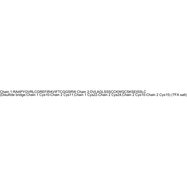
Chain 1:RAAPYGVRLCGREFIRAVIFTCGGSRW Chain 2:DVLAGLSSSCCKWGCSKSEISSLC (Disulfide bridge:Chain 1 Cys10-Chain 2 Cys11;Chain 1 Cys22-Chain 2 Cys24;Chain 2 Cys10-Chain 2 Cys15)


化学情報
-
性状 Solid
-
分子量 5500.32 (free base)
-
分子式 C237H374N70O69S6.xC2HF3O2
-
輸送条件
Room temperature in continental US; may vary elsewhere.
-
保管条件
-20°C, protect from light, stored under nitrogen
* In solvent : -80°C, 6 months; -20°C, 1 month (protect from light, stored under nitrogen)


純度とドキュメンテーション
-
データシート (287 KB)
-
SDS (252 KB)
- Français - FR (252 KB)
- Deutsch - DE (252 KB)
- Norwegian - NO (252 KB)
- Español - ES (252 KB)
- Swedish - SV (252 KB)
- Italian - IT (252 KB)
- Portuguese - PT (252 KB)
-
取扱説明書 (2659 KB)
参考文献
[1]. Liu J, et al. H3 relaxin protects against calcium oxalate crystal-induced renal inflammatory pyroptosis. Cell Prolif. 2020;53(10):e12902. [Content Brief]
[2]. Zhang X, et al. H3 relaxin inhibits the collagen synthesis via ROS- and P2X7R-mediated NLRP3 inflammasome activation in cardiac fibroblasts under high glucose. J Cell Mol Med. 2018 Mar;22(3):1816-1825. doi: 10.1111/jcmm.13464. Epub 2018 Jan 5. Erratum in: J Cell Mol Med. 2018 Jun;22(6):3264-3265. [Content Brief]
[3]. Hossain MA, et al. H3 relaxin demonstrates antifibrotic properties via the RXFP1 receptor. Biochemistry. 2011;50(8):1368-1375. [Content Brief]
[4]. Yang K, et al. H3 Relaxin Alleviates Migration, Apoptosis and Pyroptosis Through P2X7R-Mediated Nucleotide Binding Oligomerization Domain-Like Receptor Protein 3 Inflammasome Activation in Retinopathy Induced by Hyperglycemia. Front Pharmacol. 2020;11:603689. Published 2020 Dec 16. [Content Brief]
[5]. Wang J, et al. H3 relaxin ameliorates mitochondrial quality control and apoptosis in cardiomyocytes of type 2 diabetic rats via activation of the AMPK pathway. Int Immunopharmacol. 2025;144:113664. [Content Brief]

Calculators
濃度 (開始) × 体積 (開始) = 濃度 (終了) × 体積 (終了)


 Powered by Bioz