



NR160

Based on 1 Customer Validation
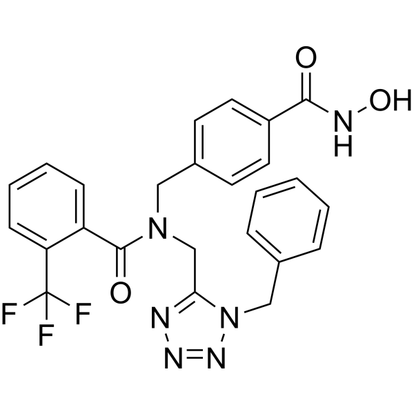
NR160 is a selective HDAC6 inhibitor with an IC50 value of 30 nM. NR160 shows low cytotoxicity against leukemia cell line. NR160 augments the apoptosis induction of Bortezomib (HY-10227) (proteasome inhibitor), Epirubicin (HY-13624) and Daunorubicin (HY-13062A) significantly.
For research use only. We do not sell to patients.
- Purity : 98.9%
- CAS No.: 2484895-50-1
- Formula: C25H21F3N6O3
- Molecular Weight:510.47
-
Storage:Powder -20°C, 3 years , 4°C, 2 years ; In solvent -80°C, 6 months , -20°C, 1 month

To place orders, for customer services and technical support, please contact: MedChemExpress USA
Tel: 609-228-6898 E-mail: [email protected] [email protected]


-
 Biological Activity
-
Chemical Information
- Protocols
- Purity & Documentation
- References
-
 Help & FAQs


-
HY-10221
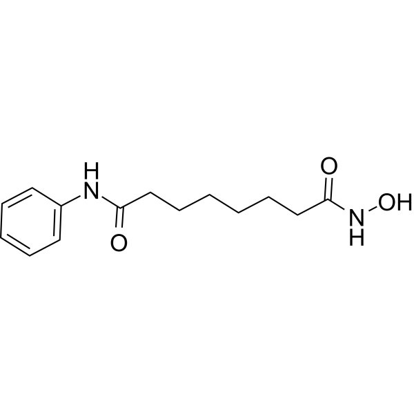
VorinostatHDAC Inhibitor -
HY-12163
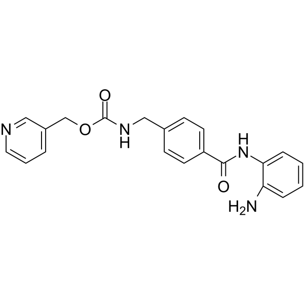
EntinostatHDAC Class I Inhibitor -
HY-126330
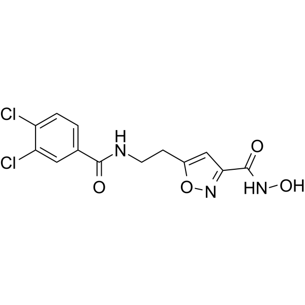
SS-208HDAC Inhibitor -
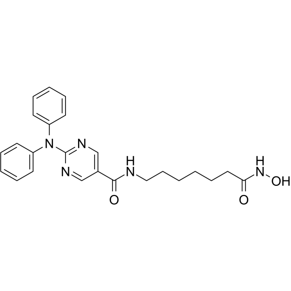
HY-16026
RicolinostatHDAC6 Inhibitor -
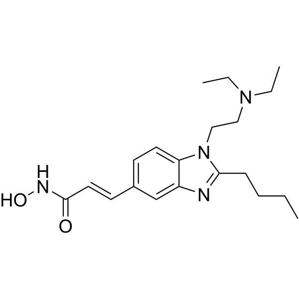
HY-13322
PracinostatHDAC/MBLAC2 Inhibitor -

HY-10221R
Vorinostat (Standard)HDAC Inhibitor -
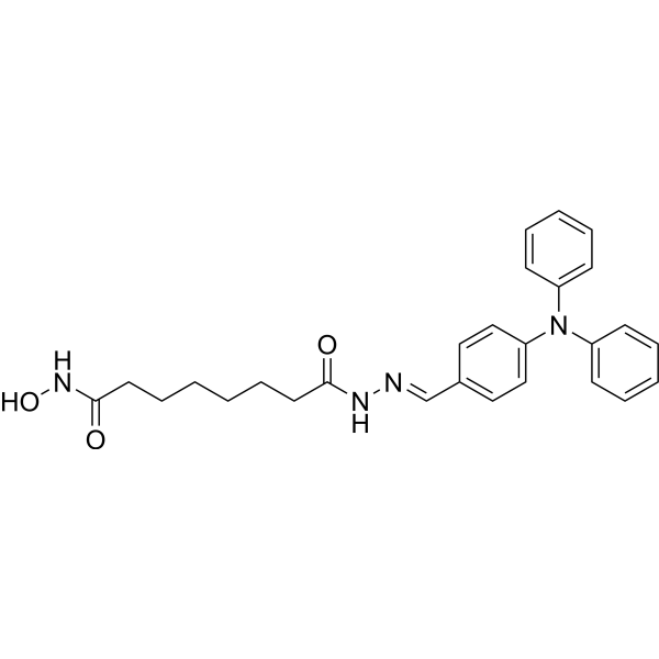
HY-100871
WT-161HDAC6 Inhibitor -
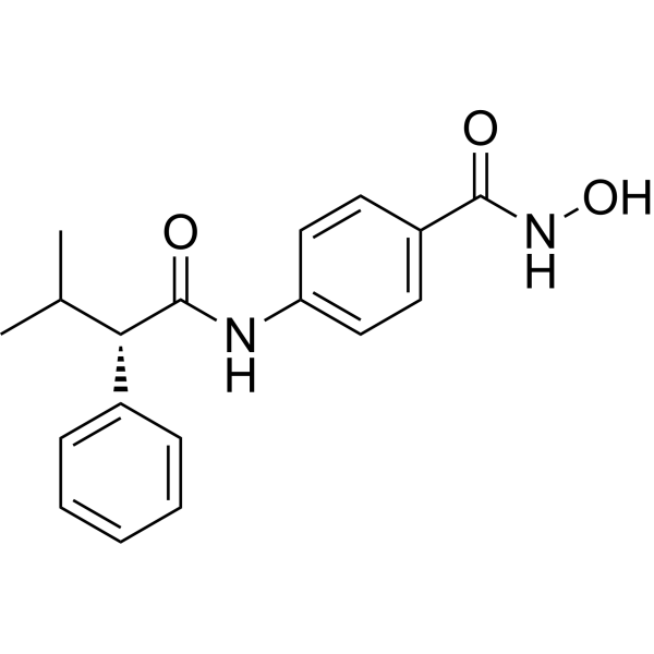
HY-13265
AR-42Pan HDAC Inhibitor -
HY-117374
HDAC3-IN-1HDAC3 Inhibitor -

HY-10221G
Vorinostat (GMP)HDAC Inhibitor -

HY-16026R
Ricolinostat (Standard)HDAC6 Inhibitor -

HY-13322R
Pracinostat (Standard)HDAC/MBLAC2 Inhibitor


Biological Activity
Description
IC50 & Target
[1]|
HDAC6 30 nM (IC50) |
Cellular Effect
|
Cell Line

|
Type | Value | Description | References |
|---|---|---|---|---|
| CCRF-HSB-2 | IC50 |
22.5 μM
Compound: 76; NR-160
|
Antiproliferative activity against human CCRF-HSB-2 cells
Antiproliferative activity against human CCRF-HSB-2 cells
|
[PMID: 37875056] |
| CCRF-HSB-2 | IC50 |
51.8 μM
Compound: 6l, NR-160
|
Cytotoxicity in human CCRF-HSB-2 cells assessed as reduction in cell viability incubated for 72 hrs by cell-Titer-Glo luminescent cell viability assay
Cytotoxicity in human CCRF-HSB-2 cells assessed as reduction in cell viability incubated for 72 hrs by cell-Titer-Glo luminescent cell viability assay
|
[PMID: 32803970] |
| HL-60 | IC50 |
22.5 μM
Compound: 76; NR-160
|
Antiproliferative activity against human HL-60 cells
Antiproliferative activity against human HL-60 cells
|
[PMID: 37875056] |
| HL-60 | IC50 |
42.9 μM
Compound: 6l, NR-160
|
Cytotoxicity in human HL-60 cells assessed as reduction in cell viability incubated for 72 hrs by cell-Titer-Glo luminescent cell viability assay
Cytotoxicity in human HL-60 cells assessed as reduction in cell viability incubated for 72 hrs by cell-Titer-Glo luminescent cell viability assay
|
[PMID: 32803970] |
| Jurkat | IC50 |
22.5 μM
Compound: 76; NR-160
|
Antiproliferative activity against human Jurkat cells
Antiproliferative activity against human Jurkat cells
|
[PMID: 37875056] |
| Jurkat | IC50 |
32.1 μM
Compound: 6l, NR-160
|
Cytotoxicity in human Jurkat cells assessed as reduction in cell viability incubated for 72 hrs by cell-Titer-Glo luminescent cell viability assay
Cytotoxicity in human Jurkat cells assessed as reduction in cell viability incubated for 72 hrs by cell-Titer-Glo luminescent cell viability assay
|
[PMID: 32803970] |
| K562 | IC50 |
22.5 μM
Compound: 76; NR-160
|
Antiproliferative activity against human K562 cells
Antiproliferative activity against human K562 cells
|
[PMID: 37875056] |
| K562 | IC50 |
41.6 μM
Compound: 6l, NR-160
|
Cytotoxicity in human K562 cells assessed as reduction in cell viability incubated for 72 hrs by cell-Titer-Glo luminescent cell viability assay
Cytotoxicity in human K562 cells assessed as reduction in cell viability incubated for 72 hrs by cell-Titer-Glo luminescent cell viability assay
|
[PMID: 32803970] |
| MOLT-4 | IC50 |
22.5 μM
Compound: 76; NR-160
|
Antiproliferative activity against human MOLT-4 cells
Antiproliferative activity against human MOLT-4 cells
|
[PMID: 37875056] |
| MOLT-4 | IC50 |
42.4 μM
Compound: 6l, NR-160
|
Cytotoxicity in human MOLT-4 cells assessed as reduction in cell viability incubated for 72 hrs by cell-Titer-Glo luminescent cell viability assay
Cytotoxicity in human MOLT-4 cells assessed as reduction in cell viability incubated for 72 hrs by cell-Titer-Glo luminescent cell viability assay
|
[PMID: 32803970] |
| SUP-B15 | IC50 |
22.5 μM
Compound: 6l, NR-160
|
Cytotoxicity in human SUP-B15 cells assessed as reduction in cell viability incubated for 72 hrs by cell-Titer-Glo luminescent cell viability assay
Cytotoxicity in human SUP-B15 cells assessed as reduction in cell viability incubated for 72 hrs by cell-Titer-Glo luminescent cell viability assay
|
[PMID: 32803970] |
| SUP-B15 | IC50 |
22.5 μM
Compound: 76; NR-160
|
Antiproliferative activity against human SUP-B15 cells
Antiproliferative activity against human SUP-B15 cells
|
[PMID: 37875056] |
| TALL-1 | IC50 |
22.5 μM
Compound: 76; NR-160
|
Antiproliferative activity against human TALL-1 cells
Antiproliferative activity against human TALL-1 cells
|
[PMID: 37875056] |
| TALL-1 | IC50 |
49.4 μM
Compound: 6l, NR-160
|
Cytotoxicity in human TALL-1 cells assessed as reduction in cell viability incubated for 72 hrs by cell-Titer-Glo luminescent cell viability assay
Cytotoxicity in human TALL-1 cells assessed as reduction in cell viability incubated for 72 hrs by cell-Titer-Glo luminescent cell viability assay
|
[PMID: 32803970] |
In Vitro
NR160 (compound 6I) (2.5 μM and 5 μM; 24 h) induces α-tubulin acetylation (ac-α-tubulin)[1].
NR160 (1-50 μM; 24 h) shows low cytotoxicity against leukemia cell lines with IC50s >20 μM[1].
NR160 (2.5 μM, 5 μM; 24 h) enhances the cytotoxicity induction of Bortezomib, Epirubicin and Daunorubicin in HL60 cells[1].
MedChemExpress (MCE) has not independently confirmed the accuracy of these methods. They are for reference only.
-
Cell Line:HL60 cells
-
Concentration:1-50 μM
-
Incubation Time:24 hours
-
Result:Inhibited leukemia cell lines with IC50s of 49.4 μM (TALL-1), 51.8 μM (HSB-2), 42.4 μM (MOLT-4), 32.1 μM (Jurkat), 42.9 μM (HL-60), 22.5 μM (SUP-B15), and 41.6 μM (K562), respectively.


Chemical Information
-
CAS No. 2484895-50-1
-
Appearance Solid
-
Molecular Weight 510.47
-
Formula C25H21F3N6O3
-
Color White to off-white
-
SMILES
O=C(N(CC1=CC=C(C(NO)=O)C=C1)CC2=NN=NN2CC3=CC=CC=C3)C4=CC=CC=C4C(F)(F)F
-
Shipping
Room temperature in continental US; may vary elsewhere.
-
Storage
Powder -20°C 3 years 4°C 2 years In solvent -80°C 6 months -20°C 1 month


Purity & Documentation
-
Data Sheet (277 KB)
-
SDS (252 KB)
- English - EN (252 KB)
- Français - FR (252 KB)
- Deutsch - DE (252 KB)
- Norwegian - NO (252 KB)
- Español - ES (252 KB)
- Swedish - SV (252 KB)
- Italian - IT (252 KB)
- Korean - KR (252 KB)
- Portuguese - PT (252 KB)
-
Handling Instructions (2659 KB)
References

Calculators
Concentration (start) × Volume (start) = Concentration (final) × Volume (final)


NR160 Related Classifications
HY-139181 Related Classifications
 Powered by Bioz