



Oligomycin B

Based on 1 Customer Validation
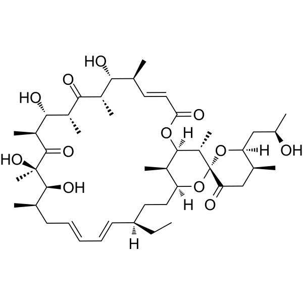
Oligomycin B is an antibiotic that acts as a non-selective inhibitor of ATP Synthase. Oligomycin B increases mitochondrial membrane potential. Oligomycin B induces apoptosis and necrosis. Oligomycin B impairs the motility of Plasmopara viticola zoospores and induces their lysis. Oligomycin B inhibits Magnaporthe oryzae (wheat blast fungus) and suppresses the development of wheat blast. Oligomycin B reduces hyphal growth and spore germination of Botrytis cinerea, and protects Arabidopsis thaliana against Botrytis cinerea infection. Oligomycin B exacerbates cytotoxic brain edema in rats with cerebral cortical contusion, increases intracranial pressure and brain water content, and aggravates mitochondrial damage in these rats. Oligomycin B is used in studies related to grape downy mildew, traumatic brain injury, wheat blast, and gray mold.
Nur für Forschungszwecke. Wir verkaufen nicht an Patienten.
- Reinheit : 97.67%
- CAS. Nr.: 11050-94-5
- Formel: C45H72O12
- Molecular Weight:805.05
-
Speicherung:
-20°C, protect from light, stored under nitrogen
* In solvent : -80°C, 6 months; -20°C, 1 month (protect from light, stored under nitrogen)

To place orders, for customer services and technical support, please contact: MedChemExpress USA
Tel: 609-228-6898 E-mail: [email protected] [email protected]


-
 Biologische Aktivität
-
Chemical Information
-
Lösungsmittel & Löslichkeit
- Protokoll
- Reinheit & Dokumentation
- Verweise
-
 Help & FAQs


-
HY-16592
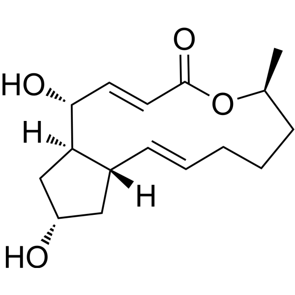
Brefeldin AProtein Transport Inhibitor -
HY-16589
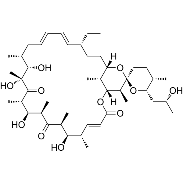
Oligomycin AMitochondrial ATP Synthase Inhibitor -
HY-D0852
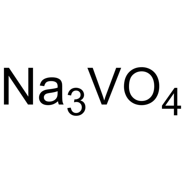
Sodium orthovanadateProtein Tyrosine/Alkaline Phosphatase Inhibitor -
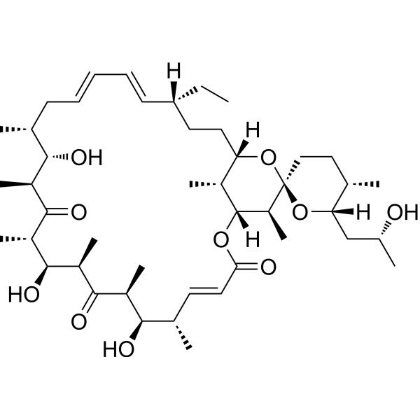
HY-N6783
Oligomycin CMacrolide Antibiotic -
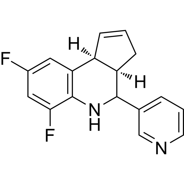
HY-100540
Golgicide AArfGEF GBF1 Inhibitor -

HY-16592R
Brefeldin A (Standard)Protein Transport Inhibitor -
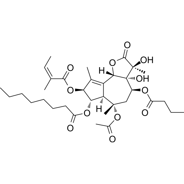
HY-13433
ThapsigarginER Stress Inducer -
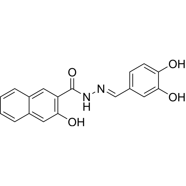
HY-15304
DynasoreAutophagy Inducer -

HY-16589R
Oligomycin A (Standard)Mitochondrial ATP Synthase Inhibitor -
HY-13441
(-)-BlebbistatinMyosin II ATPase Inhibitor -
HY-109037
MavacamtenCardiac Myosin Modulator
Alle Antibiotic Isoform-spezifische Produkte anzeigen
More

Biologische Aktivität
Beschreibung
In Vitro
Oligomycin B promotes the formation of JC-1 J-aggregates and increases the mitochondrial membrane potential of P388D1 cells[1].
Oligomycin B (20-50 μM; 24 h) induces apoptosis (at low concentrations) and predominantly necrosis (at high concentrations) in P388D1 cells, with an apoptosis rate of 25% observed at the concentration of 20 μM[1].
Oligomycin B (0-0.5 μg/mL; 0-60 min) impairs the motility of Plasmopara viticola zoospores and induces their lysis in a dose- and time-dependent manner, with corresponding IC50 values of 0.15 and 0.2 μg/mL; complete inhibition of zoospore motility and induction of lysis are achieved at concentrations as low as 0.25 μg/mL (after 60 min of treatment) or 0.5 μg/mL (after 30 min of treatment)[2].
Oligomycin B (0.05-2 μg/disk) inhibits the mycelial growth of the wheat-specialized isolate BTJP of *Magnaporthe oryzae* in a dose-dependent manner, with an inhibition rate of 8.63% at 0.05 μg/disk and 57.1% at 2 μg/disk, and also induces abnormal mycelial morphology[4].
Oligomycin B (5-100 μg/mL; 24 h) inhibits conidiation of Magnaporthe oryzae Triticum[4].
Oligomycin B (0.05 μg/mL; 6-24 h) inhibits conidial germination of Magnaporthe oryzae Triticum, induces conidial lysis and abnormal germ tube development, and completely blocks its germination and subsequent growth within 24 h[4].
Oligomycin B (10-150 μg/disc; 3 days) inhibits mycelial growth of Botrytis cinerea in the disc diffusion assay, with a minimum inhibitory concentration of 10 µg/disc[5].
Oligomycin B (5-20 µg/mL; 24 h) delays or inhibits spore germination of Botrytis cinerea in a concentration-dependent manner. It achieves complete inhibition at 20 µg/mL, while exerting a strong delaying effect at 10 µg/mL[5].
Oligomycin B (5-100 μg/mL) dose-dependently inhibits the development of wheat blast lesions on detached wheat leaves, and achieves complete inhibition at 100 μg/mL[4].
Oligomycin B (75% cell-free filtrate of Streptomyces sp. S5.1) significantly reduces the lesion size of gray mold in Arabidopsis thaliana, with average lesion sizes decreased by approximately 61% and 58%, respectively[5].
MedChemExpress (MCE) has not independently confirmed the accuracy of these methods. They are for reference only.
-
Cell Line:P388D1 cells
-
Concentration:20 μM; 50 μM
-
Incubation Time:24 h
-
Result:Induced 25% apoptosis in P388D1 cells at 20 μM.
Induced some apoptosis but primarily necrosis in P388D1 cells at 50 μM.
In Vivo
MedChemExpress (MCE) has not independently confirmed the accuracy of these methods. They are for reference only.
-
Animal Model:Sprague-Dawley (male, 250-300 g, traumatic brain injury modified impact acceleration model)[3]
-
Dosage:0.25 mg/kg
-
Administration:i.p.; two doses at 30 min and 24 h post-injury
-
Result:Increased intracranial pressure to levels 2.5-fold higher than cyclosporine A-treated rats.
Significantly increased brain water content compared to cyclosporine A-treated rats.
Caused significantly greater mitochondrial transmembrane potential loss than vehicle-treated rats.
Induced severely swollen neurons with a significantly elevated cytoplasm/nucleus area ratio, and profoundly worsened axonal damage with numerous blown-up mitochondria in severely swollen axons.
- Research Protocol for Infectious Diseases
- Mitochondrial membrane-potential fluorescent assay
- Detection of Apoptosis
- TUNEL staining for apoptotic DNA fragmentation
- Annexin V plus membrane-impermeant dye apoptosis staining
- Fluorescent plasma-membrane potential dye assay
- Mitochondrial membrane-potential and mitochondrial mass staining
- Apoptosis Solutions
- Filamentous Fungal Mold Culture and Sporulation
- Cell Viability Determination by MTT Colorimetric Assay


Chemical Information
-
CAS. Nr. 11050-94-5
-
Appearance Solid
-
Molecular Weight 805.05
-
Formel C45H72O12
-
Color White to off-white
-
SMILES
O=C([C@@H]([C@H]([C@@H](C([C@@](C)([C@H]([C@@H](C/C=C/C=C/[C@@]([H])(CC[C@]1([C@H]([C@@]([H])(O2)[C@@H]([C@]3(O1)C(C[C@@H]([C@@]([H])(O3)C[C@@H](C)O)C)=O)C)C)[H])CC)C)O)O)=O)C)O)C)[C@H]([C@@H]([C@H](/C=C/C2=O)C)O)C
-
Structure Classification
-
Initial Source
the marine Streptomyces strains B8496 and B8739
-
Versand
Room temperature in continental US; may vary elsewhere.
-
Speicherung
-20°C, protect from light, stored under nitrogen
* In solvent : -80°C, 6 months; -20°C, 1 month (protect from light, stored under nitrogen)


Lösungsmittel & Löslichkeit
In Vitro:
DMSO : 50 mg/mL (62.11 mM; Need ultrasonic; Hygroscopic DMSO has a significant impact on the solubility of product, please use newly opened DMSO)
Please refer to the solubility information to select the appropriate solvent. Once prepared, please aliquot and store the solution to prevent product inactivation from repeated freeze-thaw cycles.
Storage method and period of stock solution: -80°C, 6 months; -20°C, 1 month (protect from light, stored under nitrogen). When stored at -80°C, please use it within 6 months. When stored at -20°C, please use it within 1 month.
Please refer to the solubility information to select the appropriate solvent. Once prepared, please aliquot and store the solution to prevent product inactivation from repeated freeze-thaw cycles.
Storage method and period of stock solution: -80°C, 6 months; -20°C, 1 month (protect from light, stored under nitrogen). When stored at -80°C, please use it within 6 months. When stored at -20°C, please use it within 1 month.
Konzentration (Stammlösung) × Volumen (Stammlösung) = Konzentration (Ziellösung) × Volumen (Ziellösung)
In Vivo:
Select the appropriate dissolution method based on your experimental animal and administration route.
- For the following dissolution methods, please ensure to first prepare a clear stock solution using an In Vitro approach and then sequentially add co-solvents:
- To ensure reliable experimental results, the clarified stock solution can be appropriately stored based on storage conditions. As for the working solution for In Vivo experiments, it is recommended to prepare freshly and use it on the same day.
- The percentages shown for the solvents indicate their volumetric ratio in the final prepared solution. If precipitation or phase separation occurs during preparation, heat and/or sonication can be used to aid dissolution.
Add each solvent one by one: 10% DMSO 40% PEG300 5% Tween-80 45% Saline
Solubility: ≥ 1.25 mg/mL (1.55 mM); Clear solution
This protocol yields a clear solution of ≥ 1.25 mg/mL (saturation unknown).
Taking 1 mL working solution as an example, add 100 μL DMSO stock solution (12.5 mg/mL) to 400 μL PEG300, and mix evenly; then add 50 μL Tween-80 and mix evenly; then add 450 μL Saline to adjust the volume to 1 mL.
Preparation of Saline: Dissolve 0.9 g sodium chloride in ddH₂O and dilute to 100 mL to obtain a clear Saline solution.
In Vivo Dissolution Calculator
Please enter the basic information of animal experiments:
-
-
-
-
Recommended: Prepare an additional quantity of animals to account for potential losses during experiments.
Please enter your animal formula composition:
-
%DMSO +
Recommended: Keep the proportion of DMSO in working solution below 2% if your animal is weak.
-
%+
-
+%Tween-80 + +
-
%Saline +
The co-solvents required include: DMSO, . All of co-solvents are available by MedChemExpress (MCE). , Tween 80. All of co-solvents are available by MedChemExpress (MCE).
Working solution concentration: 0.22 mg/mL
Method for preparing stock solution: mg drug dissolved in μL DMSO. Stock solution concentration: mg/mL. * In solvent : -80°C, 6 months; -20°C, 1 month (protect from light, stored under nitrogen)
1. Take μL DMSO stock solution;
2. Add μL .
μL , mix evenly;
3. Then add μL Tween 80, mix evenly;
4. Then add μL
Please ensure that the stock solution in the first step is dissolved to a clear state, and add co-solvents in sequence. You can use ultrasonic heating (ultrasonic cleaner, recommended frequency 20-40 kHz), vortexing, etc. to assist dissolution.


Reinheit & Dokumentation
-
Data Sheet (284 KB)
-
SDS (394 KB)
- English - EN (394 KB)
- Français - FR (394 KB)
- Deutsch - DE (394 KB)
- Norwegian - NO (394 KB)
- Español - ES (394 KB)
- Swedish - SV (394 KB)
- Italian - IT (394 KB)
- Korean - KR (394 KB)
- Portuguese - PT (394 KB)
-
Handling Instructions (2659 KB)
Verweise
[1]. Moerman KL, et al. Evidence that the lichen-derived scabrosin esters target mitochondrial ATP synthase in P388D1 cells. Toxicol Appl Pharmacol. 2003;190(3):232-240. [Content Brief]
[2]. Dame ZT, et al. Oligomycins and pamamycin homologs impair motility and induce lysis of zoospores of the grapevine downy mildew pathogen, Plasmopara viticola. FEMS Microbiol Lett. 2016 Aug;363(16):fnw167. [Content Brief]
[3]. Vlodavsky E, et al. Post-traumatic cytotoxic edema is directly related to mitochondrial function. J Cereb Blood Flow Metab. 2017;37(1):166-177. [Content Brief]
[4]. Chakraborty M, et al. Oligomycins inhibit Magnaporthe oryzae Triticum and suppress wheat blast disease. PLoS One. 2020;15(8):e0233665. Published 2020 Aug 17. [Content Brief]
[5]. Louviot F, et al. Oligomycin-producing Streptomyces sp. newly isolated from Swiss soils efficiently protect Arabidopsis thaliana against Botrytis cinerea. mSphere. 2024 Jul 30;9(7):e0066723. [Content Brief]


Complete Stock Solution Preparation Table
Please refer to the solubility information to select the appropriate solvent. Once prepared, please aliquot and store the solution to prevent product inactivation from repeated freeze-thaw cycles.
Storage method and period of stock solution: -80°C, 6 months; -20°C, 1 month (protect from light, stored under nitrogen). When stored at -80°C, please use it within 6 months. When stored at -20°C, please use it within 1 month.
| Optional Solvent | Concentration Solvent Mass | 1 mg | 5 mg | 10 mg | 25 mg |
|---|---|---|---|---|---|
| DMSO | 1 mM | 1.2422 mL | 6.2108 mL | 12.4216 mL | 31.0540 mL |
| 5 mM | 0.2484 mL | 1.2422 mL | 2.4843 mL | 6.2108 mL | |
| 10 mM | 0.1242 mL | 0.6211 mL | 1.2422 mL | 3.1054 mL | |
| 15 mM | 0.0828 mL | 0.4141 mL | 0.8281 mL | 2.0703 mL | |
| 20 mM | 0.0621 mL | 0.3105 mL | 0.6211 mL | 1.5527 mL | |
| 25 mM | 0.0497 mL | 0.2484 mL | 0.4969 mL | 1.2422 mL | |
| 30 mM | 0.0414 mL | 0.2070 mL | 0.4141 mL | 1.0351 mL | |
| 40 mM | 0.0311 mL | 0.1553 mL | 0.3105 mL | 0.7763 mL | |
| 50 mM | 0.0248 mL | 0.1242 mL | 0.2484 mL | 0.6211 mL | |
| 60 mM | 0.0207 mL | 0.1035 mL | 0.2070 mL | 0.5176 mL |


Oligomycin B Related Classifications
HY-N6784 Related Classifications
 Powered by Bioz
Keywords
- Oligomycin B
- 11050-94-5
- ATP Synthase
- Bacterial
- Apoptosis
- Antibiotic
- Botrytis cinerea
- Arabidopsis thaliana
- mitochondrial ATP synthase
- wheat blast
- rats
- Plasmopara viticola
- Magnaporthe oryzae Triticum
- cerebral cytotoxic edema
- P388D1 cells
- traumatic brain injury
- antibiotic
- grape downy mildew
- gray mold
- Inhibitor
- inhibitor
- inhibit