



PI3K/mTOR Inhibitor-12
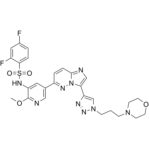
PI3K/mTOR Inhibitor-12 is a potent, orally active and selective PI3K/mTOR inhibitor with IC50 values of 0.06 nM and 3.12 nM for PI3Kα and mTOR, respectively. PI3K/mTOR Inhibitor-12 has antitumor activity. PI3K/mTOR Inhibitor-12 has lower liver toxicity.
For research use only. We do not sell to patients.
- CAS No.: 2891692-83-2
- Formula: C27H27F2N9O4S
- Molecular Weight:611.62
-
Storage:
Please store the product under the recommended conditions in the Certificate of Analysis.

To place orders, for customer services and technical support, please contact: MedChemExpress USA
Tel: 609-228-6898 E-mail: [email protected] [email protected]


-
 Biological Activity
-
Chemical Information
- Protocols
- Purity & Documentation
- References
-
 Help & FAQs




Biological Activity
Description
IC50 & Target
[1]|
PI3Kα 0.06 nM (IC50) |
mTOR 3.12 nM (IC50) |
Cellular Effect
|
Cell Line

|
Type | Value | Description | References |
|---|---|---|---|---|
| HCT-116 | IC50 |
0.54 μM
Compound: 42
|
Antiproliferative activity against human HCT-116 cells assessed as inhibition of cell growth incubated for 72 hrs by CCK-8 method
Antiproliferative activity against human HCT-116 cells assessed as inhibition of cell growth incubated for 72 hrs by CCK-8 method
|
[PMID: 36586298] |
| HT-29 | IC50 |
0.09 μM
Compound: 42
|
Antiproliferative activity against human HT-29 cells assessed as inhibition of cell growth incubated for 72 hrs by CCK-8 method
Antiproliferative activity against human HT-29 cells assessed as inhibition of cell growth incubated for 72 hrs by CCK-8 method
|
[PMID: 36586298] |
| HUVEC | IC50 |
0.68 μM
Compound: 42
|
Antiproliferative activity against human HUVEC cells assessed as inhibition of cell growth incubated for 72 hrs by CCK-8 method
Antiproliferative activity against human HUVEC cells assessed as inhibition of cell growth incubated for 72 hrs by CCK-8 method
|
[PMID: 36586298] |
| LoVo | IC50 |
0.54 μM
Compound: 42
|
Antiproliferative activity against human LoVo cells assessed as inhibition of cell growth incubated for 72 hrs by CCK-8 method
Antiproliferative activity against human LoVo cells assessed as inhibition of cell growth incubated for 72 hrs by CCK-8 method
|
[PMID: 36586298] |
| PC-3 | IC50 |
0.07 μM
Compound: 42
|
Antiproliferative activity against human PC-3 cells assessed as inhibition of cell growth incubated for 72 hrs by CCK-8 method
Antiproliferative activity against human PC-3 cells assessed as inhibition of cell growth incubated for 72 hrs by CCK-8 method
|
[PMID: 36586298] |
In Vitro
PI3K/mTOR Inhibitor-12 (compound 48; 72 h) inhibits cancer cells growth with IC50 values of 0.07, 0.09, 0.54, 0.54, and 0.68 μM for PC-3, HT-29,HCT116, LOVO, and HUVEC cells, respectively[1].
PI3K/mTOR Inhibitor-12 (0.25-1 μM; 12-48 h; HCT116 and HT-29 cells) inhibits the activation of PI3K and mTOR in the cellular context[1].
MedChemExpress (MCE) has not independently confirmed the accuracy of these methods. They are for reference only. Further protocols information, click here.
-
Cell Line:HCT116 and HT-29 cells
-
Concentration:0.25, 0.5, and 1 μM
-
Incubation Time:12, 24, and 48 hours
-
Result:Inhibited the expression levels of AKT, p70S6K and their phosphorylated forms in a dose- and time-dependent manner.
In Vivo
PI3K/mTOR Inhibitor-12 (1 and 5 mg/kg; i.v. and p.o.; male SD rats) has fast plasma clearance (1428 mL/h/kg) and short T1/2 (2.33 h) and the AUC0-∞ is 1356 h*ng/mL[1].
MedChemExpress (MCE) has not independently confirmed the accuracy of these methods. They are for reference only.
-
Animal Model:HCT116 xenografts in female BALB/c nude mice[1]
-
Dosage:20 mg/kg
-
Administration:Oral administration, daily, for 14 days
-
Result:Reduced the growth of HCT116 tumors, and the tumor growth inhibition (TGI) was 73.33%.
Had lower liver toxicity of HCT116 xenografts in female BALB/c nude mice.
-
Animal Model:Male SD rats[1]
-
Dosage:1 and 5 mg/kg
-
Administration:Intravenous injection (1 mg/kg) and oral administration (5 mg/kg)
-
Result:
1.19 Parameter I.V. (1 mg/kg) Parameter P.O. (5 mg/kg) T1/2 (h) 0.6 T1/2 (h) 2.33 CL (mL/h/kg) 1428 Cmax (ng/mL) 1218 Vdss (mL/Kg) 528 AUC0-∞ (h*ng/mL) 1356 AUC0-∞ (h*ng/mL) 760 F% 33.1


Chemical Information
-
CAS No. 2891692-83-2
-
Molecular Weight 611.62
-
Formula C27H27F2N9O4S
-
SMILES
O=S(C1=CC=C(F)C=C1F)(NC2=CC(C3=NN4C(C=C3)=NC=C4C5=CN(CCCN6CCOCC6)N=N5)=CN=C2OC)=O
-
Shipping
Room temperature in continental US; may vary elsewhere.
-
Storage
Please store the product under the recommended conditions in the Certificate of Analysis.

Protocols
-
Cell Cytotoxicity Assay
Cytotoxicity assays are usually based on the assessment of cell membrane damage, which can also be indirectly detected by measuring cell viability. Detection methods include MTT assay, CKK-8 assay, LDH assay and ATP assay, etc.
-
Hepatotoxicity Study
This protocol evaluates hepatotoxicity using complementary in vivo mouse APAP acute liver injury and in vitro hepatocyte-based cytotoxicity readouts. In vivo APAP injury is assessed by serum ALT/AST, liver histology, hepatic glutathione, protein adducts, necrosis, inflammation, and regeneration-related endpoints. In vitro hepatotoxicity is assessed by loss of viability, leakage of ALT/AST/LDH, oxidative-stress markers, mitochondrial function, nuclear morphology, intracellular calcium, and high-content imaging endpoints.
-
Subcutaneous Cell-Line-Derived Xenograft
Subcutaneous cell-line-derived xenograft (CDX) models are established by implanting cultured human cancer cell lines into immunodeficient mice, where the injected cells form localized tumors that can be monitored in vivo as a measure of tumorigenic potential, growth kinetics, and treatment response. These models are widely used in oncology research because they allow reproducible tumor formation and enable comparative assessment of tumor growth between different cell lines or genetic manipulations in a controlled in vivo microenvironment. Subcutaneous implantation of cancer cells in immunodeficient mice is a standard approach for evaluating tumor growth behavior and therapeutic response across multiple cancer types, including prostate, esophageal, pancreatic, and colon cancer models.


Purity & Documentation
References

Calculators
Concentration (start) × Volume (start) = Concentration (final) × Volume (final)


PI3K/mTOR Inhibitor-12 Related Classifications
HY-152238 Related Classifications
 Powered by Bioz