



Quisinostat hydrochloride
Based on 18 publication(s) in Google Scholar
Quisinostat (JNJ-26481585) hydrochloride is a potent and orally active pan-HDAC inhibitor (HDACi), with IC50 values ranging from 0.11 nM to 0.64 nM for HDAC1, HDAC2, HDAC4, HDAC10 and HDAC11. Quisinostat hydrochloride has a broad spectrum antitumoral activity. Quisinostat hydrochloride can induce autophagy in neuroblastoma cells.
For research use only. We do not sell to patients.
- CAS No.: 1083078-98-1
- Formula: C21H27ClN6O2
- Molecular Weight:430.93
-
Storage:
Please store the product under the recommended conditions in the Certificate of Analysis.


To place orders, for customer services and technical support, please contact: MedChemExpress USA
Tel: 609-228-6898 E-mail: [email protected] [email protected]


-
 Biological Activity
-
Chemical Information
- Protocols
- Purity & Documentation
- References
-
 Help & FAQs


Publications Citing Use of MedChemExpress (MCE) Quisinostat hydrochloride
More- Cancer Cell. 2024 Oct 14;42(10):1729-1746.e8. [Abstract]
- Nat Commun. 2024 Jul 2;15(1):5570. [Abstract]
- Nat Commun. 2020 Apr 14;11(1):1792. [Abstract]
- Exp Hematol Oncol. 2019 Nov 15:8:30. [Abstract]
- Theranostics. 2019 Jan 30;9(4):1096-1114. [Abstract]
- Adv Sci (Weinh). 2026 Jan 22:e17877. [Abstract]
- NPJ Precis Oncol. 2023 Jul 21;7(1):70. [Abstract]
- EMBO J. 2024 Nov;43(21):4954-4983. [Abstract]
- Blood Adv. 2024 Sep 24;8(18):4936-4947. [Abstract]
- Cancer Cell Int. 2025 Oct 8;25(1):340. [Abstract]
- Antimicrob Agents Chemother. 2025 Sep 3;69(9):e0181924. [Abstract]
- Toxicol Appl Pharmacol. 2021 Jan 1:410:115363. [Abstract]
- Int J Parasitol Drugs Drug Resist. 2026 May 17;31:100649.
- Patent. US20250161243A1.
- bioRxiv. 2025 January 15.
- bioRxiv. 2024 July 04.
- Albert-ludwigs-university Of Freiburg. 2019 Dec.
- Patent. US20180263995A1.
 Customer Validation & Images

-
Cell Proliferation/Viability Assay
-
IHC
-
In Vivo Efficacy Study
-
WB
-
IF


Biological Activity
Description
IC50 & Target
[1]|
HDAC1 0.11 nM (IC50) |
HDAC2 0.33 nM (IC50) |
HDAC4 0.64 nM (IC50) |
HDAC10 0.46 nM (IC50) |
HDAC11 0.37 nM (IC50) |
HDAC3 4.86 nM (EC50) |
HDAC5 3.69 nM (IC50) |
HDAC8 4.26 nM (IC50) |
HDAC9 32.1 nM (IC50) |
HDAC6 76.8 nM (IC50) |
HDAC7 119 nM (IC50) |
Cellular Effect
|
Cell Line

|
Type | Value | Description | References |
|---|---|---|---|---|
| HEK-293T | IC50 |
48 nM
Compound: 1
|
Cytotoxicity against human HEK293T cells assessed as reduction in cell viability incubated for 72 hrs by cell titer glo assay
Cytotoxicity against human HEK293T cells assessed as reduction in cell viability incubated for 72 hrs by cell titer glo assay
|
[PMID: 33541085] |
| HepG2 | IC50 |
42 nM
Compound: 1
|
Cytotoxicity against human HepG2 cells assessed as reduction in cell viability incubated for 72 hrs by cell titer glo assay
Cytotoxicity against human HepG2 cells assessed as reduction in cell viability incubated for 72 hrs by cell titer glo assay
|
[PMID: 33541085] |


Chemical Information
-
CAS No. 1083078-98-1
-
Molecular Weight 430.93
-
Formula C21H27ClN6O2
-
SMILES
O=C(NO)C1=CN=C(N=C1)N2CCC(CC2)CNCC3=CN(C4=C3C=CC=C4)C.Cl
-
Synonyms
JNJ26481585 hydrochloride
-
Shipping
Room temperature in continental US; may vary elsewhere.
-
Storage
Please store the product under the recommended conditions in the Certificate of Analysis.


Publications (18)
-
Journal Impact Factor
-
Most Recent
-
Cancer Cell
Fusobacterium nucleatum facilitates anti-PD-1 therapy in microsatellite stable colorectal cancer. [Abstract]2024 Oct 14;42(10):1729-1746.e8. PMID: 39303724 -
Nat Commun
2024 Jul 2;15(1):5570. PMID: 38956053 -
Nat Commun
Selective inhibition of cancer cell self-renewal through a Quisinostat-histone H1.0 axis. [Abstract]2020 Apr 14;11(1):1792. PMID: 32286289
Quisinostat hydrochloride purchased from MedChemExpress. Usage Cited in: Nat Commun. 2020 Apr 14;11(1):1792. [Abstract]
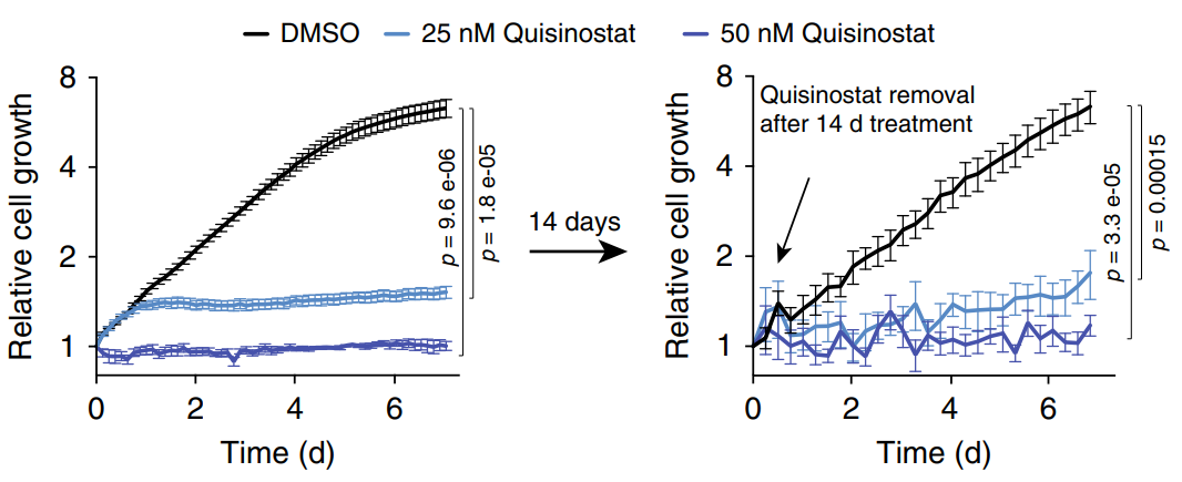
Quisinostat (25–50 nM; 7 d) blocked proliferation without inducing substantial death of HCC1569 cells.
Quisinostat hydrochloride purchased from MedChemExpress. Usage Cited in: Nat Commun. 2020 Apr 14;11(1):1792. [Abstract]
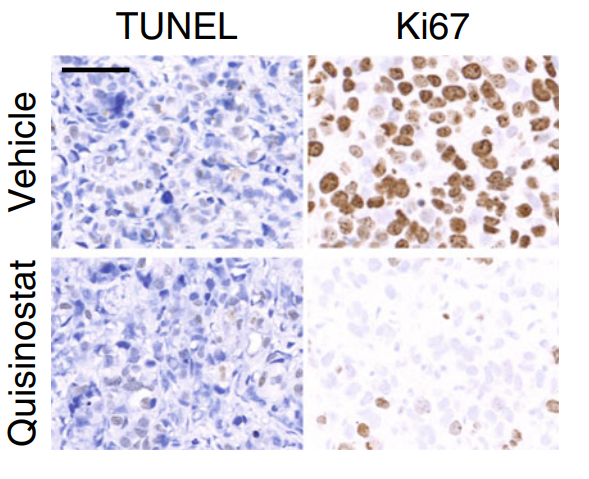
Quisinostat (4 mg/kg; i.p.; 21 d) caused reduced fractions of proliferating Ki67+ cells and no increase in apoptotic TUNEL+ cells in NSG mice to generate orthotopic xenografts, confirming that Quisinostat treatment induced cytostasis, not cell death, in vivo.
Quisinostat hydrochloride purchased from MedChemExpress. Usage Cited in: Nat Commun. 2020 Apr 14;11(1):1792. [Abstract]
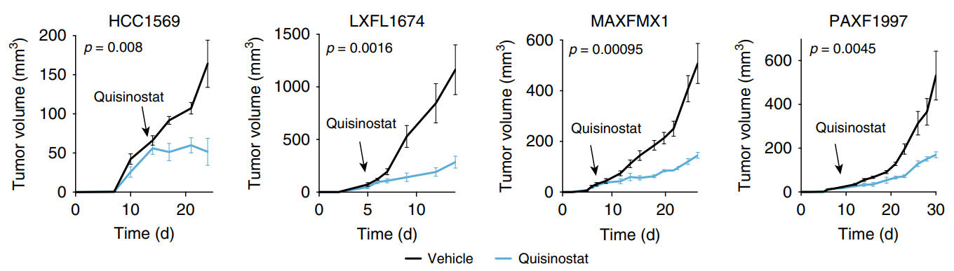
Quisinostat (4 mg/kg; i.p.; once daily) strongly inhibited tumor maintenance in the three PDX models of lung, pancreas and breast cancer.
Quisinostat hydrochloride purchased from MedChemExpress. Usage Cited in: Nat Commun. 2020 Apr 14;11(1):1792. [Abstract]
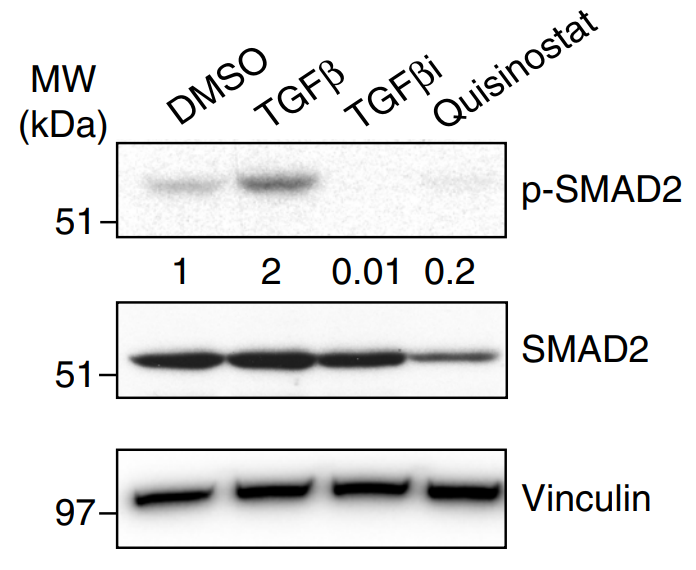
Quisinostat (100 nM; 24 h) decreased total and phosphorylated SMAD2 (p-SMAD2) in HCC1569 cells.
Quisinostat hydrochloride purchased from MedChemExpress. Usage Cited in: Nat Commun. 2020 Apr 14;11(1):1792. [Abstract]
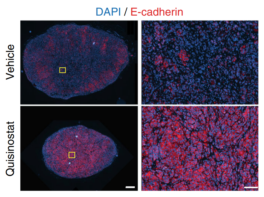
Quisinostat (4 mg/kg; i.p.; 21 d) treatment increased the levels of E-cadherin of HCC1569-induced orthotopic xenografts in NSG mice, indicating a reversion to a more epithelial phenotype.
-
Exp Hematol Oncol
A high-throughput screening identifies histone deacetylase inhibitors as therapeutic agents against medulloblastoma. [Abstract]2019 Nov 15:8:30. PMID: 31788346 -
Theranostics
Interaction between p53 and Ras signaling controls cisplatin resistance via HDAC4- and HIF-1α-mediated regulation of apoptosis and autophagy. [Abstract]2019 Jan 30;9(4):1096-1114. PMID: 30867818 -
Adv Sci (Weinh)
Macrophage TRIM21 Inhibition Ameliorates Murine Acute Pancreatitis via PHB2-Mediated Mitochondrial Stabilization. [Abstract]2026 Jan 22:e17877. PMID: 41572443 -
NPJ Precis Oncol
Tackling FGFR3-driven bladder cancer with a promising synergistic FGFR/HDAC targeted therapy. [Abstract]2023 Jul 21;7(1):70. PMID: 37479885 -
EMBO J
Acetylation of TIR domains in the TLR4-Mal-MyD88 complex regulates immune responses in sepsis. [Abstract]2024 Nov;43(21):4954-4983. PMID: 39294473 -
Blood Adv
2024 Sep 24;8(18):4936-4947. PMID: 38916861 -
Cancer Cell Int
Epigenetic potentiation of 5-fluorouracil by HDAC inhibitor quisinostat enhances antitumor effects in colorectal cancer. [Abstract]2025 Oct 8;25(1):340. PMID: 41063169 -
Antimicrob Agents Chemother
2025 Sep 3;69(9):e0181924. PMID: 40741955 -
Toxicol Appl Pharmacol
Death by histone deacetylase inhibitor quisinostat in tongue squamous cell carcinoma via apoptosis, pyroptosis, and ferroptosis. [Abstract]2021 Jan 1:410:115363. PMID: 33290780 -
-
-
-
-
-

Protocols
-
Cell Cytotoxicity Assay
Cytotoxicity assays are usually based on the assessment of cell membrane damage, which can also be indirectly detected by measuring cell viability. Detection methods include MTT assay, CKK-8 assay, LDH assay and ATP assay, etc.
-
Autophagy
Autophagy is a process in which eukaryotic cells use lysosomes to degrade their own cytoplasmic proteins and damaged organelles under the regulation of autophagy related gene (Atg). Microtubule-associated proteins light chain 3 (LC3) is recognized as autophagy marker, which transfers from cytoplasmic LC3 (LC3-I) to membrane type (LC3-II). LC3-II/I ratio could be detected by Western Blot and fluorescence microscopy.
-
Lysosome and acidic-vesicle live-cell staining
Lysosome and acidic-vesicle live-cell staining detects acidic intracellular compartments by using membrane-permeant acidotropic probes that accumulate in low-pH vesicles, including lysosomes, late endosomes, autolysosomes, and acidic phagosomes. LysoTracker staining is commonly used as an intensity-based readout of acidic lysosomal compartment abundance or enlargement, while acridine orange produces green fluorescence in less concentrated compartments and red fluorescence after concentration-dependent accumulation in acidic vesicular organelles. Loss or reduction of acridine-orange red signal can be used as a readout of lysosomal membrane permeabilization or reduced acidic-vesicle integrity. This protocol is designed for live cultured cells and can be adapted for fluorescence microscopy, high-content imaging, plate-reader readout, or flow cytometry when the selected literature supports the readout. Because these dyes report acidotropic accumulation rather than lysosome identity alone,
-
Macroautophagy Solutions
Macroautophagy is a conserved lysosome-dependent degradation pathway in which cytoplasmic material is sequestered into double-membrane autophagosomes and delivered to lysosomes for degradation and recycling. The pathway supports cellular homeostasis during nutrient limitation, organelle stress, protein-aggregate accumulation, infection, differentiation, and tissue remodeling by coupling cargo sequestration, autophagosome maturation, lysosomal fusion, and degradation of cargo-derived macromolecules. The core molecular sequence includes initiation by nutrient- and stress-regulated autophagy machinery, autophagosome nucleation, LC3/ATG8-family conjugation to autophagosomal membranes, cargo selection through receptors such as SQSTM1/p62, autophagosome-lysosome fusion, and lysosomal degradation. LC3 was identified as a mammalian homolog of yeast Atg8 that localizes to autophagosomal membranes after processing, and p62/SQSTM1 was shown to connect ubiquitinated cargo with autophagic degradati


Purity & Documentation
References
[1]. Vamsi Krishna Kommalapati, et al. Inhibition of JNJ-26481585-mediated autophagy induces apoptosis via ROS activation and mitochondrial membrane potential disruption in neuroblastoma cells. Mol Cell Biochem. 2020 May;468(1-2):21-34. [Content Brief]
[2]. Arts J, et al. JNJ-26481585, a novel "second-generation" oral histone deacetylase inhibitor, shows broad-spectrum preclinical antitumoral activity. Clin Cancer Res. 2009 Nov 15;15(22):6841-51. [Content Brief]

Calculators
Concentration (start) × Volume (start) = Concentration (final) × Volume (final)


Quisinostat hydrochloride Related Classifications
HY-15433B Related Classifications
 Powered by Bioz