



Caspase-3/CASP3 Protein, Human (His)
Based on 1 publication(s) in Google Scholar
Caspase-3/CASP3 protein is critical in the execution of apoptosis, cleaving PARP1 and activating SREBP, caspase-6, -7 and -9, initiating the cascade leading to apoptosis. In addition to apoptosis, it regulates cell adhesion, promotes inflammation, inhibits AKT1 in oxidative stress, and modulates antiviral responses by cleaving proteins such as CGAS, IRF3, and MAVS. Caspase-3/CASP3 Protein, Human (His) is the recombinant human-derived Caspase-3/CASP3 protein, expressed by E. coli , with C-His labeled tag.
- Species: Human
- Source: E. coli
-
Storage:Stored at -20°C for 2 years from date of receipt. After reconstitution, it is stable at 4°C for 1 week or -20°C for longer (with carrier protein). It is recommended to freeze aliquots at -20°C or -80°C for extended storage.


To place orders, for customer services and technical support, please contact: MedChemExpress USA
Tel: 609-228-6898 E-mail: [email protected] [email protected]


-
 Biological Activity
-
Technical Parameters
-
Product Properties
- Documentation
- Help & FAQs




Biological Activity
Description
Caspase-3/CASP3 protein is critical in the execution of apoptosis, cleaving PARP1 and activating SREBP, caspase-6, -7 and -9, initiating the cascade leading to apoptosis. In addition to apoptosis, it regulates cell adhesion, promotes inflammation, inhibits AKT1 in oxidative stress, and modulates antiviral responses by cleaving proteins such as CGAS, IRF3, and MAVS. Caspase-3/CASP3 Protein, Human (His) is the recombinant human-derived Caspase-3/CASP3 protein, expressed by E. coli , with C-His labeled tag.
Background
Caspase-3, a pivotal player in the apoptosis execution pathway, holds a central role in diverse cellular processes. During the initiation of apoptosis, it orchestrates the proteolytic cleavage of poly(ADP-ribose) polymerase PARP1, exemplifying its regulatory influence over critical cellular components. This versatile protein extends its reach by cleaving and activating sterol regulatory element-binding proteins (SREBPs), caspase-6, -7, and -9, thus participating in a cascade of events leading to apoptosis. Beyond its classical apoptotic functions, Caspase-3 exhibits involvement in cell adhesion within sympathetic neurons through RET cleavage and contributes to the inflammatory milieu by cleaving IL-1 beta. In response to oxidative stress, it inhibits the serine/threonine-protein kinase AKT1. Additionally, Caspase-3 assumes a role in regulating antiviral responses by cleaving proteins such as CGAS, IRF3, and MAVS, curbing type I interferon production. Furthermore, it plays a role in pyroptosis by cleaving and activating gasdermin-E (GSDME), as well as modulating phosphatidylserine exposure on apoptotic cell surfaces through XRCC4 and phospholipid scramblase protein cleavage. This comprehensive range of actions underscores the intricate regulatory functions of Caspase-3 in cellular homeostasis and response to diverse stimuli.
Verified Bioactivity
Measured by its ability to cleave the fluorogenic peptide substrate Ac-DEVD-AFC (HY-P1005) and the specific activity is > 5000 pmoles/min/μg. (Activation description: The proenzyme needs to be activated by Caspase-8 for an activated form.)
MCE Validation Data
-
Purity - SDS-PAGE
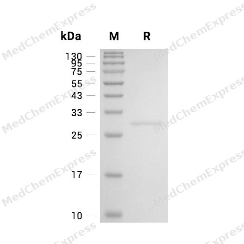
Purity - SDS-PAGE

Assay Procedure
Materials
Assay buffer: 25 mM HEPES, 0.1% (w/v) CHAPS, 10 mM dithiothreitol (DTT) , pH 7.5.
Test protein: Caspase-3/CASP3 Protein, Human (His) (HY-P72874)
Activator protein: Caspase-8 Protein, Human
Substrate: Ac-DEVD-AFC (HY-P1005) , stored as a 10 mM solution diluted in DMSO.
Standard: 7-Amino-4-(trifluoromethyl)coumarin (HY-D0981).
Procedure
1. Standard Curve Preparation:Dilute standard in assay buffer to concentrations of 0, 1.5625, 3.125, 6.25, 12.5, 25, 50, and 100 μmol/L. Add 100 μL of each dilution to a black microplate well. Measure fluorescence in kinetic mode for 5 minutes at excitation and emission wavelengths of 400 nm and 505 nm, respectively. Plot the measured RFU values on the y-axis and the standard concentrations on the x-axis to generate a standard curve and obtain the curve equation.
2. Activate 10 μg/mL of Human Caspase-3 with 10 μg/mL of Human Caspase-8 in the assay buffer.
3. Incubate at 37°C for 90 minutes.
4. Dilute the substrate to 100 μM in the assay buffer.
5. Add 50 μL of 0.4 μg/mL Human Caspase-3 to the plate and include a substrate blank containing 50 μL of assay buffer. Initiate the reaction by adding 50 μL of 100 μM substrate to the wells.
6. Read in kinetic mode for 5 minutes at excitation and emission wavelengths of 400 nm and 505 nm, respectively.
7. Calculate the specific activity:
Specific Activity (pmol/min/μg) = | Adjusted Vmax* (RFU/min) x Conversion Factor ** (pmol/RFU) |
| amount of enzyme (μg) |
*Adjusted for Control
**Derived using calibration standard


Publications (1)
-
Journal Impact Factor
-
Most Recent
-
Cells
Detection of Calpain-Mediated Beclin-1 Cleavage for Drug Discovery in Inflammatory Bowel Diseases. [Abstract]2026 May 18;15(10):917. PMID: 42193926


Technical Parameters
-
Species Human
-
Source E. coli
-
Tag C-6*His
-
Accession
P42574 (S29-H277)
-
Gene ID836
-
Molecular Construction
-
N-term
-
CASP3 (S29-H277)
Accession # P42574 -
6*His
-
C-term
-
-
Protein Length
Full Length of Mature Protein
-
Synonyms
CASP3; Protein Yama; Caspase 3; Caspase-3; Apopain; CASP-3; CPP32; CPP-32; CPP32B; SCA-1; Caspase 3, Apoptosis-Related Cysteine Peptidase; Yama; Caspase 3, Apoptosis-Related Cysteine Protease; PARP Cleavage Protease; SREBP Cleavage Activity 1; Procaspase3
-
AA Sequence
SGISLDNSYKMDYPEMGLCIIINNKNFHKSTGMTSRSGTDVDAANLRETFRNLKYEVRNKNDLTREEIVELMRDVSKEDHSKRSSFVCVLLSHGEEGIIFGTNGPVDLKKITNFFRGDRCRSLTGKPKLFIIQACRGTELDCGIETDSGVDDDMACHKIPVEADFLYAYSTAPGYYSWRNSKDGSWFIQSLCAMLKQYADKLEFMHILTRVNRKVATEFESFSFDATFHAKKQIPCIVSMLTKELYFYH
-
Predicted Molecular Mass
29.3 kDa
-
Molecular Weight
Approximately 29 kDa, based on SDS-PAGE under reducing conditions.
-
Purity
≥ 95%, as determined by reducing SDS-PAGE.


Product Properties
Lyophilized powder.
Lyophilized from a 0.22 μm filtered solution of PBS, pH 7.4, 8% trehalose.
<1 EU/μg, determined by LAL method.
It is not recommended to reconstitute to a concentration less than 100 μg/mL in ddH2O.
Stored at -20°C for 2 years from date of receipt. After reconstitution, it is stable at 4°C for 1 week or -20°C for longer (with carrier protein). It is recommended to freeze aliquots at -20°C or -80°C for extended storage.
Shipping with dry ice.


Documentation
-
Data Sheet (237 KB)
-
SDS (252 KB)
- English - EN (252 KB)
- Français - FR (252 KB)
- Deutsch - DE (252 KB)
- Norwegian - NO (252 KB)
- Español - ES (252 KB)
- Swedish - SV (252 KB)
- Italian - IT (252 KB)
- Korean - KR (252 KB)
- Portuguese - PT (252 KB)
-
Handling Instructions (2659 KB)

Calculators
Concentration (start) × Volume (start) = Concentration (final) × Volume (final)


 Powered by Bioz

