



GYPA/CD235a Protein, Human (His)

Based on 1 Customer Validation
GYPA, also known as glycoprotein A, is a major membrane protein in red blood cells and plays an important role in the integrity of cell membranes. GYPA is a receptor for influenza virus, Plasmodium falciparum erythrocyte binding Antigen 175 (EBA-175), and hepatitis A virus (HAV). GYPA/CD235a Protein, Human (His) is the recombinant human-derived GYPA/CD235a protein, expressed by E. coli , with N-6*His labeled tag.
- Species: Human
- Source: E. coli
-
Storage:Stored at -20°C for 2 years from date of receipt. After reconstitution, it is stable at 4°C for 1 week or -20°C for longer (with carrier protein). It is recommended to freeze aliquots at -20°C or -80°C for extended storage.


To place orders, for customer services and technical support, please contact: MedChemExpress USA
Tel: 609-228-6898 E-mail: [email protected] [email protected]


-
 Biological Activity
-
Technical Parameters
-
Product Properties
- Documentation
- References
- Help & FAQs




Biological Activity
Description
GYPA, also known as glycoprotein A, is a major membrane protein in red blood cells and plays an important role in the integrity of cell membranes. GYPA is a receptor for influenza virus, Plasmodium falciparum erythrocyte binding Antigen 175 (EBA-175), and hepatitis A virus (HAV). GYPA/CD235a Protein, Human (His) is the recombinant human-derived GYPA/CD235a protein, expressed by E. coli , with N-6*His labeled tag.
Background
GYPA, also known as Glycophorin A, serves as a pivotal component within the ankyrin-1 complex, a multiprotein assembly crucial for maintaining the stability and shape of the erythrocyte membrane. As the major intrinsic membrane protein of erythrocytes, GYPA contributes significantly to membrane integrity. Its N-terminal glycosylated segment, projecting beyond the erythrocyte membrane, bears MN blood group receptors and plays a vital role in erythrocyte function. GYPA is particularly essential for the optimal activity of SLC4A1, and its presence is required for the high activity of this membrane transporter. Moreover, GYPA is implicated in the translocation of SLC4A1 to the plasma membrane. Beyond its structural role, GYPA acts as a receptor for influenza virus, Plasmodium falciparum erythrocyte-binding antigen 175 (EBA-175), and Hepatitis A virus (HAV), highlighting its diverse functional repertoire. Existing as a homodimer, GYPA is an integral part of the ankyrin-1 complex, collaborating with other proteins such as ANK1, RHCE, RHAG, SLC4A1, EPB42, GYPB, and AQP1 in the erythrocyte membrane, thereby contributing to the overall structural integrity and functionality of these blood cells. The interaction with SLC4A1 results in the formation of a heterotetramer, emphasizing the cooperative nature of the erythrocyte membrane complex[1][2][3].
Verified Bioactivity
Immobilized GYPA at 2μg/mL (100μL/well) can bind GYPA Antibody. The ED50 for this effect is 1.443 μg/mL.
MCE Validation Data
-
Purity - SDS-PAGE
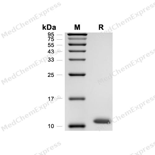
Purity - SDS-PAGE
-
Bioactivity - ELISA
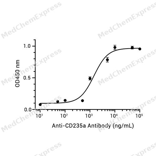
Bioactivity - ELISA


Technical Parameters
-
Species Human
-
Source E. coli
-
Tag N-6*His
-
Accession
A0A0C4DFT7 (L20-E91)
-
Molecular Construction
-
N-term
-
6*His
-
GYPA (L20-E91)
Accession # A0A0C4DFT7 -
C-term
-
-
Protein Length
Extracellular Domain
-
Synonyms
GYPA; Mi.V Glycoprotein (24 AA); Prev. MNS; Glycophorin Sta Type C; PAS-2; Glycophorin A, GPA; GPA; Glycophorin Erik; Sialoglycoprotein Alpha; Glycophorin MiI; MN Sialoglycoprotein; Glycophorin MiV; Glycophorin-A; Glycophorin SAT; CD235a; CD235a Antigen;
-
AA Sequence
LSTTEVAMHTSTSSSVTKSYISSQTNDTHKRDTYAATPRAHEVSEISVRTVYPPEEETGERVQLAHHFSEPE
-
Molecular Weight
Approximately 11 kDa, based on SDS-PAGE under reducing conditions.
-
Purity
≥ 95%, as determined by reducing SDS-PAGE.


Product Properties
Lyophilized powder.
1.Lyophilized from a 0.22 μm filtered solution of 50 mM Tris-HCl, 300 mM NaCl, pH 7.4.
Note: For SPR assay, please replace the buffer. Primary amine components (e.g., Tris, imidazole) can affect protein-coupled chips.
<1 EU/μg, determined by LAL method.
It is not recommended to reconstitute to a concentration less than 100 μg/mL in ddH2O.
Stored at -20°C for 2 years from date of receipt. After reconstitution, it is stable at 4°C for 1 week or -20°C for longer (with carrier protein). It is recommended to freeze aliquots at -20°C or -80°C for extended storage.
Room temperature in continental US;may vary elsewhere.


Documentation
-
Data Sheet (263 KB)
-
SDS (251 KB)
- English - EN (251 KB)
- Français - FR (251 KB)
- Deutsch - DE (251 KB)
- Norwegian - NO (251 KB)
- Español - ES (251 KB)
- Swedish - SV (251 KB)
- Italian - IT (251 KB)
- Korean - KR (251 KB)
- Portuguese - PT (251 KB)
-
Handling Instructions (2659 KB)


References
[1]. Suwanwootichai P, et al. Fatal haemolytic transfusion reaction due to anti-Ena and identification of a novel GYPA c.295delG variant in a Thai family. Vox Sang. 2022 Nov;117(11):1327-1331. [Content Brief]
[2]. Bigham AW, et al. Complex signatures of natural selection at GYPA. Hum Genet. 2018 Feb;137(2):151-160. [Content Brief]
[3]. Suades R, et al. Growing thrombi release increased levels of CD235a(+) microparticles and decreased levels of activated platelet-derived microparticles. Validation in ST-elevation myocardial infarction patients. J Thromb Haemost. 2015 Oct;13(10):1776-86. [Content Brief]

Calculators
Concentration (start) × Volume (start) = Concentration (final) × Volume (final)


 Powered by Bioz

