



HGFR Protein, Mouse (HEK293, His)
Based on 1 Customer Validation
HGFR Protein, a receptor tyrosine kinase, transduces signals by binding hepatocyte growth factor/HGF ligand. It regulates proliferation, scattering, morphogenesis, and cell survival. Upon ligand binding, HGFR undergoes autophosphorylation, creating docking sites for downstream molecules like PI3K, PLCG1, SRC, GRB2, STAT3, or GAB1, activating RAS-ERK, PI3K-AKT, and PLCgamma-PKC cascades. HGFR Protein, Mouse (HEK293, His) is the recombinant mouse-derived HGFR protein, expressed by HEK293 , with C-His labeled tag.
- Species: Mouse
- Source: HEK293
-
Storage:Stored at -20°C for 2 years from date of receipt. After reconstitution, it is stable at 4°C for 1 week or -20°C for longer (with carrier protein). It is recommended to freeze aliquots at -20°C or -80°C for extended storage.


To place orders, for customer services and technical support, please contact: MedChemExpress USA
Tel: 609-228-6898 E-mail: [email protected] [email protected]


-
 Biological Activity
-
Technical Parameters
-
Product Properties
- Documentation
- Help & FAQs




Biological Activity
Description
HGFR Protein, a receptor tyrosine kinase, transduces signals by binding hepatocyte growth factor/HGF ligand. It regulates proliferation, scattering, morphogenesis, and cell survival. Upon ligand binding, HGFR undergoes autophosphorylation, creating docking sites for downstream molecules like PI3K, PLCG1, SRC, GRB2, STAT3, or GAB1, activating RAS-ERK, PI3K-AKT, and PLCgamma-PKC cascades. HGFR Protein, Mouse (HEK293, His) is the recombinant mouse-derived HGFR protein, expressed by HEK293 , with C-His labeled tag.
Background
Hepatocyte growth factor receptor (HGFR), a receptor tyrosine kinase, functions as a signal transducer from the extracellular matrix by binding to hepatocyte growth factor/HGF ligand. It plays a pivotal role in regulating diverse physiological processes, including proliferation, scattering, morphogenesis, and cell survival. Upon ligand binding at the cell surface, HGFR undergoes autophosphorylation on its intracellular domain, creating docking sites for downstream signaling molecules. Upon activation by ligand, it interacts with the PI3-kinase subunit PIK3R1, PLCG1, SRC, GRB2, STAT3, or the adapter GAB1, leading to the activation of multiple signaling cascades, including RAS-ERK, PI3 kinase-AKT, and PLCgamma-PKC. RAS-ERK activation is associated with morphogenetic effects, while PI3K/AKT coordinates prosurvival effects. In embryonic development, HGFR signaling contributes to gastrulation, the development and migration of neuronal precursors, angiogenesis, and kidney formation. During skeletal muscle development, it is crucial for the migration of muscle progenitor cells and the proliferation of secondary myoblasts. In adults, it participates in wound healing, organ regeneration, tissue remodeling, and promotes the differentiation and proliferation of hematopoietic cells. Additionally, in the context of microbial infection, HGFR acts as a receptor for Listeria monocytogenes internalin InlB, mediating the entry of the pathogen into cells[1][2][3].
Verified Bioactivity
1.This product does not contain protein kinase domain.
2.Immobilized Human HGF at 2 μg/mL (100 μL/well) can bind Mouse HGFR. The ED50 for this effect is 83.3 ng/mL.
MCE Validation Data
-
Purity - SDS-PAGE
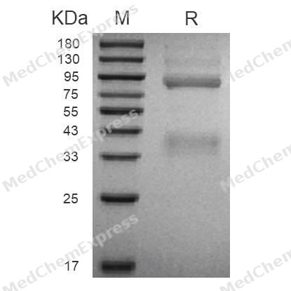
Purity - SDS-PAGE
-
Bioactivity - ELISA
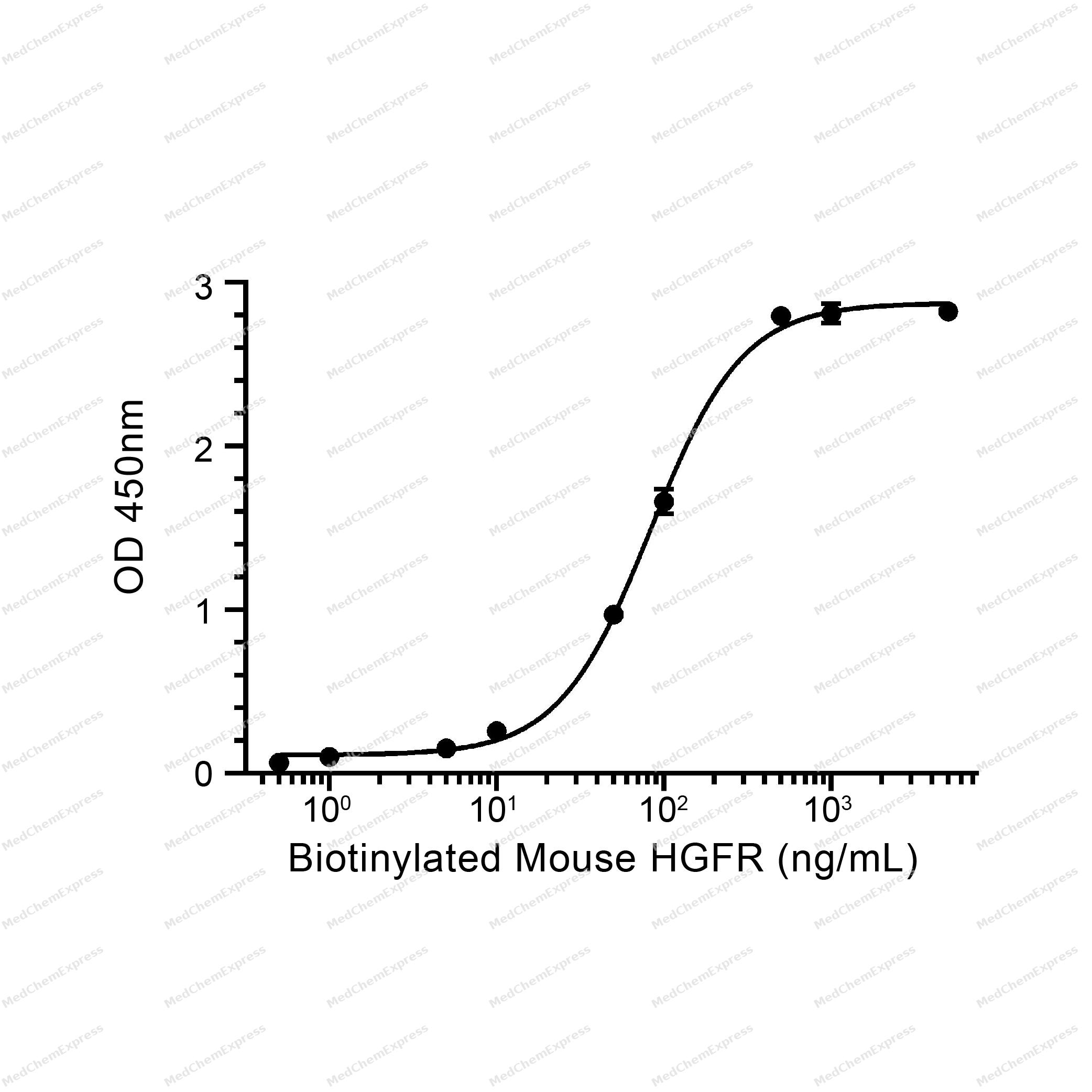
Bioactivity - ELISA


Technical Parameters
-
Species Mouse
-
Source HEK293
-
Tag C-His
-
Accession
EDL13881.1 (E25-N929)
-
Molecular Construction
-
N-term
-
HGFR (E25-N929)
Accession # EDL13881.1 -
His
-
C-term
-
-
Protein Length
Extracellular Domain
-
Synonyms
MET; Soluble MET Variant 13; MET Proto-Oncogene, Receptor Tyrosine Kinase; Soluble MET Variant 14; Hepatocyte Growth Factor Receptor; Soluble MET Variant 15; DFNB97; Soluble MET Variant 1; RCCP2; Soluble MET Variant 2; HGFR; Soluble MET Variant 3; Tyrosin
-
AA Sequence
ECKEALVKSEMNVNMKYQLPNFTAETPIQNVVLHGHHIYLGATNYIYVLNDKDLQKVSEFKTGPVLEHPDCLPCRDCSSKANSSGGVWKDNINMALLVDTYYDDQLISCGSVNRGTCQRHVLPPDNSADIQSEVHCMFSPEEESGQCPDCVVSALGAKVLLSEKDRFINFFVGNTINSSYPPGYSLHSISVRRLKETQDGFKFLTDQSYIDVLPEFQDSYPIKYIHAFESNHFIYFLTVQKETLDAQTFHTRIIRFCSVDSGLHSYMEMPLECILTEKRRKRSTREEVFNILQAAYVSKPGANLAKQIGASPSDDILFGVFAQSKPDSAEPVNRSAVCAFPIKYVNDFFNKIVNKNNVRCLQHFYGPNHEHCFNRTLLRNSSGCEARSDEYRTEFTTALQRVDLFMGRLNQVLLTSISTFIKGDLTIANLGTSEGRFMQVVLSRTAHLTPHVNFLLDSHPVSPEVIVEHPSNQNGYTLVVTGKKITKIPLNGLGCGHFQSCSQCLSAPYFIQCGWCHNQCVRFDECPSGTWTQEICLPAVYKVFPTSAPLEGGTVLTICGWDFGFRKNNKFDLRKTKVLLGNESCTLTLSESTTNTLKCTVGPAMSEHFNVSVIISNSRETTQYSAFSYVDPVITSISPRYGPQAGGTLLTLTGKYLNSGNSRHISIGGKTCTLKSVSDSILECYTPAQTTSDEFPVKLKIDLANRETSSFSYREDPVVYEIHPTKSFISGGSTITGIGKTLNSVSLPKLVIDVHEVGVNYTVACQHRSNSEIICCTTPSLKQLGLQLPLKTKAFFLLDGILSKHFDLTYVHNPVFEPFEKPVMISMGNENVVEIKGNNIDPEAVKGEVLKVGNQSCESLHWHSGAVLCTVPSDLLKLNSELNIEWKQAVSSTVLGKVIVQPDQN
-
Molecular Weight
Approximately 42 kDa & 87 kDa, based on SDS-PAGE under reducing conditions, due to the glycosylation.
-
Purity
≥ 90%, as determined by reducing SDS-PAGE.


Product Properties
Lyophilized powder.
Lyophilized from a 0.22 μm filtered solution of PBS, pH 7.4.
<1 EU/μg, determined by LAL method.
It is not recommended to reconstitute to a concentration less than 100 μg/mL in ddH2O. For long term storage it is recommended to add a carrier protein (0.1% BSA, 5% HSA, 10% FBS or 5% Trehalose).
Stored at -20°C for 2 years from date of receipt. After reconstitution, it is stable at 4°C for 1 week or -20°C for longer (with carrier protein). It is recommended to freeze aliquots at -20°C or -80°C for extended storage.
Room temperature in continental US; may vary elsewhere.


Documentation
-
Data Sheet (242 KB)
-
SDS (251 KB)
- English - EN (251 KB)
- Français - FR (251 KB)
- Deutsch - DE (251 KB)
- Norwegian - NO (251 KB)
- Español - ES (251 KB)
- Swedish - SV (251 KB)
- Italian - IT (251 KB)
- Korean - KR (251 KB)
- Portuguese - PT (251 KB)
-
Handling Instructions (2659 KB)

Calculators
Concentration (start) × Volume (start) = Concentration (final) × Volume (final)


 Powered by Bioz

