



Violaxanthin
Based on 1 publication(s) in Google Scholar
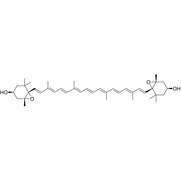
Violaxanthin is a naturally derived diepoxy xanthophyll carotenoid and light-harvesting accessory pigment with biological activities such as antioxidation and anti-inflammation. Violaxanthin transfers excitation energy to chlorophyll a under low-light conditions. Violaxanthin participates in the xanthophyll cycle and dissipates excess light energy in the form of heat, thereby protecting thylakoid membrane lipids from photo-oxidative damage. Violaxanthin exhibits activities including inhibition of lipid peroxidation, quenching of singlet oxygen, scavenging of DPPH free radicals, and scavenging of ABTS+ free radicals. Violaxanthin can be used in antioxidant-related research.
For research use only. We do not sell to patients.
- Purity : 98.00%
- CAS No.: 126-29-4
- Formula: C40H56O4
- Molecular Weight:600.87
-
Storage:
Solution, -20°C, 2 years

To place orders, for customer services and technical support, please contact: MedChemExpress USA
Tel: 609-228-6898 E-mail: [email protected] [email protected]


-
 Biological Activity
-
Chemical Information
- Protocols
- Purity & Documentation
- References
-
 Help & FAQs



Publications Citing Use of MedChemExpress (MCE) Violaxanthin
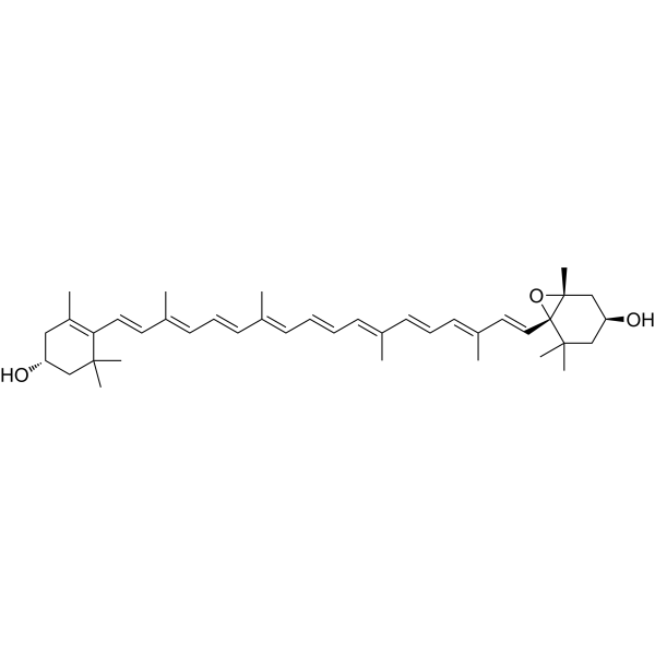
MoreAll Endogenous Metabolite Isoforms
More

Biological Activity
Description
In Vitro
Violaxanthin (HPLC≥90%) (1,500 µmol of photons·m-2·s-1; 15 min) converts into antheraxanthin and zeaxanthin under high light, and this process occurs in leaves of the Arabidopsis npq4-1 mutant; by contrast, no such conversion is observed in leaves of the npq4-1 npq1-2 double mutant, which lacks functional violaxanthin de-epoxidase[1].
Violaxanthin (HPLC≥90%) can be heterologously synthesized in Saccharomyces cerevisiae by reconstructing a β-carotene synthesis pathway synergistically enhanced by truncated ZEP, crtZ and Arabidopsis redox chaperones, with a corresponding yield of 7.3 mg/g cell dry weight. Targeting ZEP to the endoplasmic reticulum via the Cpr5p signal peptide can further increase violaxanthin yield, and the maximum violaxanthin yield of the cCrZEPer engineered strain in Saccharomyces cerevisiae strain YPH500 reaches 31 μg/g cell dry weight[2].
Violaxanthin (HPLC≥90%) is biosynthesized by zeaxanthin (HY-120318) in photosynthetic eukaryotes (including higher plants) through anther xanthin under the action of zeaxanthin cyclooxygenase (ZEP), and participates in the lutein cycle to eliminate excess light energy[2].
Violaxanthin (HPLC≥90%) (1.25-80 µg/mL) exhibits concentration-dependent reducing capacity and DPPH free radical scavenging activity in vitro, with an EC50 of 41.42 µg/mL[3].
MedChemExpress (MCE) has not independently confirmed the accuracy of these methods. They are for reference only.


Chemical Information
-
CAS No. 126-29-4
-
Appearance Liquid
-
Molecular Weight 600.87
-
Formula C40H56O4
-
Color Colorless to light yellow
-
SMILES
C[C@@](O1)(C[C@H](CC2(C)C)O)[C@@]12/C=C/C(C)=C/C=C/C(C)=C/C=C/C=C(C)/C=C/C=C(C)/C=C/[C@]34[C@](O3)(C[C@H](CC4(C)C)O)C
-
Structure Classification
-
Initial Source
-
Shipping
Room temperature in continental US; may vary elsewhere.
-
Storage
Solution, -20°C, 2 years


Publications (1)
-
Journal Impact Factor
-
Most Recent


Purity & Documentation
-
Data Sheet (267 KB)
-
SDS (251 KB)
- English - EN (251 KB)
- Français - FR (251 KB)
- Deutsch - DE (251 KB)
- Norwegian - NO (251 KB)
- Español - ES (251 KB)
- Swedish - SV (251 KB)
- Italian - IT (251 KB)
- Korean - KR (251 KB)
- Portuguese - PT (251 KB)
-
Handling Instructions (2659 KB)
References
[1]. Havaux M, et al. The violaxanthin cycle protects plants from photooxidative damage by more than one mechanism. Proceedings of the National Academy of Sciences of the United States of America. 1999 Jul 20;96(15):8762-7. [Content Brief]
[2]. Takemura M, et al. Violaxanthin: natural function and occurrence, biosynthesis, and heterologous production. Applied microbiology and biotechnology. 2021 Aug;105(16-17):6133-6142. [Content Brief]

Calculators
Concentration (start) × Volume (start) = Concentration (final) × Volume (final)


Violaxanthin Related Classifications
HY-N9625 Related Classifications
 Powered by Bioz